- Corneal transplantation
-
Corneal transplantation Intervention ICD-9-CM 11.6 MeSH D016039 Corneal transplantation, also known as corneal grafting, is a surgical procedure where a damaged or diseased cornea is replaced by donated corneal tissue (the graft) in its entirety (penetrating keratoplasty) or in part (lamellar keratoplasty). The graft has been removed from a recently deceased individual with no known diseases or other factors that may affect the viability of the donated tissue or the health of the recipient. The cornea is the transparent front part of the eye that covers the iris, pupil and anterior chamber. The surgical procedure is performed by ophthalmologists, medical doctors who specialize in eyes, and is often done on an outpatient basis. Donors can be of any age as was Janis Babson, who donated her eyes at age 10.
Contents
Indications
Indications for corneal transplantation include the following:
- Optical: To improve visual acuity by replacing the opaque or distorted host tissue by clear healthy donor tissue. The most common indication in this category is pseudophakic bullous keratopathy, followed by keratoconus, corneal degeneration, keratoglobus and dystrophy, as well as scarring due to keratitis and trauma.
- Tectonic/reconstructive: To preserve corneal anatomy and integrity in patients with stromal thinning and descemetoceles, or to reconstruct the anatomy of the eye, e.g. after corneal perforation.
- Therapeutic: To remove inflamed corneal tissue unresponsive to treatment by antibiotics or anti-virals.
- Cosmetic: To improve the appearance of patients with corneal scars that have given a whitish or opaque hue to the cornea.
Pre-operative examination
In most instances, the patient will meet with their ophthalmologist for an examination in the weeks or months preceding the surgery. During the exam, the ophthalmologist will examine the eye and diagnose the condition. The doctor will then discuss the condition with the patient, including the different treatment options available. The doctor will also discuss the risks and benefits of the various options. If the patient elects to proceed with the surgery, the doctor will have the patient sign an informed consent form. The doctor might also perform a physical examination and order lab tests, such as blood work, X-rays, or an EKG.
The surgery date and time will also be set, and the patient will be told where the surgery will take place. The surgery only takes place when the best corresponding donor tissue is found. This can take weeks and months.
Procedure
On the day of the surgery, the patient arrives to either a hospital or an outpatient surgery center, where the procedure will be performed. The patient is given a brief physical examination by the surgical team and is taken to the operating room. In the OR, the patient lies down on an operating table and is either given general anesthesia, or local anesthesia and a sedative.
With anesthesia induced, the surgical team prepares the eye to be operated on and drapes the face around the eye. An eyelid speculum is placed to keep the lids open, and some lubrication is placed on the eye to prevent drying. In children, a metal ring is stitched to the sclera which will provide support of the sclera during the procedure.
Penetrating keratoplasty
A trephine (a circular cutting device) is then placed over the cornea and is used by the surgeon to cut the host cornea, which removes a circular disc of the patient cornea. The trephine is then removed and the surgeon cuts a circular graft (a "button") from the donor cornea. Once this is done, the surgeon returns to the patient's eye and removes the host cornea.
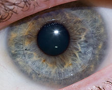
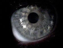
The donor cornea is then brought into the surgical field and maneuvered into place with forceps. Once in place, the surgeon will fasten the cornea to the eye with a running stitch (as used in the upper image above) or a multiple interrupted stitches (as in the lower image). The surgeon then reforming the anterior chamber with a sterile solution injected by a cannula, then testing that it's watertight by placing a dye on the wound exterior.
Antibiotic eyedrops placed, the eye is patched, and the patient is taken to a recovery area while the effects of the anesthesia wear off. The patient typically goes home following this and sees the doctor the following day for the first post operative appointment.
Lamellar keratoplasty
This procedure consists in leaving just the patient's own Descemet membrane and endothelium, while transplanting approximately 95% of the cornea. The great advantage of this technique is the virtually "no rejection" post-op. The main disadvantage is that the visual acuity is not as sharp as it is with the full cornea transplantation penetrating keratoplasty). The final visual acuity is usually around 20/40.
Risks
While the cornea is avascular, there is still a potential for some blood loss, usually from suturing the metal ring to the sclera. Any blood loss is typically less than 2 ml (0.07 imp fl oz; 0.07 US fl oz).
There is also a risk of infection. Since the cornea has no blood vessels (it takes its nutrients from the aqueous humor) it heals much more slowly than a cut on the skin. While the wound is healing, it is possible that it might become infected by various microorganisms. This risk is minimized by antibiotic prophylaxis (using antibiotic eyedrops, even when no infection exists).
Graft failure can occur at any time after the cornea has been transplanted, even years or decades later. The causes can vary, though it is usually due to new injury or illness. Treatment can be either medical or surgical, depending on the individual case. An early, technical cause of failure, may be an excessively tight stitch cheesewiring through the sclera.
Prognosis
When the primary purpose of a cornea transplant is to improve visual acuity, the prognosis is dependent upon whether the rest of the eye is healthy. If it is, then it should be possible to recover normal vision.
History
The first cornea transplant was performed in 1905 by Eduard Zirm (Olomouc Eye Clinic, now Czech Republic), making it one of the first types of transplant surgery successfully performed. Another pioneer of the operation was Ramon Castroviejo. Russian eye surgeon Vladimir Filatov's attempts at tranplanting cornea started with the first try in 1912 and were continued, gradually improving until at 6 May 1931 he successfully grafted a patient using corneal tissue from a deceased person.[1] He widely reported of another transplant in 1936, disclosing his technique in full detail.[2] In 1936, Castroviejo did a first transplantation in an advanced case of keratoconus, achieving significant improvement in patient's vision.[3][4]
Advances in operating microscopes enabled surgeons to have a more magnified view of the surgical field, while advances in materials science enabled them to use sutures finer than a human hair.
Instrumental in the success of cornea transplants were the establishment of eye banks. These are organizations located throughout the world to coordinate the distribution of donated corneas to surgeons, as well as providing eyes for research. Some eye banks also distribute other anatomical gifts.
Synthetic corneas
Main article: KeratoprosthesisBoston keratoprosthesis
The Boston keratoprosthesis is the most widely used synthetic cornea to date with over 900 procedures performed worldwide in 2008. The Boston KPro was developed at the Massachusetts Eye and Ear Infirmary under the leadership of Claes Dohlman, MD, PhD.[5]
AlphaCor
In cases where there have been several graft failures or the risk for keratoplasty is high, synthetic corneas can substitute successfully for donor corneas. Such a device contains a peripheral skirt and a transparent central region. These two parts are connected on a molecular level by an interpenetrating polymer network, made from poly-2-hydroxyethyl methacrylate (pHEMA). AlphaCor is an FDA-approved type of synthetic cornea measuring 7.0 mm in diameter and 0.5 mm in thickness. The main advantages of synthetic corneas are that they are biocompatible, and the network between the parts and the device prevents complications that could arise at their interface. The probability of retention in one large study was estimated at 62% at 2 years follow-up.[6]
References
- ^ Vladimir Filatov, peoples.ru
- ^ Eye repair - TIME, Monday, Apr. 13, 1936
- ^ Castroviejo R (1948). "Keratoplasty for the Treatment of Keratoconus". Trans Am Ophthalmol Soc 46: 127–53. PMC 1312756. PMID 16693468. http://www.pubmedcentral.nih.gov/articlerender.fcgi?tool=pmcentrez&artid=1312756.
- ^ Castroviejo, R.: International Abstract of Surgery, 65:5, December, 1937.
- ^ "The Boston Keratoprosthesis". Cataract & Refractive Surgery Today. September 2009. http://bmctoday.net/crstoday/2009/09/article.asp?f=CRST0909_16.php.
- ^ Hicks CR, Crawford GJ, Dart JK, Grabner G, Holland EJ, Stulting RD, Tan DT, Bulsara M (October 2006). "AlphaCor: Clinical outcomes". Cornea 25 (9): 1034–42. doi:10.1097/01.ico.0000229982.23334.6b. PMID 17133049. http://meta.wkhealth.com/pt/pt-core/template-journal/lwwgateway/media/landingpage.htm?an=00003226-200610000-00007.
Categories:- Tissue transplants
- Eye surgery
Wikimedia Foundation. 2010.


