- Symphysis menti
-
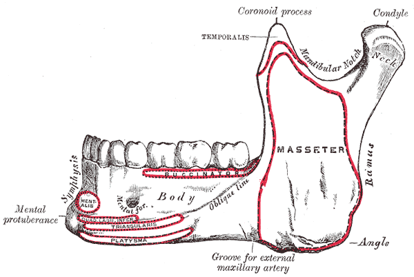
Bone: Symphysis menti 
Mandible. Outer surface. Side view. (Symphysis menti visible at left). Latin symphysis mandibulae Gray's subject #44 172 The external surface of the mandible is marked in the median line by a faint ridge, indicating the symphysis menti, mandibular symphysis, or line of junction of the two pieces of which the bone is composed at an early period of life.
This ridge divides below and encloses a triangular eminence, the mental protuberance, the base of which is depressed in the center but raised on either side to form the mental tubercle.
It serves as the origin for the Geniohyoid and the Genioglossus.
See also
Additional images
-
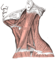
Muscles of the neck. Lateral view.
-

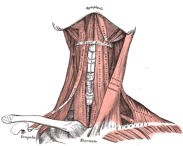
Muscles of the neck. Anterior view.
This article was originally based on an entry from a public domain edition of Gray's Anatomy. As such, some of the information contained within it may be outdated.
Bones of head and neck: the facial skeleton of the skull (TA A02.1.08–15, GA 2.156–177) Maxilla SurfacesProcessesOtherZygomatic Palatine FossaePlatesProcessesMandible external surface (Symphysis menti, Lingual foramen, Mental protuberance, Mental foramen, Mandibular incisive canal) · internal surface (Mental spine, Mylohyoid line, Sublingual fovea, Submandibular fovea) · Alveolar part of mandibleMinor/
noseNasal bone: Internasal suture · Nasal foramina
Inferior nasal concha: Ethmoidal process · Maxillary process
Vomer: Vomer anterior · Synostosis vomerina · Vomer posterior (Wing)
Lacrimal: Posterior lacrimal crest · Lacrimal groove · Lacrimal hamulusCategories:- Bones of the head and neck
- Musculoskeletal system stubs
-

Wikimedia Foundation. 2010.