- Condyloid process
-
Bone: Condyloid process 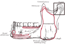
Mandible. Outer surface. Side view. (Condyle and neck labeled at upper right.) Latin processus condylaris mandibulae Gray's subject #44 174 The condyloid process is part of the mandible and is thicker than the coronoid, and consists of two portions: the condyle, and the constricted portion which supports it, the neck.
Contents
Condyle
The condyle presents an articular surface for articulation with the articular disk of the temporomandibular joint; it is convex from before backward and from side to side, and extends farther on the posterior than on the anterior surface.
Its long axis is directed medialward and slightly backward, and if prolonged to the middle line will meet that of the opposite condyle near the anterior margin of the foramen magnum.
At the lateral extremity of the condyle is a small tubercle for the attachment of the temporomandibular ligament.
The articular surface of the condyle is covered by fibrous tissue, and interfaces with an articular disk (or meniscus) of avascular, non-innervated fibrous tissue (collagen, fibroblasts). When the mouth is closed the meniscus is bordered medially and superiorly by the glenoid fossa of the petrous portion of the temporal bone. When the mouth is opened maximally, the meniscus is distracted anteriorly and inferiorly along the slope of the inferior portion of the temporal bone towards the tubercle, or articular eminence, in order to remain interposed between the condyle and the temoporal bone in all jaw positions.
Neck
The neck is flattened from before backward, and strengthened by ridges which descend from the forepart and sides of the condyle.
Its posterior surface is convex; its anterior presents a depression for the attachment of the Pterygoideus externus (lateral pterygoid muscle).
See also
- Ramus mandibulae
Additional images
-

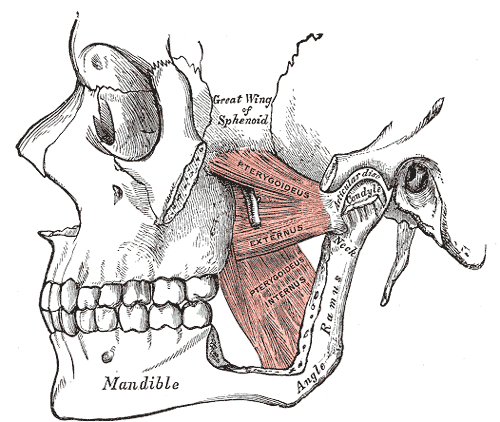
The Pterygoidei; the zygomatic arch and a portion of the ramus of the mandible have been removed.
-

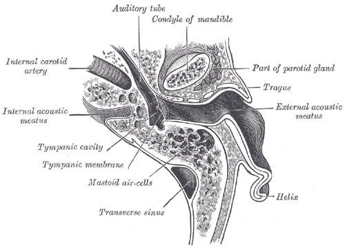
Horizontal section through left ear; upper half of section.
External links
- lesson1 at The Anatomy Lesson by Wesley Norman (Georgetown University)
- SUNY Labs 22:os-1001 - "Osteology of the Skull: Mandible of Intact Skull"
- MeSH Mandibular+condyle
- Roche Lexicon - illustrated navigator, at Elsevier 34256.000-2
This article was originally based on an entry from a public domain edition of Gray's Anatomy. As such, some of the information contained within it may be outdated.
Bones of head and neck: the facial skeleton of the skull (TA A02.1.08–15, GA 2.156–177) Maxilla SurfacesProcessesOtherZygomatic Palatine FossaePlatesProcessesMandible external surface (Symphysis menti, Lingual foramen, Mental protuberance, Mental foramen, Mandibular incisive canal) · internal surface (Mental spine, Mylohyoid line, Sublingual fovea, Submandibular fovea) · Alveolar part of mandibleMylohyoid groove (Mandibular canal, Lingula) · Mandibular foramen · Angle
Coronoid process · Mandibular notch · Condyloid process · Pterygoid foveaMinor/
noseNasal bone: Internasal suture · Nasal foramina
Inferior nasal concha: Ethmoidal process · Maxillary process
Vomer: Vomer anterior · Synostosis vomerina · Vomer posterior (Wing)
Lacrimal: Posterior lacrimal crest · Lacrimal groove · Lacrimal hamulusCategories:- Bones of the head and neck
- Musculoskeletal system stubs

Wikimedia Foundation. 2010.