- Cavernous plexus
Infobox Nerve
Name = PAGENAME
Latin = plexus cavernosus
GraySubject = 215
GrayPage = 978
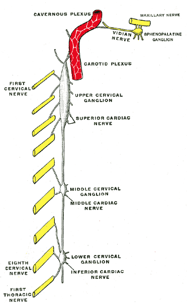
Caption = Diagram of the cervical sympathetic. (Cavernous plexus labeled at top.)
Caption2 =
Innervates =
BranchFrom =
BranchTo =
MeshName =
MeshNumber =
The cavernous plexus is situated below and medial to that part of theinternal carotid artery which is placed by the side of thesella turcica in thecavernous sinus , and is formed chiefly by themedial division of theinternal carotid nerve .It communicates with the
oculomotor , thetrochlear , theophthalmic and theabducent nerves, and with theciliary ganglion , and distributes filaments to the wall of theinternal carotid artery .The branch of communication with the oculomotor nerve joins that nerve at its point of division; the branch to the trochlear nerve joins it as it lies on the lateral wall of the cavernous sinus; other filaments are connected with the under surface of the ophthalmic nerve; and a second filament joins the abducent nerve.
=Additional
Wikimedia Foundation. 2010.