- Osteopontin
-
Secreted phosphoprotein 1 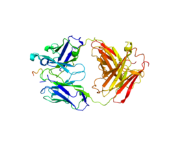
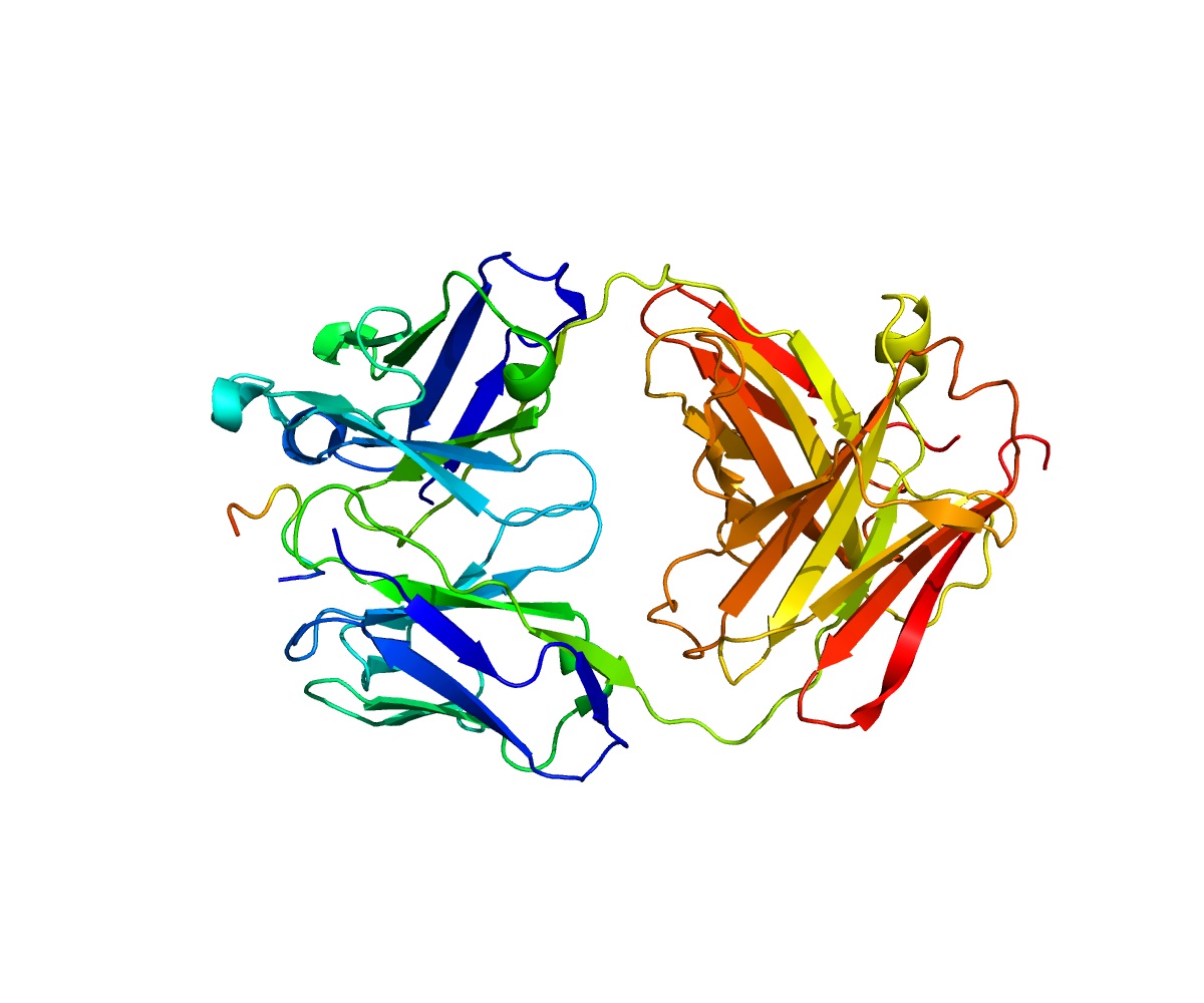
Rendering based on PDB 3CXD.Available structures PDB 3CXD, 3DSF Identifiers Symbols SPP1; BNSP; BSPI; ETA-1; MGC110940; OPN External IDs OMIM: 166490 MGI: 98389 HomoloGene: 20156 GeneCards: SPP1 Gene Gene Ontology Molecular function • cytokine activity
• extracellular matrix bindingCellular component • extracellular region
• extracellular region
• extracellular space
• cytoplasm
• apical part of cellBiological process • ossification
• cell adhesion
• embryo implantation
• positive regulation of cell-substrate adhesion
• biomineral tissue development
• response to vitamin D
• decidualizationSources: Amigo / QuickGO RNA expression pattern 
More reference expression data Orthologs Species Human Mouse Entrez 6696 20750 Ensembl ENSG00000118785 ENSMUSG00000029304 UniProt P10451 Q3TND2 RefSeq (mRNA) NM_000582.2 NM_009263 RefSeq (protein) NP_000573.1 NP_033289 Location (UCSC) Chr 4:
88.9 – 88.9 MbChr 5:
104.86 – 104.87 MbPubMed search [1] [2] Osteopontin (OPN), also known as bone sialoprotein I (BSP-1 or BNSP), early T-lymphocyte activation (ETA-1), secreted phosphoprotein 1 (SPP1), 2ar and Rickettsia resistance (Ric), is a human gene product,[1] which is also conserved in other species. Osteopontin is a SIBLING glycoprotein that was first identified in 1986 in osteoblasts.
The prefix of the word "osteo" indicates that the protein is expressed in bone, although it is also expressed in other tissues. The suffix "-pontin" is derived from "pons," the Latin word for bridge, and signifies osteopontin's role as a linking protein. Osteopontin is an extracellular structural protein and therefore an organic component of bone. Synonyms for this protein include sialoprotein I and 44K BPP (bone phosphoprotein).
The gene has 7 exons, spans 5 kilobases in length and in humans it is located on the long arm of chromosome 4 region 13 (4q13). The protein is composed of ~300 amino acids residues and has ~30 carbohydrate residues attached including 10 sialic acid residues, which are attached to the protein during post-translational modification in the Golgi apparatus. The protein is rich in acidic residues: 30-36% are either aspartic or glutamic acid.
Contents
Structure
General structure
OPN is a highly negatively charged, extracellular matrix protein that lacks an extensive secondary structure.[2] It is composed of about 300 amino acids (297 in mouse; 314 in human) and is expressed as a 33-kDa nascent protein; there are also functionally important cleavage sites. OPN can go through posttranslational modifications which increase its apparent molecular weight to about 44 kDa.[3] The OPN gene is composed of 7 exons, 6 of which contain coding sequence.[4][5] The first two exons contain the 5' untranslated region (5' UTR).[6] Exons 2, 3, 4, 5, 6, and 7 code for 17, 13, 27, 14, 108 and 134 amino acids, respectively.[6] All intron-exon boundaries are of the phase 0 type, thus alternative exon splicing maintains the reading frame of the OPN gene.
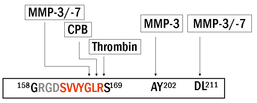
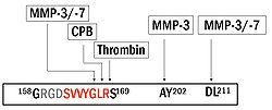 Figure 1. Proteolytic cleavage sites for full length osteopontin (OPN-FL). Thrombin exposes the cleaved epitope SVVYGLR (OPN-R), and then CPB removes the c-terminal arginine from OPN-R. The cleaved epitope has a non-RGD domain, which binds to integrin receptors (α4β1, α9β1, and α9β4). Next to the cleaved epitope, there is a RGD domain which interacts with other integrin receptors (αvβ1,3,5, and α5β1).
Isoforms
Full length OPN (OPN-FL) can be modified by thrombin cleavage, which exposes a cryptic sequence, SVVYGLR on the cleaved form of the protein known as OPN-R (Fig. 1). This thrombin-cleaved OPN (OPN-R) exposes an epitope for integrin receptors of α4β1, α9β1, and α9β4.[7][8] These integrin receptors are present on a number of immune cells such as mast cells,[9] neutrophils,[10] and T cells. It is also expressed by monocytes and macrophages.[11] Upon binding these receptors, cells use several signal transduction pathways to elicit immune responses in these cells (See Section 3 for more detail). OPN-R can be further cleaved by Carboxypeptidase B (CPB) by removal of C-terminal arginine and become OPN-L (Fig. 2). The function of OPN-L is largely unknown.
It appears an intracellular variant of OPN (iOPN) is involved in a number of cellular processes including migration, fusion and motility.[12][13][14][15] Intracellular OPN is generated using an alternative translation start site on the same mRNA species used to generate the extracellular isoform.[16] This alternative translation start site is downstream of the N-terminal endoplasmic reticulum-targeting signal sequence, thus allowing cytoplasmic translation of OPN.
Various human cancers, including breast cancer, have been observed to express splice variants of OPN.[17][18] The cancer specific splice variants are osteopontin-a, osteopontin-b and osteopontin-c. Exon 5 is lacking from osteopontin-b, whereas osteopontin-c lacks exon 4.[17] Osteopontin-c has been suggested to facilitate the anchorage-independent phenotype of some human breast cancer cells due to its inability to associate with the extracellular matrix.[17]
Biosynthesis
Osteopontin is biosynthesized by a variety of tissue types including fibroblasts[19] preosteoblasts, osteoblasts, osteocytes, odontoblasts, some bone marrow cells, hypertrophic chondrocytes, dendritic cells, macrophages,[20] smooth muscle,[21] skeletal muscle myoblasts,[22] endothelial cells, and extraosseous (non-bone) cells in the inner ear, brain, kidney, deciduum, and placenta. Synthesis of osteopontin is stimulated by calcitriol (1,25-dihydroxy-vitamin D3).
Regulation
Regulation of the osteopontin gene is incompletely understood. Different cell types may differ in their regulatory mechanisms of the OPN gene. OPN expression in bone predominantly occurs by osteoblasts and osteocyctes (bone-forming cells) as well as osteoclasts (bone-resorbing cells).[23] Runx2 (aka Cbfa1) and osterix (Osx) transcription factors are required for the expression of OPN [24] Runx2 and Osx bind promoters of osteoblast-specific genes such as Col1α1, Bsp, and Opn and upregulate transcription.[25]
Hypocalcemia and hypophosphatemia (instances that stimulate kidney proximal tubule cells to produce calcitriol (1α,25-dihydroxyvitamin D3)) lead to increases in OPN transcription, translation and secretion.[26] This is due to the presence of a high-specificity vitamin D response element (VDRE) in the OPN gene promoter.[27][28][29]
Extracellular inorganic phosphate (ePi) has also been identified as a modulator of OPN expression.[30]
Stimulation of OPN expression also occurs upon exposure of cells to pro-inflammatory cytokines,[31] classical mediators of acute inflammation (e.g. tumour necrosis factor α [TNFα], infterleukin-1β [IL-1β]), angiotensin II, transforming growth factor β (TGFβ) and parathyroid hormone (PTH),[32][33] although a detailed mechanistic understanding of these regulatory pathways are not yet known. Hyperglycemia and hypoxia are also known to increase OPN expression.[32][34][35]
Biological function
Role in bone remodeling
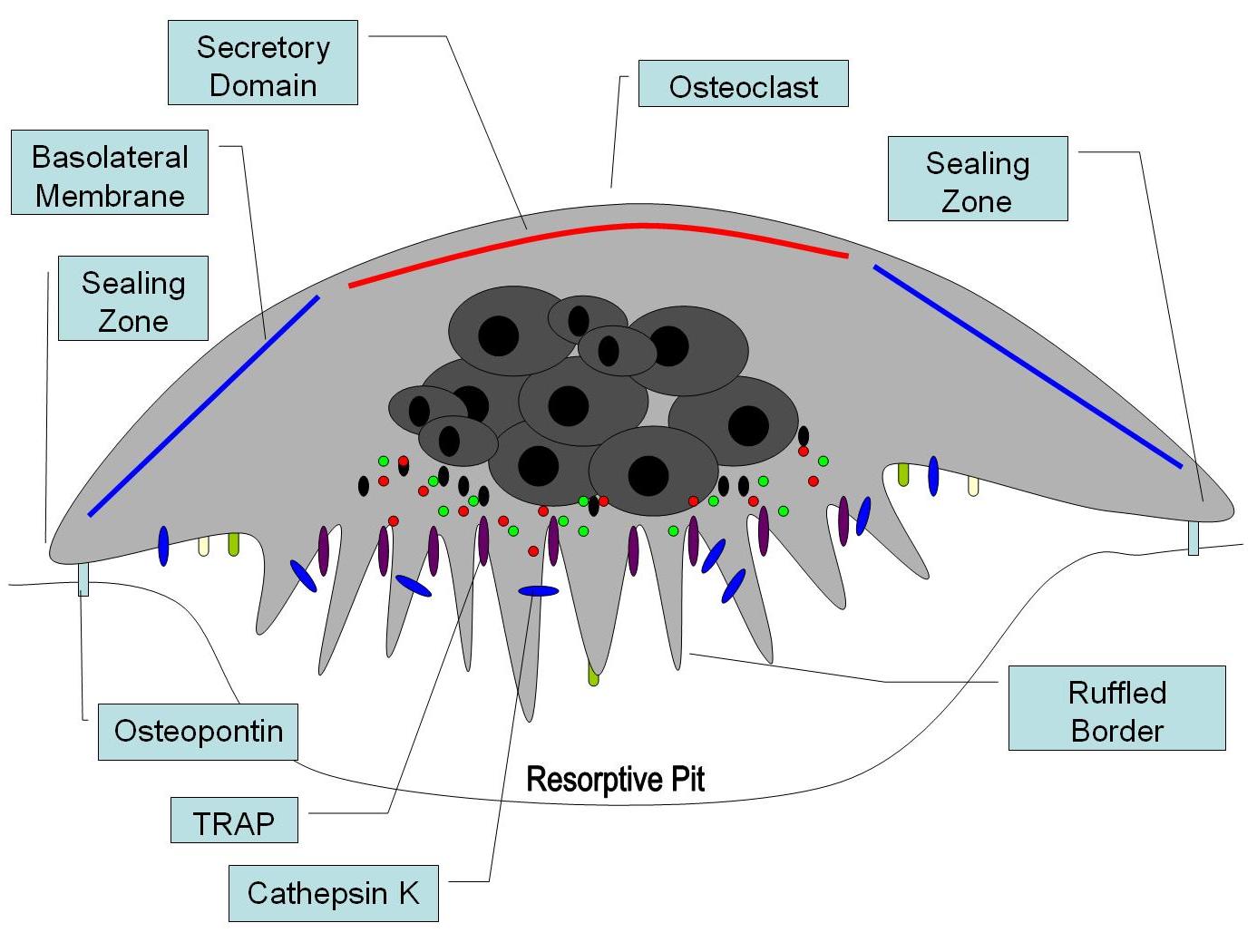
Osteopontin has been implicated as an important factor in bone remodeling.[36] Specifically, research suggests it plays a role in anchoring osteoclasts to the mineral matrix of bones.[9] The organic part of bone is about 20% of the dry weight, and counts in, other than osteopontin, collagen type I, osteocalcin, osteonectin, bone sialo protein and alkaline phosphatase. Collagen type I counts for 90% of the protein mass. The inorganic part of bone is the mineral hydroxyapatite, Ca10(PO4)6(OH)2. Loss of this mineral may lead to osteoporosis, as the bone is depleted for calcium if this is not supplied in the diet.
OPN serves to initiate the process by which osteoclasts develop their ruffled borders to begin bone resorption. It is also found in urine, where it inhibits kidney stone formation.
Role in immune functions
As discussed, OPN binds to several integrin receptors including α4β1, α9β1, and α9β4 expressed by leukocytes. These receptors have been well-established to function in cell adhesion, migration, and survival in these cells. Therefore, recent research efforts have focused on the role of OPN in mediating such responses.
Osteopontin (OPN) is expressed in a range of immune cells, including macrophages, neutrophils, dendritic cells, and T and B cells, with varying kinetics. OPN is reported to act as an immune modulator in a variety of manners.[2] Firstly, it has chemotactic properties, which promote cell recruitment to inflammatory sites. It also functions as an adhesion protein, involved in cell attachment and wound healing. In addition, OPN mediates cell activation and cytokine production, as well as promoting cell survival by regulating apoptosis.[2] The following examples are found.[2]
Chemotaxis
OPN plays an important role in neutrophil recruitment in alcoholic liver disease.[10][37] OPN is important for the migration of neutrophil in vitro.[38] In addition, OPN recruits inflammatory cells to arthritis joints in the collagen-induced arthritis model of rheumatoid arthritis.[39][40] A recent in vitro study in 2008 has found that OPN plays a role in mast cell migration.[41] Here OPN knock-out mast cells were cultured and they observed a decreased level of chemotaxis in these cells compared to wildtype mast cells. OPN was also found to act as a macrophage chemotactic factor.[42] In this study, researchers looked at the accumulation of macrophages in the brain of rhesus monkeys and found that OPN prevented macrophages from leaving the accumulation site, indicating an increased level of chemotaxis.
Cell activation
Activated T cells are promoted by IL-12 to differentiate towards the Th1 type, producing cytokines including IL-12 and IFNγ. OPN inhibits production of the Th2 cytokine IL-10, which leads to enhanced Th1 response. OPN influences cell-mediated immunity and has Th1 cytokine functions. It enhances B cell immunoglobulin production and proliferation.[2] Recent studies in 2008 suggest that OPN also induces mast cell degranulation.[41] The researchers here observed that IgE-mediated anaphylaxis was significantly reduced in OPN knock-out mice compared to wild type mice. The role of OPN in activation of macrophages has also been implicated in a cancer study, when researchers discovered that OPN-producing tumors were able to induce macrophage activation compared to OPN-deficient tumors.[43]
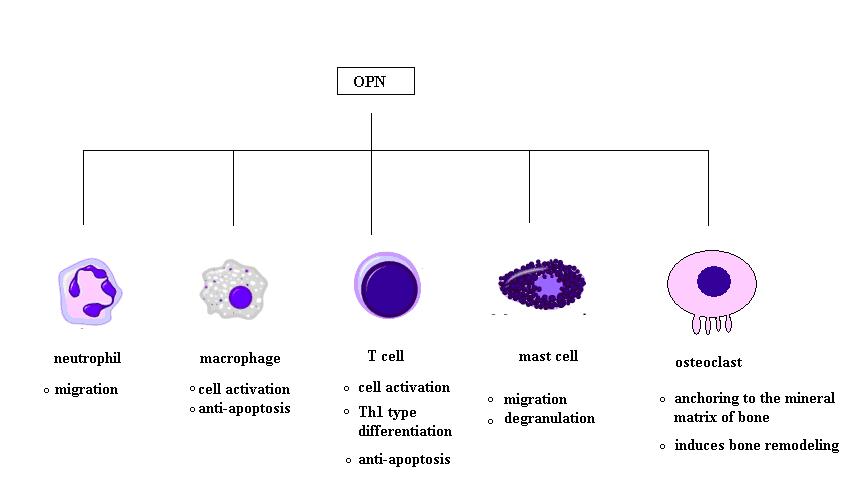
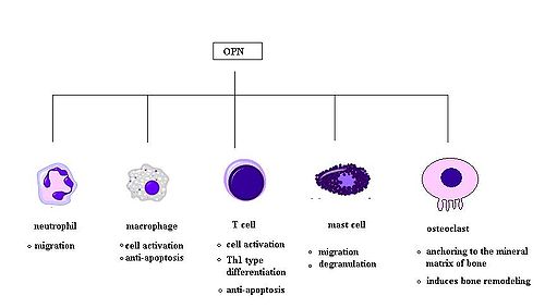 Fig 2. Known immunologic functions of OPN. OPN binds to several integrin receptors including α4β1, α9β1, and α9β4 expressed by leukocytes and are known to induce cell adhesion, migration, and survival in immune cells including neutrophils, macrophages, T cells, mast cells, and osteoclasts.
Apoptosis
OPN is an important anti-apoptotic factor in many circumstances. OPN blocks the activation-induced cell death of macrophages and T cells as well as fibroblasts and endothelial cells exposed to harmful stimuli.[44][45] OPN prevents non-programmed cell death in inflammatory colitis.[46]
Potential clinical application
The fact that OPN interacts with multiple cell surface receptors which are ubiquitously expressed makes it an active player in many physiological and pathological processes including wound healing, bone turnover, tumorigenesis, inflammation, ischemia and immune responses1. Therefore, manipulation of plasma OPN levels may be useful in the treatment of autoimmune diseases, cancer metastasis, osteoporosis and some forms of stress.[2]
Role in autoimmune diseases
OPN has been implicated in pathogenesis of rheumatoid arthritis. For instance, researchers found that OPN-R, the thrombin-cleaved form of OPN, was elevated in the rheumatoid arthritis joint16. However, the role of OPN in rheumatoid arthritis is still unclear. One group found that OPN knock-out mice were protected against arthritis.[47] while others were not able to reproduce this observation.[48] OPN has been found to play a role in other autoimmune diseases including autoimmune hepatitis, allergic airway disease, and multiple sclerosis.[49]
Role in cancers and inflammatory diseases
It has been shown that OPN drives IL-17 production;[50] OPN is overexpressed in a variety of cancers, including lung cancer, breast cancer, colorectal cancer, stomach cancer, ovarian cancer, papillary thyroid carcinoma, melanoma and pleural mesothelioma; OPN contributes both glomerulonephritis and tubulointerstitial nephritis; and OPN is found in atheromatous plaques within arteries. Thus, manipulation of plasma OPN levels may be useful in the treatment of autoimmune diseases, cancer metastasis, osteoporosis and some forms of stress.[2]
Research has implicated osteopontin in excessive scar-forming and a gel has been developed to inhibit its effect.[51]
Role in allergy and asthma
Osteopontin has recently been associated with allergic inflammation and asthma. Using a murine model of allergic inflammation, it was demonstrated that OPN-s, the secreted form of OPN, exerts opposing effects on mouse Th2 effector responses and subsequent allergic airway disease: pro-inflammatory at primary systemic sensitization, and anti-inflammatory during secondary pulmonary antigenic challenge, mainly through the regulation of different dendritic cell subsets.[52] OPN deficiency was also reported to protect against remodeling and bronchial hyperresponsiveness (BHR), again using a chronic allergen-challenge model of airway remodeling.[53] Furthermore, it was recently demonstrated that OPN expression is upregulated in human asthma, is associated with remodeling changes and its subepithelial expression correlates to disease severity.[54]
Role in muscle disease and injury
Evidence is accumulating that suggests that osteopontin plays a number of roles in diseases of skeletal muscle, such as Duchenne muscular dystrophy. Osteopontin has been described as a component of the inflammatory environment of dystrophic and injured muscles [55][56] [57] [22], and has also been shown to increase scarring of diaphragm muscles of aged dystrophic mice [58]. A recent study has identified osteopontin as a determinant of disease severity in patients with Duchenne muscular dystrophy[59]. This study found that a mutation in the osteopontin gene promoter, known to cause low levels of osteopontin expression, is associated with a decrease in age to loss of ambulation and muscle strength in patients with Duchenne muscular dystrophy.
References
- ^ "Entrez Gene: SPP1 secreted phosphoprotein 1". http://www.ncbi.nlm.nih.gov/sites/entrez?db=gene&cmd=retrieve&dopt=default&list_uids=6696&rn=1.
- ^ a b c d e f g Wang KX, Denhardt DT (2008). "Osteopontin: role in immune regulation and stress responses". Cytokine Growth Factor Rev. 19 (5-6): 333–45. doi:10.1016/j.cytogfr.2008.08.001. PMID 18952487.
- ^ Rangaswami H, Bulbule A, Kundu GC (February 2006). "Osteopontin: role in cell signaling and cancer progression". Trends Cell Biol. 16 (2): 79–87. doi:10.1016/j.tcb.2005.12.005. PMID 16406521.
- ^ Young. M.F., Kerr, J.M., Termine, J.D., Wewer, U.M., Wang, M.G., McBride, O.W., Fisher, L.W. (August 1990). "cDNA cloning, mRNA distribution and heterogeneity, chromosomal location, and RFLP analysis of human osteopontin (OPN)". Genomics. 7 (1): 491–502. doi:10.1016/0888-7543(90)90191-V. PMID 1974876.
- ^ Kiefer, M.C., Bauer, D.M., Barr, P.J. (April 1989). "The cDNA and derived amino acid sequence for human osteopontin.". Nucleic Acids Res. 17 (1): 3306. doi:10.1093/nar/17.8.3306. PMC 317745. PMID 2726470. http://www.pubmedcentral.nih.gov/articlerender.fcgi?tool=pmcentrez&artid=317745.
- ^ a b Crosby, A.H., Edwards, S.J., Murray, J.C., Dixon, M.J. (May 1995). "Genomic organization of the human osteopontin gene: exclusion of the locus from a causative role in the pathogenesis of dentinogenesis imperfecta type II". Genomics 27 (1): 155–160. doi:10.1006/geno.1995.1018. PMID 7665163.
- ^ Laffón A, García-Vicuña R, Humbría A, et al. (August 1991). "Upregulated expression and function of VLA-4 fibronectin receptors on human activated T cells in rheumatoid arthritis". J. Clin. Invest. 88 (2): 546–52. doi:10.1172/JCI115338. PMC 295383. PMID 1830891. http://www.pubmedcentral.nih.gov/articlerender.fcgi?tool=pmcentrez&artid=295383.
- ^ Seiffge D (December 1996). "Protective effects of monoclonal antibody to VLA-4 on leukocyte adhesion and course of disease in adjuvant arthritis in rats". J. Rheumatol. 23 (12): 2086–91. PMID 8970045.
- ^ a b Reinholt FP, Hultenby K, Oldberg A, Heinegård D (June 1990). "Osteopontin--a possible anchor of osteoclasts to bone". Proc. Natl. Acad. Sci. U.S.A. 87 (12): 4473–5. doi:10.1073/pnas.87.12.4473. PMC 54137. PMID 1693772. http://www.pubmedcentral.nih.gov/articlerender.fcgi?tool=pmcentrez&artid=54137.
- ^ a b Banerjee A, Apte UM, Smith R, Ramaiah SK (March 2006). "Higher neutrophil infiltration mediated by osteopontin is a likely contributing factor to the increased susceptibility of females to alcoholic liver disease". J. Pathol. 208 (4): 473–85. doi:10.1002/path.1917. PMID 16440289.
- ^ Sodek J, Batista Da Silva AP, Zohar R (May 2006). "Osteopontin and mucosal protection". J. Dent. Res. 85 (5): 404–15. doi:10.1177/154405910608500503. PMID 16632752. http://jdr.iadrjournals.org/cgi/pmidlookup?view=long&pmid=16632752.
- ^ Zohar R, Suzuki N, Suzuki K, Arora P, Glogauer M, McCulloch CA, Sodek J (July 2000). "Intracellular osteopontin is an integral component of the CD44-ERM complex involved in cell migration". J Cell Physiol 184 (1): 118–130. doi:10.1002/(SICI)1097-4652(200007)184:1<118::AID-JCP13>3.0.CO;2-Y. PMID 10825241.
- ^ Suzuki K, Zhu B, Rittling SR, Denhardt DT, Goldberg HA, McCulloch CA, Sodek J (August 2002). "Colocalization of intracellular osteopontin with CD44 is associated with migration, cell fusion, and resorption in osteoclasts". J Bone Miner Res 17 (1): 1486–1497. doi:10.1359/jbmr.2002.17.8.1486. PMID 12162503.
- ^ Zhu B, Suzuki K, Goldberg HA, Rittling SR, Denhardt DT, McCulloch CA, Sodek J (January 2004). "Osteopontin modulates CD44-dependent chemotaxis of peritoneal macrophages through G-protein-coupled receptors: evidence of a role for an intracellular form of osteopontin". Journal of Cellular Physiology 198 (1): 155–167. doi:10.1002/jcp.10394. PMID 14584055.
- ^ Junaid A, Moon MC, Harding GE, Zahradka P (February 2007). "Osteopontin localizes to the nucleus of 293 cells and associates with polo-like kinase-1". Am J Physiol Cell Physiol 292 (1): 919–926. doi:10.1152/ajpcell.00477.2006. PMID 17005603.
- ^ Shinohara ML, Kim HJ, Kim JH, Garcia VA, Cantor H (May 2008). "Alternative translation of osteopontin generates intracellular and secreted isoforms that mediate distinct biological activities in dendritic cells". Proc Natl Acad Sci USA 105 (1): 7235–7239. doi:10.1073/pnas.0802301105. PMC 2438233. PMID 18480255. http://www.pubmedcentral.nih.gov/articlerender.fcgi?tool=pmcentrez&artid=2438233.
- ^ a b c He B, Mirza M, Weber GF (April 2006). "An osteopontin splice variant induces anchorage independence in human breast cancer cells". Oncogene 25 (1): 2192–2202. doi:10.1038/sj.onc.1209248. PMID 16288209.
- ^ Mirza M, Shaughnessy E, Hurley JK, Vanpatten KA, Pestano GA, He B, Weber GF (February 2008). "Osteopontin-c is a selective marker of breast cancer". Int J Cancer 122 (1): 889–897. doi:10.1002/ijc.23204. PMID 17960616.
- ^ Ashizawa N, Graf K, Do YS, et al. (November 1996). "Osteopontin is produced by rat cardiac fibroblasts and mediates A(II)-induced DNA synthesis and collagen gel contraction". J. Clin. Invest. 98 (10): 2218–27. doi:10.1172/JCI119031. PMC 507670. PMID 8941637. http://www.pubmedcentral.nih.gov/articlerender.fcgi?tool=pmcentrez&artid=507670.
- ^ Murry CE, Giachelli CM, Schwartz SM, Vracko R (December 1994). "Macrophages express osteopontin during repair of myocardial necrosis". Am. J. Pathol. 145 (6): 1450–62. PMC 1887495. PMID 7992848. http://www.pubmedcentral.nih.gov/articlerender.fcgi?tool=pmcentrez&artid=1887495.
- ^ Ikeda T, Shirasawa T, Esaki Y, Yoshiki S, Hirokawa K (December 1993). "Osteopontin mRNA is expressed by smooth muscle-derived foam cells in human atherosclerotic lesions of the aorta". J. Clin. Invest. 92 (6): 2814–20. doi:10.1172/JCI116901. PMC 288482. PMID 8254036. http://www.pubmedcentral.nih.gov/articlerender.fcgi?tool=pmcentrez&artid=288482.
- ^ a b Uaesoontrachoon K, Yoo HJ, Tudor E, Pike RN, Mackie EJ, Pagel CN (April 2008). "Osteopontin and skeletal muscle myoblasts: Association with muscle regeneration and regulation of myoblast function in vitro". Int. J. Biochem. Cell Biol. 40 (10): 2303–14. doi:10.1016/j.biocel.2008.03.020. PMID 18490187.
- ^ Merry, K., Dodds, R., Littlewood, A., Gowen, M. (April 1993). "Expression of Osteopontin mRNA by osteoclasts and osteoblasts in modelling adult human bone". J Cell Sci 104 (4): 1013–1020. PMID 8314886.
- ^ Nakashima, K., Zhou, X., Kunkel, G., Zhang, Z., Deng, J.M., Behringer, R.R., de Crombrugghe, B. (January 2002). "The novel zinc finger-containing transcription factor osterix is required for osteoblast differentiation and bone formation". Cell 108 (1): 17–29. doi:10.1016/S0092-8674(01)00622-5. PMID 11792318.
- ^ Ducy, P., Zhang, R., Geoffroy, V., Ridall, A.L., Karsenty, G. (May 1997). "Osf2/Cbfa1: a transcriptional activator of osteoblast differentiation". Cell 89 (1): 747–754. doi:10.1016/S0092-8674(00)80257-3. PMID 9182762.
- ^ Yucha, C., Guthrie, D. (December 2003). "Renal homeostasis of calcium". Nephrol Nurs J 30 (1): 755–764. PMID 14730782.
- ^ Prince, C.W., Butler, W.T. (September 1987). "1,25-Dihydroxyvitamin D3 regulates the biosyntheis of osteopontin, a bone-derived cell attachment protein, in clonal osteoblast-like osteosarcoma cells". Coll Relat Res 7 (1): 305–313. PMID 14730782.
- ^ Oldberg, A., Jirskog-Hed, B., Alexsson, S., Heinegard, D. (December 1989). "Regulation of bone sialoprotein mRNA by steroid hormones". J Cell Biol 109 (1): 3183–3186. doi:10.1083/jcb.109.6.3183. PMC 2115918. PMID 2592421. http://www.pubmedcentral.nih.gov/articlerender.fcgi?tool=pmcentrez&artid=2115918.
- ^ Chang, P.L., Prince, C.W. (May 1991). "1 alpha,25-Dihydroxyvitamin D3 enhances 12-O-tetradecanoylphorbol-13-acetate- induced tumorigenic transformation and osteopontin expression in mouse JB6 epidermal cells". Cancer Res 51 (1): 2144–2150. PMID 8485706.
- ^ Fatherazi S, Matsa-Dunn D, Foster BL, Rutherford RB, Somerman MJ, Presland RB (January 2009). "Phosphate regulates osteopontin gene transcription". J Dent Res 88 (1): 39–44. doi:10.1177/0022034508328072. PMID 19131315.
- ^ Guo H, Cai CQ, Schroeder RA, Kuo PC (January 2001). "Osteopontin is a negative feedback regulator of nitric oxide synthesis in murine macrophages". J Immunol 166 (1): 1079–1086. PMID 11145688.
- ^ a b Ricardo SD, Franzoni DF, Roesener CD, Crisman JM, Diamond JR (May 2000). "Angiotensinogen and AT(1) antisense inhibition of osteopontin translation in rat proximal tubular cells". Am J Physiol Renal Physiol 278 (1): 708–716. PMID 10807582.
- ^ Noda M, Rodan GA (February 1989). "Transcriptional regulation of osteopontin production in rat osteoblast-like cells by parathyroid hormone". J Cell Biol 108 (1): 713–718. doi:10.1083/jcb.108.2.713. PMC 2115413. PMID 2465299. http://www.pubmedcentral.nih.gov/articlerender.fcgi?tool=pmcentrez&artid=2115413.
- ^ Hullinger TG, Pan Q, Viswanathan HL, Somerman MJ (January 2001). "TGFbeta and BMP-2 activation of the OPN promoter: roles of smad- and hox-binding elements". Exp Cell Res 262 (1): 69–74. doi:10.1006/excr.2000.5074. PMID 11120606.
- ^ Sodhi CP, Phadke SA, Batlle D, Sahai A (April 2001). "Hypoxia and high glucose cause exaggerated mesangial cell growth and collagen synthesis: role of osteopontin". Am J Physiol Renal Physiol 280 (1): 667–674. PMID 11249858.
- ^ Choi ST, Kim JH, Kang EJ, Lee SW, Park MC, Park YB, Lee SK (December 2008). "Osteopontin might be involved in bone remodelling rather than in inflammation in ankylosing spondylitis". Rheumatology (Oxford) 47 (12): 1775–9. doi:10.1093/rheumatology/ken385. PMID 18854347.
- ^ Apte UM, Banerjee A, McRee R, Wellberg E, Ramaiah SK (August 2005). "Role of osteopontin in hepatic neutrophil infiltration during alcoholic steatohepatitis". Toxicol. Appl. Pharmacol. 207 (1): 25–38. doi:10.1016/j.taap.2004.12.018. PMID 15885730.
- ^ Koh A, da Silva AP, Bansal AK, Bansal M, Sun C, Lee H, Glogauer M, Sodek J, Zohar R (December 2007). "Role of osteopontin in neutrophil function". Immunology 122 (4): 466–75. doi:10.1111/j.1365-2567.2007.02682.x. PMC 2266047. PMID 17680800. http://www.pubmedcentral.nih.gov/articlerender.fcgi?tool=pmcentrez&artid=2266047.
- ^ Ohshima S, Kobayashi H, Yamaguchi N, Nishioka K, Umeshita-Sasai M, Mima T, Nomura S, Kon S, Inobe M, Uede T, Saeki Y (April 2002). "Expression of osteopontin at sites of bone erosion in a murine experimental arthritis model of collagen-induced arthritis: possible involvement of osteopontin in bone destruction in arthritis". Arthritis Rheum. 46 (4): 1094–101. doi:10.1002/art.10143. PMID 11953989.
- ^ Sakata M, Tsuruha JI, Masuko-Hongo K, Nakamura H, Matsui T, Sudo A, Nishioka K, Kato T (July 2001). "Autoantibodies to osteopontin in patients with osteoarthritis and rheumatoid arthritis". J. Rheumatol. 28 (7): 1492–5. PMID 11469452.
- ^ a b Nagasaka A, Matsue H, Matsushima H, et al. (February 2008). "Osteopontin is produced by mast cells and affects IgE-mediated degranulation and migration of mast cells". Eur. J. Immunol. 38 (2): 489–99. doi:10.1002/eji.200737057. PMID 18200503.
- ^ Burdo TH, Wood MR, Fox HS (June 2007). "Osteopontin prevents monocyte recirculation and apoptosis". J. Leukoc. Biol. 81 (6): 1504–11. doi:10.1189/jlb.1106711. PMC 2490714. PMID 17369493. http://www.pubmedcentral.nih.gov/articlerender.fcgi?tool=pmcentrez&artid=2490714.
- ^ Crawford HC, Matrisian LM, Liaw L (November 1998). "Distinct roles of osteopontin in host defense activity and tumor survival during squamous cell carcinoma progression in vivo". Cancer Res. 58 (22): 5206–15. PMID 9823334.
- ^ Denhardt DT, Noda M, O'Regan AW, Pavlin D, Berman JS (May 2001). "Osteopontin as a means to cope with environmental insults: regulation of inflammation, tissue remodeling, and cell survival". J. Clin. Invest. 107 (9): 1055–61. doi:10.1172/JCI12980. PMC 209291. PMID 11342566. http://www.pubmedcentral.nih.gov/articlerender.fcgi?tool=pmcentrez&artid=209291.
- ^ Standal T, Borset M, Sundan A (September 2004). "Role of osteopontin in adhesion, migration, cell survival and bone remodeling". Exp. Oncol. 26 (3): 179–84. PMID 15494684.
- ^ Da Silva AP, Pollett A, Rittling SR, Denhardt DT, Sodek J, Zohar R (September 2006). "Exacerbated tissue destruction in DSS-induced acute colitis of OPN-null mice is associated with downregulation of TNF-alpha expression and non-programmed cell death". J. Cell. Physiol. 208 (3): 629–39. doi:10.1002/jcp.20701. PMID 16741956.
- ^ Yumoto K, Ishijima M, Rittling SR, et al. (April 2002). "Osteopontin deficiency protects joints against destruction in anti-type II collagen antibody-induced arthritis in mice". Proc. Natl. Acad. Sci. U.S.A. 99 (7): 4556–61. doi:10.1073/pnas.052523599. PMC 123686. PMID 11930008. http://www.pubmedcentral.nih.gov/articlerender.fcgi?tool=pmcentrez&artid=123686.
- ^ Jacobs JP, Pettit AR, Shinohara ML, et al. (August 2004). "Lack of requirement of osteopontin for inflammation, bone erosion, and cartilage damage in the K/BxN model of autoantibody-mediated arthritis". Arthritis Rheum. 50 (8): 2685–94. doi:10.1002/art.20381. PMID 15334485.
- ^ Chabas D, Baranzini SE, Mitchell D, et al. (November 2001). "The influence of the proinflammatory cytokine, osteopontin, on autoimmune demyelinating disease". Science 294 (5547): 1731–5. doi:10.1126/science.1062960. PMID 11721059.
- ^ Steinman L (February 2007). "A brief history of T(H)17, the first major revision in the T(H)1/T(H)2 hypothesis of T cell-mediated tissue damage". Nat. Med. 13 (2): 139–45. doi:10.1038/nm1551. PMID 17290272.
- ^ "Gel 'to speed up wound healing'". Health. BBC NEWS. 2008-01-22. http://news.bbc.co.uk/2/hi/health/7199897.stm. Retrieved 2009-01-26.
- ^ Xanthou G, Alissafi T, Semitekolou M, Simoes DC, Economidou E, Gaga M, Lambrecht BN, Lloyd CM, Panoutsakopoulou V (May 2007). "Osteopontin has a crucial role in allergic airway disease through regulation of dendritic cell subsets". Nat. Med. 13 (5): 570–9. doi:10.1038/nm1580. PMID 17435770.
- ^ Simoes DC, Xanthou G, Petrochilou K, Panoutsakopoulou V, Roussos C, Gratziou C (May 2009). "Osteopontin deficiency[disambiguation needed
 ] protects against airway remodeling and hyperresponsiveness in chronic asthma". Am J Respir Crit Care Med. 179 (10): 894–902. doi:10.1164/rccm.200807-1081OC. PMID 19234104.
 - ^ Samitas K, Zervas E, Vittorakis S, Semitekolou M, Alissafi T, Bossios A, Gogos H, Economidou E, Lötvall J, Xanthou G, Panoutsakopoulou V, Gaga M (2010). "Osteopontin expression and relation to disease severity in human asthma.". Eur. Respir. J. 37 (2): 331–41. doi:10.1183/09031936.00017810. PMID 20562127.
- ^ Porter JD, Khanna S, Kaminski HJ, Rao JS, Merriam AP, Richmonds CR, Leahy P, Li J, Guo W, Andrade FH (May 2002). "A chronic inflammatory response dominates the skeletal muscle molecular signature in dystrophin-deficient mdx mice". Hum Mol Genet 11 (3): 263–72. doi:10.1093/hmg/11.3.263. PMID 11823445.
- ^ Haslett JN, Sanoudou D, Kho AT, Bennett RR, Greenberg SA, Kohane IS, Beggs AH, Kunkel LM (2002). "Gene expression comparison of biopsies from Duchenne muscular dystrophy (DMD) and normal skeletal muscle". Proc Natl Acad Sci U S A 99 (23): 15000–15005. doi:10.1073/pnas.192571199. PMC 137534. PMID 12415109. http://www.pubmedcentral.nih.gov/articlerender.fcgi?tool=pmcentrez&artid=137534.
- ^ Hirata A, Masuda S, Tamura T, Kai K, Ojima K, Fukase A, Motoyoshi K, Kamakura K, Miyagoe-Suzuki Y, Takeda S (2003). "Expression profiling of cytokines and related genes in regenerating skeletal muscle after cardiotoxin injection: a role for osteopontin". Am J Pathol 163 (1): 203–215. doi:10.1016/S0002-9440(10)63644-9. PMC 1868192. PMID 12819025. http://www.pubmedcentral.nih.gov/articlerender.fcgi?tool=pmcentrez&artid=1868192.
- ^ Vetrone SA, Montecino-Rodriguez E, Kudryashova E, Kramerova I, Hoffman EP, Liu SD, Miceli MC, Spencer MJ (2009). "Osteopontin promotes fibrosis in dystrophic mouse muscle by modulating immune cell subsets and intramuscular TGF-beta". J Clin Invest 119 (6): 1583–1594. doi:10.1172/JCI37662. PMC 2689112. PMID 19451692. http://www.pubmedcentral.nih.gov/articlerender.fcgi?tool=pmcentrez&artid=2689112.
- ^ Pegoraro E, Hoffman EP, Piva L, Gavassini BF, Cagnin S, Ermani M, Bello L, Soraru G, Pacchioni B, Bonifati MD, Lanfranchi G, Angelini C, Kesari A, Lee I, Gordish-Dressman H, Devaney JM, McDonald CM (2011). "SPP1 genotype is a determinant of disease severity in Duchenne muscular dystrophy". Neurology 76 (3): 219–226. doi:10.1212/WNL.0b013e318207afeb. PMC 3034396. PMID 21178099. http://www.pubmedcentral.nih.gov/articlerender.fcgi?tool=pmcentrez&artid=3034396.
Additional images
Further reading
- Fujisawa R (2002). "[Recent advances in research on bone matrix proteins]". Nippon Rinsho. 60 Suppl 3: 72–8. PMID 11979972.
- Denhardt DT, Mistretta D, Chambers AF, et al. (2003). "Transcriptional regulation of osteopontin and the metastatic phenotype: evidence for a Ras-activated enhancer in the human OPN promoter". Clin. Exp. Metastasis 20 (1): 77–84. doi:10.1023/A:1022550721404. PMID 12650610.
- Yeatman TJ, Chambers AF (2003). "Osteopontin and colon cancer progression". Clin. Exp. Metastasis 20 (1): 85–90. doi:10.1023/A:1022502805474. PMID 12650611.
- O'Regan A (2004). "The role of osteopontin in lung disease". Cytokine Growth Factor Rev. 14 (6): 479–88. doi:10.1016/S1359-6101(03)00055-8. PMID 14563350.
- Wai PY, Kuo PC (2004). "The role of Osteopontin in tumor metastasis". J. Surg. Res. 121 (2): 228–41. doi:10.1016/j.jss.2004.03.028. PMID 15501463.
- Konno S, Hizawa N, Nishimura M, Huang SK (2007). "Osteopontin: a potential biomarker for successful bee venom immunotherapy and a potential molecule for inhibiting IgE-mediated allergic responses". Allergology international : official journal of the Japanese Society of Allergology 55 (4): 355–9. doi:10.2332/allergolint.55.355. PMID 17130676.
- Rodrigues LR, Teixeira JA, Schmitt FL, et al. (2007). "The role of osteopontin in tumor progression and metastasis in breast cancer". Cancer Epidemiol. Biomarkers Prev. 16 (6): 1087–97. doi:10.1158/1055-9965.EPI-06-1008. PMID 17548669.
- Ramaiah SK, Rittling S (2007). "Role of osteopontin in regulating hepatic inflammatory responses and toxic liver injury". Expert opinion on drug metabolism & toxicology 3 (4): 519–26. doi:10.1517/17425225.3.4.519. PMID 17696803.
External links
Cell signaling: cytokines By family CCLCXCLCX3CLXCLOtherIL-10 familyIL-17 familyOtherBy function/
cellCategories:- Human proteins
- Glycoproteins
- Carbohydrates
- Extracellular matrix proteins

Wikimedia Foundation. 2010.