- Craniopharyngioma
-
Craniopharyngioma Classification and external resources 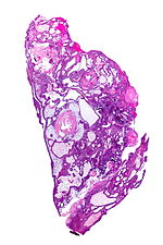
Micrograph of a craniopharyngioma (adamantinomatous). HPS stain.ICD-10 D44.4 ICD-9 237.0 ICD-O: 9350/1 DiseasesDB 3153 MedlinePlus 000345 eMedicine radio/196 MeSH D003397 Craniopharyngioma is a type of brain tumor derived from pituitary gland embryonic tissue,[1] that occurs most commonly in children but also in men and women in their 50s and 60s.[2] It arises from nests of odontogenic (tooth-forming) epithelium within the suprasellar/diencephalic region and, therefore, contains deposits of calcium, which are evident on an x-ray. Histologically, craniopharyngiomas resemble adamantinomas (the most common tumors of the tooth). Patients may present with bitemporal inferior quadrantanopia leading to bitemporal hemianopia, as the tumor may compress the optic chiasm.
It has a point prevalence of approximately 2/100,000.[3]
Craniopharyngiomas are also known as Rathke pouch tumors, hypophyseal duct tumors, or adamantinomas.
Contents
Presentation
 CT scan showing a craniopharyngioma.
Craniopharyngiomas are typically very slow growing tumors. They arise from the cells along the pituitary stalk. They are classified by histology as benign;[4] however, as with many brain tumors, their treatment can be difficult, and significant morbidities are associated with both the tumor and treatment.
Craniopharyngioma is a rare, usually suprasellar[5] neoplasm, which may be cystic, that develops from rests of epithelium derived from Rathke's pouch.[6][7] Rathke's pouch is an embryonic precursor of the anterior pituitary.
Histology
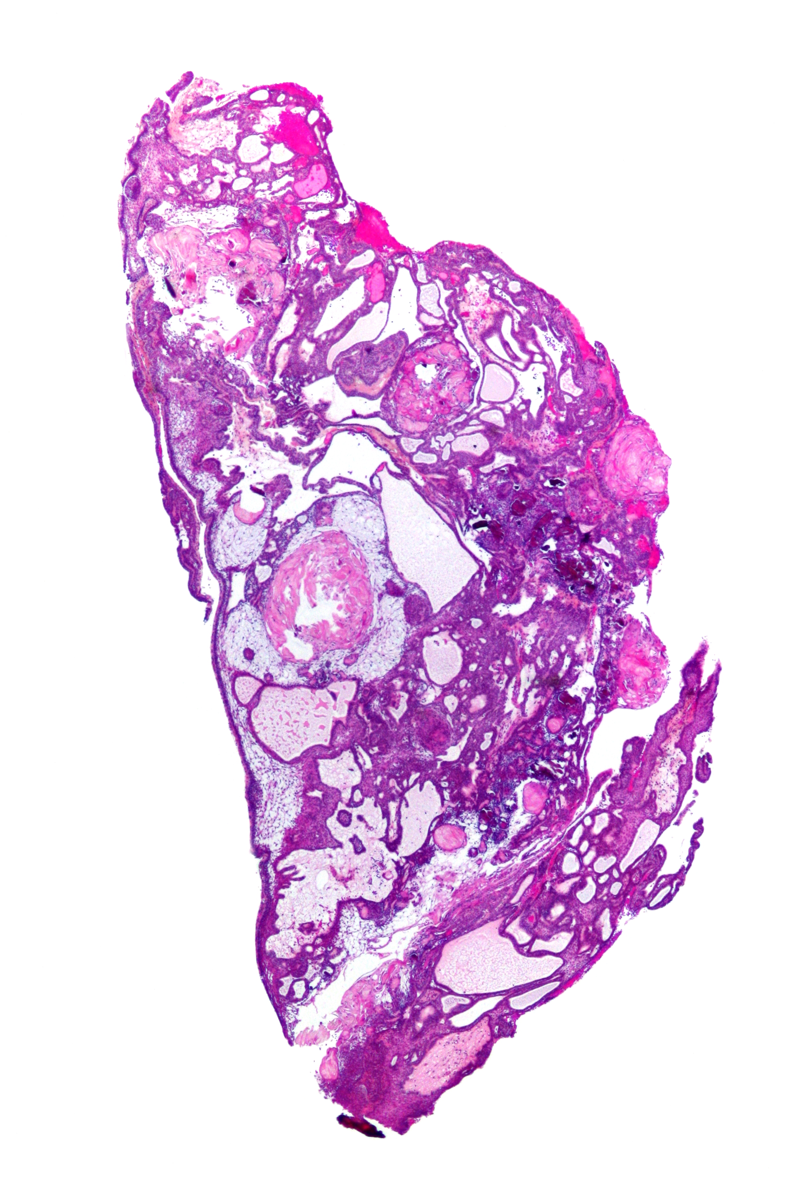
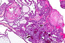 Micrograph showing the characteristic features of an adamantinomatous craniopharyngioma - cystic spaces, calcifications, and "wet" keratin. HPS stain.
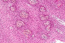
The histologic pattern consists of nesting of squamous epithelium bordered by radially arranged cells. It is frequently accompanied by calcium deposition and may have a microscopic papillary architecture.
Two distinct types are recognized:[8][9]
- Adamantinomatous craniopharyngioma and,
- Papillary craniopharyngioma.
In the adamantinomatous type, calcifications are visible on neuroimaging and are helpful in diagnosis. The papillary type rarely calcifies.
On macroscopic examination, craniopharyngiomas are cystic or partially-cystic with solid areas. On light microscopy, the cysts are seen to be lined by stratified squamous epithelium. Keratin pearls may also be seen. The cysts are usually filled with a yellow, viscous fluid which is rich in cholesterol crystals. In addition to a long list of possible symptoms, the most common presentation include: headaches, growth failure, and bitemporal hemianopsia.
The histologic pattern consists of nesting of squamous epithelium bordered by radially arranged cells. It is frequently accompanied by calcium deposition and may have a microscopic papillary architecture.
Prognosis
Craniopharyngiomas are generally benign but are known to recur after resection.
Treatment
Treatment generally consists of subfrontal or transsphenoidal excision. Adjuvant radiotherapy is also used if total removal is not possible. Due to the morbidities associated with damage to the pituitary and hypothalmus from surgical removal and radiation, experimental therapies using intracavitary P32, yttrium or bleomycin delivered via an external reservoir are frequently employed, especially in young patients.[10]
Possible symptoms
- Anorexia
- Balance disorder
- Dry skin
- Fatigue
- Fever
- Headache (obstructive hydrocephalus)
- Lethargy
- Myxedema
- Nausea
- Short stature
- Polydipsia
- Polyuria (diabetes insipidus)
- Vision loss (bitemporal hemianopia)
- Vomiting
- Weight gain
- Amenorrhea
Additional images
-

Enhanced T1 weighted MRI's of craniopharyngiomas.
See also
- Rathke's cleft cyst
References
- ^ "craniopharyngioma" at Dorland's Medical Dictionary
- ^ Hamid R, Sarkar S, Hossain MA, Mazumder U, Akanda NI, Parvin R (2007). "Clinical picture of craniopharyngioma in childhood". Mymensingh medical journal : MMJ 16 (2): 123–6. PMID 17703145.
- ^ Garnett MR, Puget S, Grill J, Sainte-Rose C (2007). "Craniopharyngioma". Orphanet journal of rare diseases 2: 18. doi:10.1186/1750-1172-2-18. PMC 1855047. PMID 17425791. http://www.pubmedcentral.nih.gov/articlerender.fcgi?tool=pmcentrez&artid=1855047.
- ^ Garrè ML, Cama A (2007). "Craniopharyngioma: modern concepts in pathogenesis and treatment". Curr. Opin. Pediatr. 19 (4): 471–9. doi:10.1097/MOP.0b013e3282495a22. PMID 17630614.
- ^ Rodriguez FJ, Scheithauer BW, Tsunoda S, Kovacs K, Vidal S, Piepgras DG (2007). "The spectrum of malignancy in craniopharyngioma". Am. J. Surg. Pathol. 31 (7): 1020–8. doi:10.1097/PAS.0b013e31802d8a96. PMID 17592268.
- ^ Moore, Kraig D. (January 15, 2000). "41. Craniopharyngioma". In Bernstein, Mark; Berger, Mitchel S.. Neuro-oncology: the essentials. Endorsed by the Joint Tumor Section of the American Association of Neurological Surgeons & the Congress of Neurological Surgeons. Thieme. pp. 409–418. ISBN 9780865778801. http://books.google.com/books?id=gbwSC43Eia4C&pg=PA409. Retrieved August 8, 2011.
- ^ "Endocrine Pathology". http://library.med.utah.edu/WebPath/ENDOHTML/ENDO115.html. Retrieved 2009-05-08.
- ^ Sekine S, Shibata T, Kokubu A, et al (December 2002). "Craniopharyngiomas of adamantinomatous type harbor beta-catenin gene mutations". Am. J. Pathol. 161 (6): 1997–2001. PMC 1850925. PMID 12466115. http://ajp.amjpathol.org/cgi/pmidlookup?view=long&pmid=12466115.
- ^ Sekine S, Takata T, Shibata T, et al (December 2004). "Expression of enamel proteins and LEF1 in adamantinomatous craniopharyngioma: evidence for its odontogenic epithelial differentiation". Histopathology 45 (6): 573–9. doi:10.1111/j.1365-2559.2004.02029.x. PMID 15569047. http://www3.interscience.wiley.com/resolve/openurl?genre=article&sid=nlm:pubmed&issn=0309-0167&date=2004&volume=45&issue=6&spage=573.
- ^ Wisoff JH (February 2008). "Craniopharyngioma". J Neurosurg Pediatr 1 (2): 124–5; discussion 125. doi:10.3171/PED/2008/1/2/124. PMID 18352780.
External links
- CINN
- Cancer.Net: Craniopharyngioma, Childhood
- Boston Neurosurgical Foundation
- Craniopharyngioma Online Support Group
- Adamantinomatous Craniopharyngioma
Nervous tissue tumors/NS neoplasm/Neuroectodermal tumor (ICD-O 9350–9589) (C70–C72, D32–D33, 191–192/225) Endocrine/
sellar (9350–9379)sellar: Craniopharyngioma · Pituicytomaother: PinealomaCNS
(9380–9539)Astrocytoma (Pilocytic astrocytoma, Pleomorphic xanthoastrocytoma, Fibrillary (also diffuse or lowgrade) astrocytomas, Anaplastic astrocytoma, Glioblastoma multiforme)Ependymoma · SubependymomaMultiple/unknownMature
neuronNeuroblastoma (Esthesioneuroblastoma, Ganglioneuroblastoma) · Medulloblastoma · Atypical teratoid rhabdoid tumorPrimitiveMeningiomas
(meninges)HematopoieticPNS: NST
(9540–9579)cranial and paraspinal nerves: Neurofibroma (Neurofibrosarcoma, Neurofibromatosis) · Neurilemmoma/Schwannoma (Acoustic neuroma) · Malignant peripheral nerve sheath tumornote: not all brain tumors are of nervous tissue, and not all nervous tissue tumors are in the brain (see brain metastases)
Tumors: endocrine gland neoplasia (C73–C75/D34–D35, 193–194/226–227) Pancreas/
islets of LangerhansHypothalamic/
pituitary axes
+parathyroidPituitaryCraniopharyngiomaThyroidThyroid cancer (malignant): epithelial cell /carcinoma (Papillary, Follicular/Hurthle cell) · parafollicular cell (Medullary) · AnaplasticBenign: Thyroid adenoma · Struma ovariiParathyroidGonadssee genital neoplasiaPinealoma Pinealoblastoma · PineocytomaMEN Categories:- Nervous system neoplasia


Wikimedia Foundation. 2010.