- Ependymoma
-
Ependymoma Classification and external resources 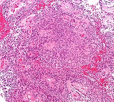
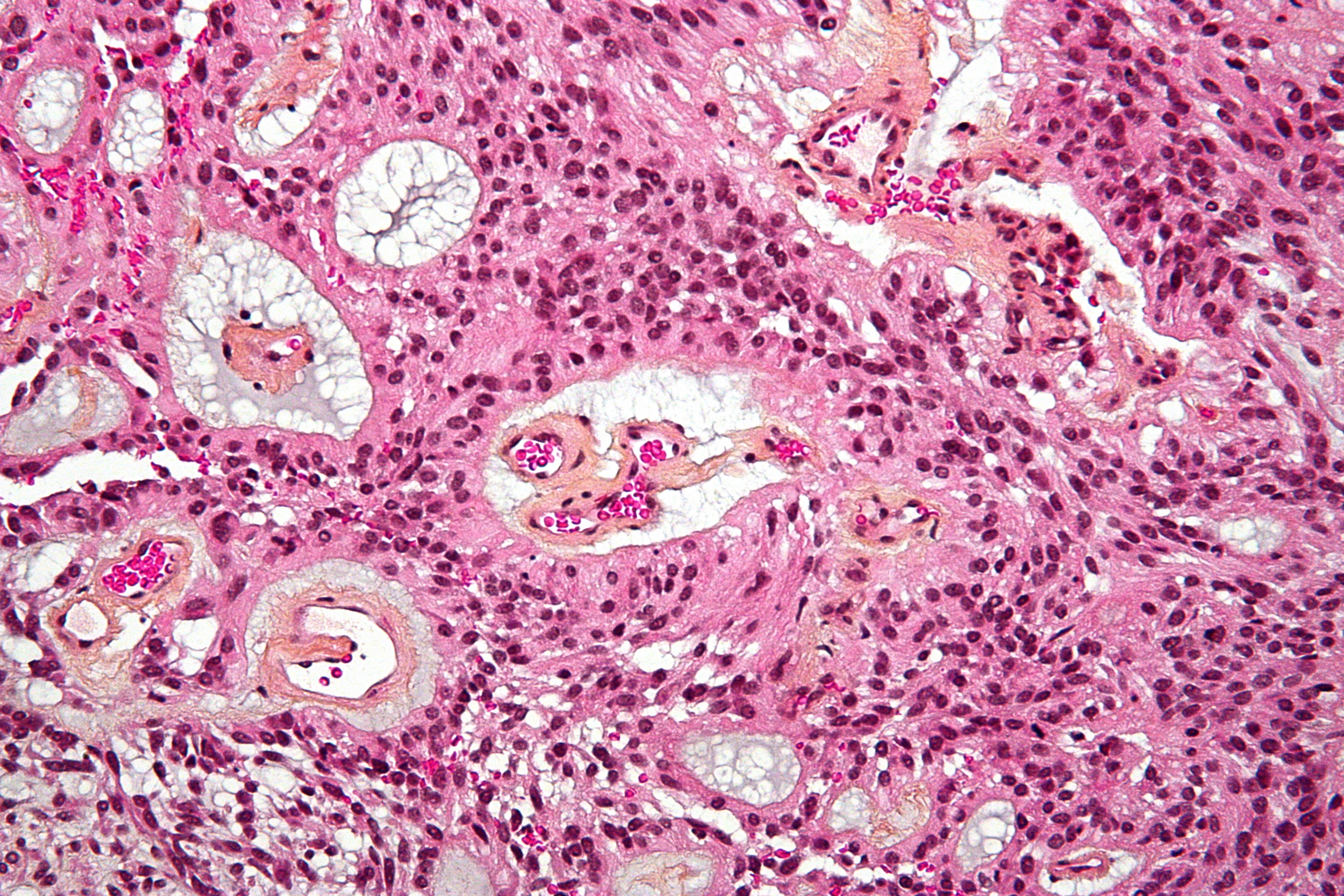
Micrograph of an ependymoma. H&E stain.ICD-10 C71 ICD-9 191.9, 225.0, 237.5 ICD-O: M9391/3-9394/1 DiseasesDB 29452 eMedicine med/700 MeSH D004806  Ependymoma of 4.ventricle in MRI.
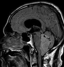 Ependymoma of 4.ventricle in MRI.
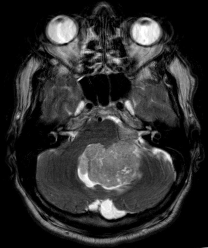
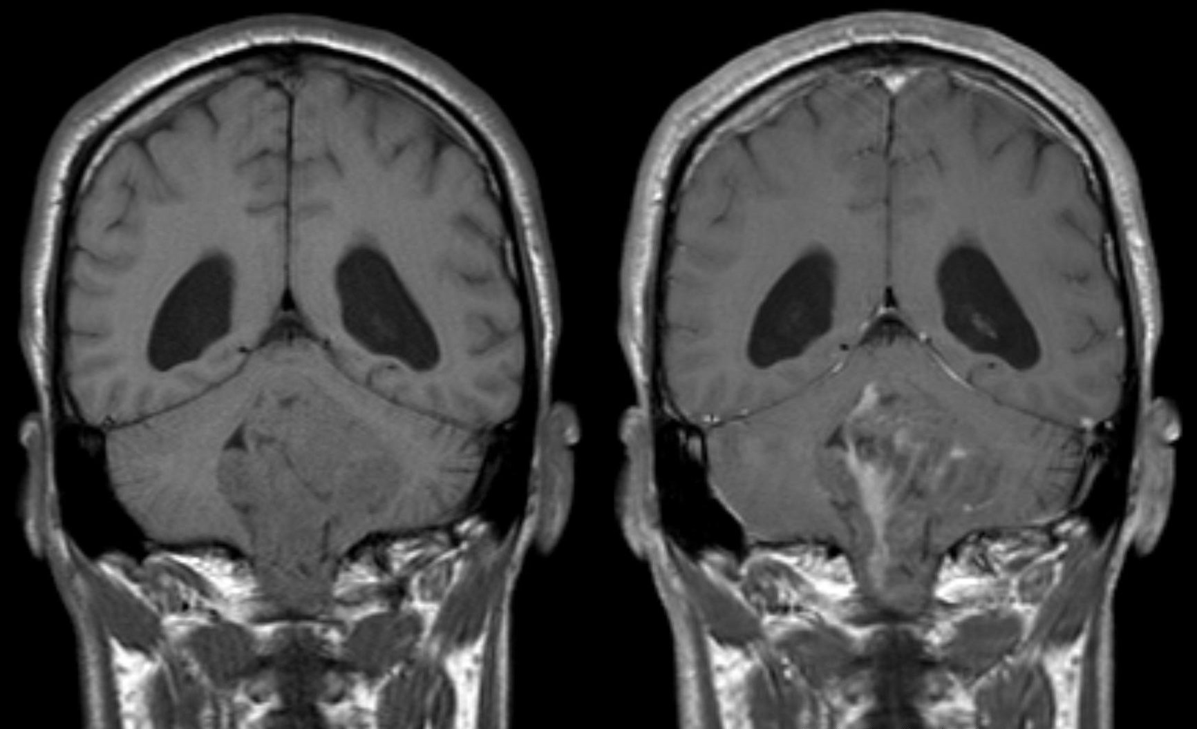
 Ependymoma of 4.ventricle in MRI. Left without, right with contrast-enhancement.
Ependymoma is a tumor that arises from the ependyma, a tissue of the central nervous system. Usually, in pediatric cases the location is intracranial, while in adults it is spinal. The common location of intracranial ependymoma is the fourth ventricle. Rarely, ependymoma can occur in the pelvic cavity.
Syringomyelia can be caused by an ependymoma. Ependymomas are also seen with neurofibromatosis type II.
Contents
Symptoms
- severe headache
- visual loss
- vomiting
- bilateral Babinski sign
- drowsiness (after several hours of the above symptoms)
Morphology
Ependymomas are composed of cells with regular, round to oval nuclei. There is a variably dense fibrillary background. Tumor cells may form gland-like round or elongated structures that resemble the embryologic ependymal canal, with long, delicate processes extending into the lumen; more frequently present are perivascular pseudorosettes in which tumor cells are arranged around vessels with an intervening zone consisting of thin ependymal processes directed toward the wall of the vessel.[2]
It has been suggested that ependymomas are derived from radial glia.[3]
Ependymoma tumors
Ependymomas make up about 5% of adult intracranial gliomas and up to 10% of childhood tumors of the central nervous system (CNS). Their occurrence seems to peak at age 5 years and then again at age 35. They develop from cells that line both the hollow cavities of the brain and the canal containing the spinal cord, but they usually arise from the floor of the fourth ventricle, situated in the lower back portion of the brain, where they may produce headache, nausea and vomiting by obstructing the flow of cerebrospinal fluid. This obstruction may also cause hydrocephalus. Other symptoms can include (but are not limited to): loss of appetite, difficulty sleeping, temporary inability to distinguish colors, uncontrollable twitching, seeing vertical or horizontal lines when in bright light, and temporary memory loss. It should be remembered that these symptoms also are prevalent in many other illnesses not associated with ependymoma.
About 85% of ependymomas are benign myxopapillary ependymoma (MPE)[citation needed]. MPE is a localized and slowly growing, low-grade tumor. Although some ependymomas are of a more anaplastic and malignant type, most of them are not anaplastic. For this reason, well-differentiated ependymomas are usually treated with radiation therapy only. For other ependymomas, total surgical removal is the preferred treatment and those that cannot be totally removed also require radiation therapy. The malignant (anaplastic) varieties of this tumor, malignant ependymoma and the ependymoblastoma, are treated similarly to medulloblastoma but the prognosis is much less favorable. Malignant ependymomas may be treated with a combination of radiation therapy and chemotherapy. Ependymoblastomas, which occur in infants and children younger than 5 years of age, may spread through the cerebrospinal fluid and usually require radiation therapy. The subependymoma, a variant of the ependymoma, is apt to arise in the fourth ventricle but may occur in the septum pellucidum and the cervical spinal cord. It usually affects people over 40 years of age and more often affects men than women.
Extraspinal ependymoma (EEP), also known as extradural ependymoma, may be an unusual form of teratoma[4] or may be confused with a sacrococcygeal teratoma.[5]
References
- ^ PRITE 2010 Part II q.13
- ^ Cotran, Ramzi S.; Kumar, Vinay; Fausto, Nelson; Nelso Fausto; Robbins, Stanley L.; Abbas, Abul K. (2005). "Ch. 28 The central nervous system". Robbins and Cotran pathologic basis of disease (7th ed.). St. Louis, Mo: Elsevier Saunders. ISBN 0-7216-0187-1.
- ^ Poppleton H, Gilbertson RJ (January 2007). "Stem cells of ependymoma". Br. J. Cancer 96 (1): 6–10. doi:10.1038/sj.bjc.6603519. PMC 2360214. PMID 17179988. http://www.pubmedcentral.nih.gov/articlerender.fcgi?tool=pmcentrez&artid=2360214.
- ^ Aktuğ T, Hakgüder G, Sarioğlu S, Akgür FM, Olguner M, Pabuçcuoğlu U (March 2000). "Sacrococcygeal extraspinal ependymomas: the role of coccygectomy". J. Pediatr. Surg. 35 (3): 515–8. doi:10.1016/S0022-3468(00)90228-8. PMID 10726703. http://linkinghub.elsevier.com/retrieve/pii/S0022346800129197.
- ^ Hany MA, Bouvier R, Ranchère D, Bergeron C, Schell M, Chappuis JP, Philip T, Frappaz D (1998). "Case report: a preterm infant with an extradural myxopapillary ependymoma component of a teratoma and high levels of alpha-fetoprotein". Pediatric hematology and oncology 15 (5): 437–41. doi:10.3109/08880019809016573. PMID 9783311.
External links
- Brain and Spinal Tumors: Hope Through Research (National Institute of Neurological Disorders and Stroke)
- Collaborative Ependymoma Research Network (CERN)
- Cancer.Net: Ependymoma, Childhood
Images and technical information
- Adult Ependymoma Images
- Pediatric Ependymoma Images
- Ependymoma Topics — MedPix
- Ependymoma Information from ABTA (PDF) for Completely Resected, Differentiated, Supratentorial Ependymoma]
- St. Jude Ependymoma Information
- Cancer Back Up — Ependymoma
- Walid MS, Parveen T, Grigorian AA, Robinson J (2007). "Chronic Back Pain: Clinical Quiz". The Internet Journal of Neurosurgery 4 (1). http://www.ispub.com/ostia/index.php?xmlPrinter=true&xmlFilePath=journals/ijns/vol4n1/myxo.xml.
- Spinal Cord Tumor Association
Treatment Information
- Treatment Information — from the CERN Foundation
- Treatment for Adults
- Pediatric Treatment
- Ependymoma-Specific Clinical Trials
- Ependymoma Outcomes Survey Project
Nervous tissue tumors/NS neoplasm/Neuroectodermal tumor (ICD-O 9350–9589) (C70–C72, D32–D33, 191–192/225) Endocrine/
sellar (9350–9379)other: PinealomaCNS
(9380–9539)Astrocytoma (Pilocytic astrocytoma, Pleomorphic xanthoastrocytoma, Fibrillary (also diffuse or lowgrade) astrocytomas, Anaplastic astrocytoma, Glioblastoma multiforme)Ependymoma · SubependymomaMultiple/unknownNeuroblastoma (Esthesioneuroblastoma, Ganglioneuroblastoma) · Medulloblastoma · Atypical teratoid rhabdoid tumorPrimitiveHematopoieticPNS: NST
(9540–9579)cranial and paraspinal nerves: Neurofibroma (Neurofibrosarcoma, Neurofibromatosis) · Neurilemmoma/Schwannoma (Acoustic neuroma) · Malignant peripheral nerve sheath tumornote: not all brain tumors are of nervous tissue, and not all nervous tissue tumors are in the brain (see brain metastases)
Categories:- Rare cancers
- Brain tumor


Wikimedia Foundation. 2010.