- Sacrococcygeal teratoma
Infobox_Disease
Name = Sacrococcygeal teratoma
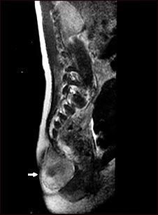
Caption =MRI in thesagittal plane of an adult, oriented head up and facing right, with a sacrococcygeal teratoma (arrow) at the base of the spine
DiseasesDB =
ICD10 =
ICD9 =
ICDO =
OMIM =
MedlinePlus =
eMedicineSubj = med
eMedicineTopic = 2248
MeshID =Sacrococcygeal teratoma (SCT) is a
teratoma (a kind of tumor) located at the base of thecoccyx (tailbone). It is thought to be a derivative of theprimitive streak .Natural history
SCT is seen in 1 in every 35,000 live births, and is the most common tumor
presenting in newborn humans. Most SCTs are found in babies and children, but SCTs have been reported in adults [Killen DA, Jackson LM (1964) "Sacrococcygeal teratoma in the adult"Archives of Surgery 88(3):425-433.] and the increasingly routine use of prenatal ultrasound exams has dramatically increased the number of diagnosed SCTs presenting in fetuses. Like other teratomas, an SCT can grow very large. Unlike other teratomas, an SCT sometimes grows larger than the rest of the fetus.Sacrococcygeal teratomas are the most common type of
germ cell tumor s (bothbenign andmalignant ) diagnosed inneonates ,infants , andchildren younger than 4 years. [ [http://www.cancer.gov/cancertopics/pdq/treatment/extracranial-germ-cell/HealthProfessional/page6 (PDQ) Sacrococcygeal Tumors in Children] ] SCTs occur more often in girls than in boys; ratios of 3:1 to 4:1 have been reported. [Rescorla FJ, Sawin RS, Coran AG, et al.: Long-term outcome for infants and children with sacrococcygeal teratoma: a report from the Childrens Cancer Group. J Pediatr Surg 33 (2): 171-6, 1998. [http://www.ncbi.nlm.nih.gov/entrez/query.fcgi?cmd=Retrieve&db=PubMed&list_uids=9498381&dopt=Abstract PUBMED Abstract] ]Historically, sacrococcygeal teratomas present in 2 clinical patterns related to the child’s age, tumor location, and likelihood of tumor malignancy. With the advent of routine prenatal ultrasound examinations, a third clinical pattern is emerging.
* Fetal tumors present during prenatal ultrasound exams, with or without maternal symptoms. SCTs found during routine exams tend to be small and partly or entirely external. The internal SCTs are not easily seen via ultrasound, unless they are large enough to reveal their presence by the abnormal position of the fetal
urinary bladder and other organs, but large fetal SCTs frequently produce maternal complications which necessitate non-routine, investigative ultrasounds.* Neonatal tumors present at birth protruding from the sacral site and are usually mature or immature teratomas.
*Among infants and young children, the tumor presents as a palpable mass in the sacropelvic region compressing the bladder or rectum. [Rescorla FJ: Pediatric germ cell tumors. Semin Surg Oncol 16 (2): 144-58, 1999. [http://www.ncbi.nlm.nih.gov/entrez/query.fcgi?cmd=Retrieve&db=PubMed&list_uids=9988869&dopt=Abstract PUBMED Abstract] ] These pelvic tumors have a greater likelihood of being malignant. An early survey found that the rate of tumor malignancy was 48% for girls and 67% for boys older than 2 months at the time of sacrococcygeal tumor diagnosis, compared with a malignant tumor incidence of 7% for girls and 10% for boys younger than 2 months at the time of diagnosis. The pelvic site of the
primary tumor has been reported to be an adverse prognostic factor, most likely caused by a higher rate of incomplete resection.*In older children and adults, the tumor may be mistaken for a
pilonidal sinus , or it may be found during a rectal exam or other evaluation.Diagnosis
During
prenatal ultrasound , an SCT having an external component may appear as a fluid-filled cyst or a solid mass sticking out from the fetus' body. Fetal SCTs that are entirely internal may be undetected if they are small; detection (or at least suspicion) is possible when the fetal bladder is seen in an abnormal position, due to the SCT pushing other organs out of place.At
birth , the usual presentation is a visible lump or mass under the skin at the top of the buttocks crease. If not visible, it can sometimes be felt; gently prodded, it feels somewhat like a hardboiled egg. A small SCT, if it is entirely inside the body, may not present for years, until it grows large enough to cause pain, constipation and other symptoms of a large mass inside thepelvis , or until it begins to extend out of the pelvis. Even a relatively large SCT may be missed, if it is internal, because the bonypelvis conceals and protects it.Mediastinal tumor s, including teratomas, are similarly concealed and protected by therib cage .Some SCTs are discovered when a child begins to talk at about age 2 years and complains of their bottom hurting or feeling "poopy" when they ride in a car seat.
Other tumors can occur in the sacrococcygeal and/or presacral regionscite journal
author = Bale PM
title = Sacrococcygeal developmental abnormalities and tumors in children.
journal = Perspectives in pediatric pathology
volume = 8
issue = 1
pages = 9–56
year = 1984
pmid = 6366733
doi =
issn = ] and hence must be ruled out to obtain adifferential diagnosis . These include extraspinal ependymomacite journal
author = Aktuğ T, Hakgüder G, Sarioğlu S, Akgür FM, Olguner M, Pabuçcuoğlu U
title = Sacrococcygeal extraspinal ependymomas: the role of coccygectomy.
journal = J. Pediatr. Surg.
volume = 35
issue = 3
pages = 515–8
year = 2000
pmid = 10726703
doi =
issn = ] ,ependymoblastoma ,cite journal
author = Santi M, Bulas D, Fasano R, "et al"
title = Congenital ependymoblastoma arising in the sacrococcygeal soft tissue: a case study
journal = Clin. Neuropathol.
volume = 27
issue = 2
pages = 78–82
year = 2008
pmid = 18402386
doi =
url =
issn = ]neuroblastoma andrhabdomyosarcoma .Smaller SCTs with an external component, seen in prenatal ultrasounds or at birth, often are mistaken for
spina bifida .Fact|date=August 2008 Cystic SCT and terminal myelocystocele are especially difficult to distinguish; for more accurate diagnosis, MRI has been recommended.cite journal
author = Yu JA, Sohaey R, Kennedy AM, Selden NR
title = Terminal myelocystocele and sacrococcygeal teratoma: a comparison of fetal ultrasound presentation and perinatal risk
journal = AJNR Am J Neuroradiol
volume = 28
issue = 6
pages = 1058–60
year = 2007
pmid = 17569957
doi = 10.3174/ajnr.A0502
url =
issn = ]Treatment
The preferred first treatment for SCT is complete surgical removal (ie, complete resection). The preferred approach to a small SCT is through the
perineum ; a large SCT may require an additional approach through theabdomen . Resection should include the coccyx and may also include portions of thesacrum . The surgery should include reattachment of the small muscles and ligaments formerly attached to the coccyx, in effect reconstructing the posterior perineum. If not, there is an increased risk ofperineal hernia later in life.SCTs are classified morphologically according to their relative extent outside and inside the body:
*Altman type I — entirely outside, sometimes attached to the body only by a narrow stalk
*Altman type II — mostly outside
*Altman type III — mostly inside
*Altman type IV — entirely inside; this is also known as a presacral teratoma or retrorectal teratomaThe Altman type is significant in the contexts of management of labor and delivery, surgical approach, and complications of SCT. Serial ultrasound and MRI monitoring of SCTs in fetuses
in utero has demonstrated that the Altman type can change over time. As the tumor grows, it can push between other organs and through the perineum to the body surface where the tumor appears as a bulge covered only by skin. Sometimes, the tumor bulge later slips back inside the perineum.Like all
teratoma s, a sacrococcygeal teratoma has the potential to be malignant, and the standard of care requires long-term followup by anoncologist .Management of fetal SCTs
Management of most fetal SCTs involves
watchful waiting prior to any treatment. An often useddecision tree is as follows:*Perform detailed ultrasound exam including fetal
echocardiogram andDoppler flow analysis
**If fetalhigh output failure ,placentomegaly , orhydrops
***If fetus not mature, performpregnancy termination orfetal intervention
***Else fetus mature, perform emergencyCesarean section
**Else no emergent problems, perform serialnon-stress test s and ultrasoundbiophysical profile s and plan delivery, as follows
***If emergent problems develop, return to top of decision tree
***Else if SCT over 5–10cm orpolyhydramnios , perform early (37 weeks gestation) elective Cesarean section
***Else SCT small and no complications, permit termspontaneous vaginal delivery Emergent problems include maternal
mirror syndrome ,polyhydramnios , andpreterm labor . Poor management decisions, including interventions that are either premature or delayed, can have dire consequences.cite journal
author = Mazneĭkova V, Dimitrova V
title = [Prenatal ultrasonographic diagnosis of four cases of sacrococcygeal teratoma]
language = Bulgarian
journal = Akusherstvo i ginekologii͡a
volume = 38
issue = 1
pages = 64–9
year = 1999
pmid = 11965727
doi =
issn = ] cite journal
author = Sheil AT, Collins KA
title = Fatal birth trauma due to an undiagnosed abdominal teratoma: case report and review of the literature
journal = The American journal of forensic medicine and pathology : official publication of the National Association of Medical Examiners
volume = 28
issue = 2
pages = 121–7
year = 2007
pmid = 17525561
doi = 10.1097/01.paf.0000257373.91126.0d
issn = ] A very small retrospective study of 9 babies with SCTs greater than 10 cm diameter reported slightly higher survivorship in babies remaining in utero slightly longer.cite journal
author = Holcroft CJ, Blakemore KJ, Gurewitsch ED, Driggers RW, Northington FJ, Fischer AC
title = Large fetal sacrococcygeal teratomas: could early delivery improve outcome?
journal = Fetal. Diagn. Ther.
volume = 24
issue = 1
pages = 55–60
year = 2008
pmid = 18504383
doi = 10.1159/000132408
url =
issn = ]In many cases, a fetus with a small SCT (under 5 or 10 cm) may be delivered vaginally.cite journal
author = Anteby EY, Yagel S
title = Route of delivery of fetuses with structural anomalies
journal = Eur. J. Obstet. Gynecol. Reprod. Biol.
volume = 106
issue = 1
pages = 5–9
year = 2003
pmid = 12475573
doi =
issn = ] cite journal
author = Ruangtrakool R, Nitipon A, Laohapensang M, "et al"
title = Sacrococcygeal teratoma: 25 year experience
journal = Journal of the Medical Association of Thailand = Chotmaihet thangphaet
volume = 84
issue = 2
pages = 265–73
year = 2001
pmid = 11336088
doi =
issn = ] cite journal
author = McCurdy CM, Seeds JW
title = Route of delivery of infants with congenital anomalies
journal = Clinics in perinatology
volume = 20
issue = 1
pages = 81–106
year = 1993
pmid = 8458172
doi =
issn = ] cite journal
author = Kainer F, Winter R, Hofmann HM, Karpf EF
title = [Sacrococcygeal teratoma. Prenatal diagnosis and prognosis]
language = German
journal = Zentralblatt für Gynäkologie
volume = 112
issue = 10
pages = 609–16
year = 1990
pmid = 2205995
doi =
issn = ] Prior to the advent of prenatal detection and hence scheduled C-section, 90% of babies diagnosed with SCT were born full term.cite journal
author = Gonzalez-Crussi F, Winkler RF, Mirkin DL
title = Sacrococcygeal teratomas in infants and children: relationship of histology and prognosis in 40 cases.
journal = Arch. Pathol. Lab. Med.
volume = 102
issue = 8
pages = 420–5
year = 1978
pmid = 580884
doi =
issn = ]Management of adult SCTs
SCTs are very rare in adults, and as a rule these tumors are benign and have extremely low potential for malignancy. This estimation of potential is based on the idea that because the tumor existed for decades prior to diagnosis, without becoming malignant, it has little or no potential to ever become malignant. For this reason, and because coccygectomy in adults has greater risks than in babies, some surgeons prefer not to remove the coccyx of adult survivors of SCT. There are case reports of good outcomes.cite journal
author = Jucá M, de Oliveira FF, Gomes EG, Le Campion E
title = Sacrococcycygeal Teratoma in Adult: Report of a Case
journal = Int J Gastrointest Cancer
volume = 37
issue = 2-3
pages = 91–93
year = 2006
month = September
pmid = 17827528
doi = 10.1007/s12029-007-0004-6
url =
issn = ]Complications
Maternal complications of pregnancy may include
mirror syndrome .cite journal
author = Finamore PS, Kontopoulos E, Price M, Giannina G, Smulian JC
title = Mirror syndrome associated with sacrococcygeal teratoma: a case report
journal = The Journal of reproductive medicine
volume = 52
issue = 3
pages = 225–7
year = 2007
pmid = 17465292
doi =
issn = ] Maternal complications of delivery may include aCesarean section or, alternatively, a vaginal delivery with mechanicaldystocia .cite journal
author = Nalbanski B, Markov D, Brankov O
title = [Sacrococcygeal teratoma--a case report and literature review]
language = Bulgarian
journal = Akusherstvo i ginekologii͡a
volume = 46
issue = 2
pages = 41–5
year = 2007
pmid = 17469451
doi =
issn = ]Complications of the mass effect of a teratoma in general are addressed on the
teratoma page. Complications of the mass effect of a large SCT may include hip dysplasia,bowel obstruction ,urinary obstruction ,hydronephrosis andhydrops fetalis . Even a small SCT can produce complications of mass effect, if it is presacral (Altman Type IV).cite journal
author = Galili O, Mogilner J
title = Type IV sacrococcygeal teratoma causing urinary retention: a rare presentation.
journal = J. Pediatr. Surg.
volume = 40
issue = 2
pages = E18–20
year = 2005
pmid = 15750911
doi = 10.1016/j.jpedsurg.2004.10.003
issn = ] In the fetus, severe hydronephrosis may contribute to inadequate lung development. Also in the fetus and newborn, theanus may be imperforate.Later complications of the mass effect and/or surgery may include
neurogenic bladder , other forms ofurinary incontinence ,fecal incontinence , and other chronic problems resulting from accidental damage to or sacrifice of nerves and muscles within the pelvis.cite journal
author = Engelskirchen R, Holschneider AM, Rhein R, Hecker WC, Höpner F
title = [Sacral teratomas in childhood. An analysis of long-term results in 87 children]
language = German
journal = Zeitschrift für Kinderchirurgie : organ der Deutschen, der Schweizerischen und der Osterreichischen Gesellschaft für Kinderchirurgie = Surgery in infancy and childhood
volume = 42
issue = 6
pages = 358–61
year = 1987
pmid = 3439358
doi =
issn = ] Removal of the coccyx may include additional complications. In one review of 25 patients, [ [http://www.ncbi.nlm.nih.gov/sites/entrez?cmd=Retrieve&db=PubMed&list_uids= 16373161&dopt=AbstractPlus PubMed] ] however, the most frequent complication was an unsatisfactory appearance of the surgical scar.Late effects
Late effects are of two kinds: consequences of the tumor itself, and consequences of surgery and other treatments for the tumor.
Complications of not removing the coccyx may include both recurrence of the teratomacite journal
author = Lahdenne P, Heikinheimo M, Nikkanen V, Klemi P, Siimes MA, Rapola J
title = Neonatal benign sacrococcygeal teratoma may recur in adulthood and give rise to malignancy.
journal = Cancer
volume = 72
issue = 12
pages = 3727–31
year = 1993
pmid = 8252490
doi =
issn = Synopsis: 45 survivors of infant SCT were followed up. Two reported recurrent benign teratoma and one reported metastatic adenocarcinoma originating from the residual coccyx. They were aged 21-43 at diagnosis.] and metastatic cancer.cite journal
author = Lack EE, Glaun RS, Hefter LG, Seneca RP, Steigman C, Athari F
title = Late occurrence of malignancy following resection of a histologically mature sacrococcygeal teratoma. Report of a case and literature review.
journal = Arch. Pathol. Lab. Med.
volume = 117
issue = 7
pages = 724–8
year = 1993
pmid = 8323438
doi =
issn = Synopsis: A 40 year old man has widely metastaticadenocarcinoma arising from the residual coccyx remaining after surgical removal of an apparently benign SCT at age 2 months.] Late malignancies usually involve incomplete excision of the coccyx and areadenocarcinoma .Although functional disability in survivors is common,cite journal
author = Derikx JP, De Backer A, van de Schoot L, "et al"
title = Long-term functional sequelae of sacrococcygeal teratoma: a national study in The Netherlands
journal =J. Pediatr. Surg.
volume = 42
issue = 6
pages = 1122–6
year = 2007
pmid = 17560233
doi = 10.1016/j.jpedsurg.2007.01.050
url =
issn = ] a small comparative studycite journal
author = Cozzi F, Schiavetti A, Zani A, Spagnol L, Totonelli G, Cozzi DA
title = The functional sequelae of sacrococcygeal teratoma: a longitudinal and cross-sectional follow-up study
journal =J. Pediatr. Surg.
volume = 43
issue = 4
pages = 658–61
year = 2008
pmid = 18405712
doi = 10.1016/j.jpedsurg.2007.10.066
url =
issn = ] found a nonsignificant difference between SCT survivors and a matched control group.In rare cases, pelvic scarring may necessitate that a pregnant woman who is a SCT survivor deliver her baby by
Cesarean section .cite journal
author = Kohlberger P, Helbich T, Schaller A
title = [Delivery following surgically treated sacrococcygeal teratoma in the mother]
language = German
journal =Z Geburtshilfe Neonatol
volume = 201
issue = 4
pages = 148–51
year = 1997
pmid = 9410520
doi =
url =
issn = ]ee also
*
Currarino syndrome
*Teratoma
*Sacrococcygeal symphysis References
External links
* [http://www.thedoctorsdoctor.com/diseases/sacrococcygeal_teratoma.htm The Doctor's Doctor page on SCT]
Wikimedia Foundation. 2010.