- ACTG1
-
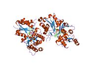
Actin, gamma 1, also known as ACTG1, is a gene.
Actins are highly conserved proteins that are involved in various types of cell motility, and maintenance of the cytoskeleton. In vertebrates, three main groups of actin isoforms, alpha, beta and gamma have been identified. The alpha actins are found in muscle tissues and are a major constituent of the contractile apparatus. The beta and gamma actins co-exist in most cell types as components of the cytoskeleton, and as mediators of internal cell motility. Actin, gamma 1, encoded by this gene, is a cytoplasmic actin found in nonmuscle cells.[1]
Contents
Interactions
ACTG1 has been shown to interact with TMSB4X[2][3] and CAP1.[4]
See also
References
- ^ "Entrez Gene: ACTG1 actin, gamma 1". http://www.ncbi.nlm.nih.gov/sites/entrez?Db=gene&Cmd=ShowDetailView&TermToSearch=71.
- ^ Hertzog, Maud; van Heijenoort Carine, Didry Dominique, Gaudier Martin, Coutant Jérôme, Gigant Benoît, Didelot Gérard, Préat Thomas, Knossow Marcel, Guittet Eric, Carlier Marie-France (May. 2004). "The beta-thymosin/WH2 domain; structural basis for the switch from inhibition to promotion of actin assembly". Cell (United States) 117 (5): 611–23. doi:10.1016/S0092-8674(04)00403-9. ISSN 0092-8674. PMID 15163409.
- ^ Van Troys, M; Dewitte D, Goethals M, Carlier M F, Vandekerckhove J, Ampe C (Jan. 1996). "The actin binding site of thymosin beta 4 mapped by mutational analysis". EMBO J. (ENGLAND) 15 (2): 201–10. ISSN 0261-4189. PMC 449934. PMID 8617195. http://www.pubmedcentral.nih.gov/articlerender.fcgi?tool=pmcentrez&artid=449934.
- ^ Hubberstey, A; Yu G, Loewith R, Lakusta C, Young D (Jun. 1996). "Mammalian CAP interacts with CAP, CAP2, and actin". J. Cell. Biochem. (UNITED STATES) 61 (3): 459–66. doi:10.1002/(SICI)1097-4644(19960601)61:3<459::AID-JCB13>3.0.CO;2-E. ISSN 0730-2312. PMID 8761950.
Further reading
- Snásel J, Pichová I (1997). "The cleavage of host cell proteins by HIV-1 protease.". Folia Biol. (Praha) 42 (5): 227–30. doi:10.1007/BF02818986. PMID 8997639.
- Rodríguez Del Castillo A, Vitale ML, Trifaró JM (1992). "Ca2+ and pH determine the interaction of chromaffin cell scinderin with phosphatidylserine and phosphatidylinositol 4,5,-biphosphate and its cellular distribution during nicotinic-receptor stimulation and protein kinase C activation.". J. Cell Biol. 119 (4): 797–810. doi:10.1083/jcb.119.4.797. PMC 2289683. PMID 1331119. http://www.pubmedcentral.nih.gov/articlerender.fcgi?tool=pmcentrez&artid=2289683.
- Adams LD, Tomasselli AG, Robbins P, et al. (1992). "HIV-1 protease cleaves actin during acute infection of human T-lymphocytes.". AIDS Res. Hum. Retroviruses 8 (2): 291–5. doi:10.1089/aid.1992.8.291. PMID 1540415.
- Dawson SJ, White LA (1992). "Treatment of Haemophilus aphrophilus endocarditis with ciprofloxacin.". J. Infect. 24 (3): 317–20. doi:10.1016/S0163-4453(05)80037-4. PMID 1602151.
- Tomasselli AG, Hui JO, Adams L, et al. (1991). "Actin, troponin C, Alzheimer amyloid precursor protein and pro-interleukin 1 beta as substrates of the protease from human immunodeficiency virus.". J. Biol. Chem. 266 (22): 14548–53. PMID 1907279.
- Shoeman RL, Kesselmier C, Mothes E, et al. (1991). "Non-viral cellular substrates for human immunodeficiency virus type 1 protease.". FEBS Lett. 278 (2): 199–203. doi:10.1016/0014-5793(91)80116-K. PMID 1991513.
- Erba HP, Eddy R, Shows T, et al. (1988). "Structure, chromosome location, and expression of the human gamma-actin gene: differential evolution, location, and expression of the cytoskeletal beta- and gamma-actin genes.". Mol. Cell. Biol. 8 (4): 1775–89. PMC 363338. PMID 2837653. http://www.pubmedcentral.nih.gov/articlerender.fcgi?tool=pmcentrez&artid=363338.
- Vandekerckhove J, Schering B, Bärmann M, Aktories K (1988). "Botulinum C2 toxin ADP-ribosylates cytoplasmic beta/gamma-actin in arginine 177.". J. Biol. Chem. 263 (2): 696–700. PMID 3335520.
- Chou CC, Davis RC, Fuller ML, et al. (1987). "Gamma-actin: unusual mRNA 3'-untranslated sequence conservation and amino acid substitutions that may be cancer related.". Proc. Natl. Acad. Sci. U.S.A. 84 (9): 2575–9. doi:10.1073/pnas.84.9.2575. PMC 304700. PMID 3472224. http://www.pubmedcentral.nih.gov/articlerender.fcgi?tool=pmcentrez&artid=304700.
- Hesterberg LK, Weber K (1986). "Isolation of a domain of villin retaining calcium-dependent interaction with G-actin, but devoid of F-actin fragmenting activity.". Eur. J. Biochem. 154 (1): 135–40. doi:10.1111/j.1432-1033.1986.tb09368.x. PMID 3510866.
- Erba HP, Gunning P, Kedes L (1986). "Nucleotide sequence of the human gamma cytoskeletal actin mRNA: anomalous evolution of vertebrate non-muscle actin genes.". Nucleic Acids Res. 14 (13): 5275–94. doi:10.1093/nar/14.13.5275. PMC 311540. PMID 3737401. http://www.pubmedcentral.nih.gov/articlerender.fcgi?tool=pmcentrez&artid=311540.
- Fuchs E, Kim KH, Hanukoglu I, Tanese N (1984). "The evolution and complexity of the genes encoding the cytoskeletal proteins of human epidermal cells.". Curr. Probl. Dermatol. 11: 27–44. PMID 6686106.
- Gunning P, Ponte P, Okayama H, et al. (1983). "Isolation and characterization of full-length cDNA clones for human alpha-, beta-, and gamma-actin mRNAs: skeletal but not cytoplasmic actins have an amino-terminal cysteine that is subsequently removed.". Mol. Cell. Biol. 3 (5): 787–95. PMC 368601. PMID 6865942. http://www.pubmedcentral.nih.gov/articlerender.fcgi?tool=pmcentrez&artid=368601.
- Bretscher A, Weber K (1980). "Villin is a major protein of the microvillus cytoskeleton which binds both G and F actin in a calcium-dependent manner.". Cell 20 (3): 839–47. doi:10.1016/0092-8674(80)90330-X. PMID 6893424.
- Pedrotti B, Colombo R, Islam K (1995). "Microtubule associated protein MAP1A is an actin-binding and crosslinking protein.". Cell Motil. Cytoskeleton 29 (2): 110–6. doi:10.1002/cm.970290203. PMID 7820861.
- Pope B, Maciver S, Weeds A (1995). "Localization of the calcium-sensitive actin monomer binding site in gelsolin to segment 4 and identification of calcium binding sites.". Biochemistry 34 (5): 1583–8. doi:10.1021/bi00005a014. PMID 7849017.
- Jesaitis AJ, Erickson RW, Klotz KN, et al. (1993). "Functional molecular complexes of human N-formyl chemoattractant receptors and actin.". J. Immunol. 151 (10): 5653–65. PMID 8228254.
- Hawkins M, Pope B, Maciver SK, Weeds AG (1993). "Human actin depolymerizing factor mediates a pH-sensitive destruction of actin filaments.". Biochemistry 32 (38): 9985–93. doi:10.1021/bi00089a014. PMID 8399167.
- Yu FX, Lin SC, Morrison-Bogorad M, et al. (1993). "Thymosin beta 10 and thymosin beta 4 are both actin monomer sequestering proteins.". J. Biol. Chem. 268 (1): 502–9. PMID 8416954.
- Jalaguier S, Mornet D, Mesnier D, et al. (1996). "Human mineralocorticoid receptor interacts with actin under mineralocorticoid ligand modulation.". FEBS Lett. 384 (2): 112–6. doi:10.1016/0014-5793(96)00295-5. PMID 8612804.
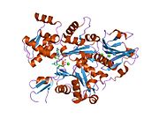
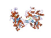
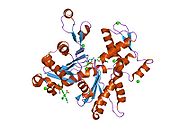
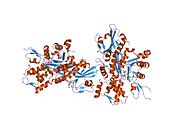
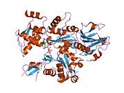
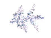
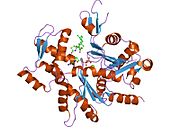
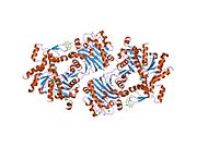
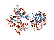
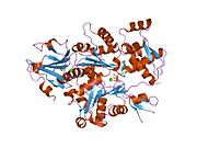
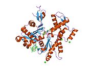
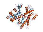
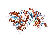
PDB gallery 1atn: Atomic structure of the actin:DNASE I complex1c0f: CRYSTAL STRUCTURE OF DICTYOSTELIUM CAATP-ACTIN IN COMPLEX WITH GELSOLIN SEGMENT 11c0g: CRYSTAL STRUCTURE OF 1:1 COMPLEX BETWEEN GELSOLIN SEGMENT 1 AND A DICTYOSTELIUM/TETRAHYMENA CHIMERA ACTIN (MUTANT 228: Q228K/T229A/A230Y/E360H)1d4x: Crystal Structure of Caenorhabditis Elegans Mg-ATP Actin Complexed with Human Gelsolin Segment 1 at 1.75 A resolution.1dej: CRYSTAL STRUCTURE OF A DICTYOSTELIUM/TETRAHYMENA CHIMERA ACTIN (MUTANT 646: Q228K/T229A/A230Y/A231K/S232E/E360H) IN COMPLEX WITH HUMAN GELSOLIN SEGMENT 11eqy: COMPLEX BETWEEN RABBIT MUSCLE ALPHA-ACTIN: HUMAN GELSOLIN DOMAIN 11esv: COMPLEX BETWEEN LATRUNCULIN A:RABBIT MUSCLE ALPHA ACTIN:HUMAN GELSOLIN DOMAIN 11h1v: GELSOLIN G4-G6/ACTIN COMPLEX1hlu: STRUCTURE OF BOVINE BETA-ACTIN-PROFILIN COMPLEX WITH ACTIN BOUND ATP PHOSPHATES SOLVENT ACCESSIBLE1ijj: THE X-RAY CRYSTAL STRUCTURE OF THE COMPLEX BETWEEN RABBIT SKELETAL MUSCLE ACTIN AND LATRUNCULIN A AT 2.85 A RESOLUTION1j6z: UNCOMPLEXED ACTIN1kxp: CRYSTAL STRUCTURE OF HUMAN VITAMIN D-BINDING PROTEIN IN COMPLEX WITH SKELETAL ACTIN1lcu: Polylysine Induces an Antiparallel Actin Dimer that Nucleates Filament Assembly: Crystal Structure at 3.5 A Resolution1lot: CRYSTAL STRUCTURE OF THE COMPLEX OF ACTIN WITH VITAMIN D-BINDING PROTEIN1m8q: Molecular Models of Averaged Rigor Crossbridges from Tomograms of Insect Flight Muscle1ma9: Crystal structure of the complex of human vitamin D binding protein and rabbit muscle actin1mdu: Crystal structure of the chicken actin trimer complexed with human gelsolin segment 1 (GS-1)1mvw: MOLECULAR MODELS OF AVERAGED RIGOR CROSSBRIDGES FROM TOMOGRAMS OF INSECT FLIGHT MUSCLE1nlv: Crystal Structure Of Dictyostelium Discoideum Actin Complexed With Ca ATP And Human Gelsolin Segment 11nm1: Crystal Structure of D. Dicsoideum Actin Complexed With Gelsolin Segment 1 and Mg ATP at 1.8 A Resolution1nmd: Crystal Structure of D. Discoideum Actin-Gelsolin Segment 1 Complex Crystallized In Presence Of Lithium ATP1nwk: CRYSTAL STRUCTURE OF MONOMERIC ACTIN IN THE ATP STATE1o18: MOLECULAR MODELS OF AVERAGED RIGOR CROSSBRIDGES FROM TOMOGRAMS OF INSECT FLIGHT MUSCLE1o19: MOLECULAR MODELS OF AVERAGED RIGOR CROSSBRIDGES FROM TOMOGRAMS OF INSECT FLIGHT MUSCLE1o1a: MOLECULAR MODELS OF AVERAGED RIGOR CROSSBRIDGES FROM TOMOGRAMS OF INSECT FLIGHT MUSCLE1o1b: MOLECULAR MODELS OF AVERAGED RIGOR CROSSBRIDGES FROM TOMOGRAMS OF INSECT FLIGHT MUSCLE1o1c: MOLECULAR MODELS OF AVERAGED RIGOR CROSSBRIDGES FROM TOMOGRAMS OF INSECT FLIGHT MUSCLE1o1d: MOLECULAR MODELS OF AVERAGED RIGOR CROSSBRIDGES FROM TOMOGRAMS OF INSECT FLIGHT MUSCLE1o1e: MOLECULAR MODELS OF AVERAGED RIGOR CROSSBRIDGES FROM TOMOGRAMS OF INSECT FLIGHT MUSCLE1o1f: MOLECULAR MODELS OF AVERAGED RIGOR CROSSBRIDGES FROM TOMOGRAMS OF INSECT FLIGHT MUSCLE1o1g: MOLECULAR MODELS OF AVERAGED RIGOR CROSSBRIDGES FROM TOMOGRAMS OF INSECT FLIGHT MUSCLE1p8z: Complex Between Rabbit Muscle alpha-Actin: Human Gelsolin Residues Val26-Glu1561qz5: Structure of rabbit actin in complex with kabiramide C1qz6: Structure of rabbit actin in complex with jaspisamide A1rdw: Actin Crystal Dynamics: Structural Implications for F-actin Nucleation, Polymerization and Branching Mediated by the Anti-parallel Dimer1rfq: Actin Crystal Dynamics: Structural Implications for F-actin Nucleation, Polymerization and Branching Mediated by the Anti-parallel Dimer1rgi: Crystal structure of gelsolin domains G1-G3 bound to actin1s22: Absolute Stereochemistry of Ulapualide A1sqk: CRYSTAL STRUCTURE OF CIBOULOT IN COMPLEX WITH SKELETAL ACTIN1t44: Structural basis of actin sequestration by thymosin-B4: Implications for arp2/3 activation1wua: The structure of Aplyronine A-actin complex1y64: Bni1p Formin Homology 2 Domain complexed with ATP-actin1yxq: Crystal structure of actin in complex with swinholide A2a3z: Ternary complex of the WH2 domain of WASP with Actin-DNAse I2a40: Ternary complex of the WH2 domain of WAVE with Actin-DNAse I2a41: Ternary complex of the WH2 Domain of WIP with Actin-DNAse I2a42: Actin-DNAse I Complex2a5x: Crystal Structure of a Cross-linked Actin Dimer2asm: Structure of Rabbit Actin In Complex With Reidispongiolide A2aso: Structure of Rabbit Actin In Complex With Sphinxolide B2asp: Structure of Rabbit Actin In Complex With Reidispongiolide C2btf: THE STRUCTURE OF CRYSTALLINE PROFILIN-BETA-ACTIN2d1k: Ternary complex of the WH2 domain of mim with actin-dnase I2ff3: Crystal structure of Gelsolin domain 1:N-wasp V2 motif hybrid in complex with actin2ff6: Crystal structure of Gelsolin domain 1:ciboulot domain 2 hybrid in complex with actin2fxu: X-ray Structure of Bistramide A- Actin Complex at 1.35 A resolution.2gwj: SpvB ADP-ribosylated actin: hexagonal crystal form2gwk: SpvB ADP-ribosylated actin: orthorhombic crystal form2hf3: Crystal structure of monomeric Actin in the ADP bound state2hf4: Crystal structure of Monomeric Actin in its ATP-bound state2hmp: Uncomplexed actin cleaved with protease ECP322oan: Structure of oxidized beta-actin2q1n: Actin Dimer Cross-linked Between Residues 41 and 3742q31: Actin Dimer Cross-linked Between Residues 41 and 374 and proteolytically cleaved by subtilisin between residues 47 and 48.2q36: Actin Dimer Cross-linked between Residues 191 and 374 and complexed with Kabiramide CProteins of the cytoskeleton Human I (MYO1A, MYO1B, MYO1C, MYO1D, MYO1E, MYO1F, MYO1G, MYO1H) · II (MYH1, MYH2, MYH3, MYH4, MYH6, MYH7, MYH7B, MYH8, MYH9, MYH10, MYH11, MYH13, MYH14, MYH15, MYH16) · III (MYO3A, MYO3B) · V (MYO5A, MYO5B, MYO5C) · VI (MYO6) · VII (MYO7A, MYO7B) · IX (MYO9A, MYO9B) · X (MYO10) · XV (MYO15A) · XVIII (MYO18A, MYO18B) · LC (MYL1, MYL2, MYL3, MYL4, MYL5, MYL6, MYL6B, MYL7, MYL9, MYLIP, MYLK, MYLK2, MYLL1)OtherOtherEpithelial keratins
(soft alpha-keratins)Ungrouped alphaNot alphaType 3Type 4Type 5OtherOtherNonhuman Categories:- Human proteins
- Chromosome 17 gene stubs
Wikimedia Foundation. 2010.


