- Crescentin
-
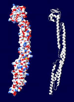 Space-filling model and ribbon diagram of Caulobacter vibrioides crescentin, created from homology model available at Q6IET3
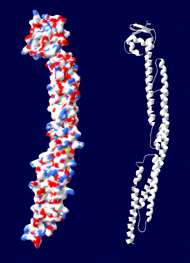
Crescentin is a protein which is a bacterial relative of the intermediate filaments found in eukaryotic cells. Just as tubulins and actins, the other major cytoskeletal proteins, have prokaryotic homologs in, respectively, the FtsZ and MreB proteins, intermediate filaments are linked to the crescentin protein.
Contents
Role in cell shape
Crescentin was recently discovered in the prokaryote Caulobacter crescentus, an aquatic bacterium which uses its crescent-shaped cells for enhanced motility.[1] The crescentin protein is located on the concave face of these cells and appears to be necessary for their shape, as mutants lacking the protein form rod-shaped cells.[2] To influence the shape of the Caulobacter cells, the helices of crescentin filaments associate with the cytoplasmic side of the cell membrane on one lateral side of the cell. This induces a curved cell shape in younger cells, which are shorter than the helical pitch of crescentin, but induces a spiral shape in older, longer cells.[3]
Protein structure
Like eukaryotic intermediate filaments, crescentin organizes into filaments and is present in a helical structure in the cell. Crescentin is necessary for both shapes of the Caulobacter prokaryote (vibroid/crescent-shape and helical shape, which it may adopt after a long stationary phase). The crescentin protein has 430 residues; its sequence mostly consists of a pattern of 7 repeated residues which form a coiled-coil structure. The DNA sequence of the protein has sections very similar to the eukaryotic keratin and lamin proteins, mostly involving the coiled-coil structure. Researchers Ausmees et al. recently proved that, like animal intermediate filament proteins, crescentin has a central rod made up of four coiled-coil segments.[4] Both intermediate filament and crescentin proteins have a primary sequence including four α-helical segments along with non-α-helical linker domains. An important difference between crescentin and animal intermediate filament proteins is that crescentin lacks certain consensus sequence elements at the ends of the rod domain which are conserved in animal lamin and keratin proteins.[5]
Assembly of filaments
Eukaryotic intermediate filament proteins assemble into filaments of 8-15 nm within the cell without the need for energy input, that is, no need for ATP or GTP. Ausmees et al. continued their crescentin research by testing whether the protein could assemble into filaments in this manner in vitro. They found that crescentin proteins were indeed able to form filaments about 10 nm wide, and that some of these filaments organized laterally into bundles, just as eukaryotic intermediate filaments do.[4] The similarity of crescentin protein to intermediate filament proteins suggests an evolutionary linkage between these two cytoskeletal proteins.
References
- ^ Biosciences, Eurekah.com, 2000-2005. Accessed 19 February. 2006 through PubMed.
- ^ Møller-Jensen, Jakob, and Löwe, Jan. “Increasing complexity of the bacterial cytoskeleton.” Curr. Opin. Cell. Biol. 2005 Feb.; 17(1): 75-81.
- ^ Margolin, William. “Bacterial shapes: concave coiled coils curve Caulobacter.” Curr. Biol. 2004 Mar.; 14: 242-244.
- ^ a b Ausmees, N. et al. “The bacterial cytoskeleton: an intermediate filament-like function in cell shape.” Cell. 2003 12 December;115(6):705-13.
- ^ Herrmann, Harald and Aebi, Ueli. “Intermediate filaments: Molecular structure, assembly mechanism, and integration into functionally distinct intracellular scaffolds.” Ann. Rev. Biochem. 73: 749-789.
Proteins of the cytoskeleton Human I (MYO1A, MYO1B, MYO1C, MYO1D, MYO1E, MYO1F, MYO1G, MYO1H) · II (MYH1, MYH2, MYH3, MYH4, MYH6, MYH7, MYH7B, MYH8, MYH9, MYH10, MYH11, MYH13, MYH14, MYH15, MYH16) · III (MYO3A, MYO3B) · V (MYO5A, MYO5B, MYO5C) · VI (MYO6) · VII (MYO7A, MYO7B) · IX (MYO9A, MYO9B) · X (MYO10) · XV (MYO15A) · XVIII (MYO18A, MYO18B) · LC (MYL1, MYL2, MYL3, MYL4, MYL5, MYL6, MYL6B, MYL7, MYL9, MYLIP, MYLK, MYLK2, MYLL1)OtherOtherEpithelial keratins
(soft alpha-keratins)Hair keratins
(hard alpha-keratins)Ungrouped alphaNot alphaType 3Type 4Type 5OtherOtherNonhuman see also cytoskeletal defects
B strc: edmb (perx), skel (ctrs), epit, cili, mito, nucl (chro)Categories:
Wikimedia Foundation. 2010.