- Esophagogastroduodenoscopy
-
"EGD" redirects here. For the computer software Entropy Gathering Daemon, see /dev/random.
For other expansions of the initialism "OGD", see the disambiguation page.
Esophagogastroduodenoscopy Intervention 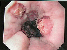
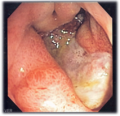
Endoscopic still of esophageal ulcers seen after banding of esophageal varices, at time of esophagogastroduodenosocopyICD-9-CM 45.13 MeSH D016145 OPS-301 code: 1-631, 1-632 In medicine (gastroenterology), esophagogastroduodenoscopy is a diagnostic endoscopic procedure that visualizes the upper part of the gastrointestinal tract up to the duodenum. It is considered a minimally invasive procedure since it does not require an incision into one of the major body cavities and does not require any significant recovery after the procedure (unless sedation or anesthesia has been used). However, a sore throat is common.[1][2][3]
Contents
Alternative names
Esophagogastroduodenoscopy may be abbreviated EGD, or OGD if one uses the British spelling oesophagogastroduodenoscopy. It is also called upper GI endoscopy (UGIE), gastroscopy or simply endoscopy (since it is the most commonly performed type of endoscopy, the ambiguous term 'endoscopy' refers to EGD by default).
Indications
Diagnostic
- Unexplained anemia (usually along with a colonoscopy)
- Upper gastrointestinal bleeding as evidenced by hematemesis or melena
- Persistent dyspepsia in patients over the age of 45 years
- Heartburn and chronic acid reflux - this can lead to a precancerous lesion called Barrett's esophagus
- Persistent vomiting
- Dysphagia - difficulty in swallowing
- Odynophagia - painful swallowing
Surveillance
- Surveillance of Barrett's esophagus
- Surveillance of gastric ulcer or duodenal ulcer
- Occasionally after gastric surgery
Confirmation of diagnosis/biopsy
- Abnormal barium swallow or barium meal
- Confirmation of celiac disease (via biopsy)
Therapeutic
- Treatment (banding/sclerotherapy) of esophageal varices
- Injection therapy (e.g. epinephrine in bleeding lesions)
- Cutting off of larger pieces of tissue with a snare device (e.g. polyps, endoscopic mucosal resection)
- Application of cautery to tissues
- Removal of foreign bodies (e.g. food) that have been ingested
- Tamponade of bleeding esophageal varices with a balloon
- Application of photodynamic therapy for treatment of esophageal malignancies
- Endoscopic drainage of pancreatic pseudocyst
- Tightening the lower esophageal sphincter
- Dilating or stenting of stenosis or achalasia
- Percutaneous endoscopic gastrostomy (feeding tube placement)
- Endoscopic retrograde cholangiopancreatography (ERCP) combines EGD with fluoroscopy
- Endoscopic ultrasound (EUS) combines EGD with 5–12 MHz ultrasound imaging
Newer interventions
- Endoscopic trans-gastric laparoscopy
- Placement of gastric balloons in bariatric surgery
Equipment
- Endoscope
- Non-coaxial optic fiber system to carry light to the tip of the endoscope
- A chip camera at the tip of the endoscope - this has now replaced the coaxial optic fibers of older scopes that were prone to damage and consequent loss of picture quality
- Irrigation channel to clean the lens
- Suction/Insufflation/Working channels - these may be in the form of one or more channels
- Control handle - this houses the controls
- Stack
- Light source
- Insufflator
- Suction
- Electrosurgical unit
- Video recorder/photo printer
- Instruments
- Biopsy forceps
- Snares
- Injecting needles
Procedure
The patient is kept NPO (Nil per os) or NBM (Nothing By Mouth) that is, told not to eat, for at least 4–6 hours before the procedure. Most patients tolerate the procedure with only topical anesthesia of the oropharynx using lidocaine spray. However, some patients may need sedation and the very anxious/agitated patient may even need a general anesthetic. Informed consent is obtained before the procedure. The main risks are bleeding and perforation. The risk is increased when a biopsy or other intervention is performed.
The patient lies on his/her left side with the head resting comfortably on a pillow. A mouth-guard is placed between the teeth to prevent the patient from biting on the endoscope. The endoscope is then passed over the tongue and into the oropharynx. This is the most uncomfortable stage for the patient. Quick and gentle manipulation under vision guides the endoscope into the esophagus. The endoscope is gradually advanced down the esophagus making note of any pathology. Excessive insufflation of the stomach is avoided at this stage. The endoscope is quickly passed through the stomach and through the pylorus to examine the first and second parts of the duodenum. Once this has been completed, the endoscope is withdrawn into the stomach and a more thorough examination is performed including a J-maneuver. This involves retroflexing the tip of the scope so it resembles a 'J' shape in order to examine the fundus and gastroesophageal junction. Any additional procedures are performed at this stage. The air in the stomach is aspirated before removing the endoscope. Still photographs can be made during the procedure and later shown to the patient to help explain any findings.
In its most basic use, the endoscope is used to inspect the internal anatomy of the digestive tract. Often inspection alone is sufficient, but biopsy is a very valuable adjunct to endoscopy. Small biopsies can be made with a pincer (biopsy forceps) which is passed through the scope and allows sampling of 1 to 3 mm pieces of tissue under direct vision. The intestinal mucosa heals quickly from such biopsies.
Biopsy allows the pathologist to render an opinion on later histologic examination of the biopsy tissue with light microscopy and/or immunohistochemistry. Biopsied material can also be tested on urease to identify Helicobacter pylori.
Complications
The complication rate is about 1 in 1000. They include:
- aspiration, causing aspiration pneumonia
- bleeding
- perforation
- cardiopulmonary problems
Limitations
Problems of gastrointestinal function are usually not well diagnosed by endoscopy since motion or secretion of the gastrointestinal tract are not easily inspected by EGD. Nonetheless, findings such as excess fluid or poor motion of gut during endoscopy can be suggestive of disorders of function. Irritable bowel syndrome and functional dyspepsia is not diagnosed with EGD, but EGD may be helpful in excluding other diseases that mimic these common disorders.
Additional images
-
Endoscopic image of adenocarcinoma of duodenum seen in the post-bulbar duodenum.
-
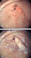
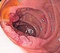
Endoscopic image of gastric antral vascular ectasia seen as a radial pattern around the pylorus before (top) and after (bottom) treatment with argon plasma coagulation
-
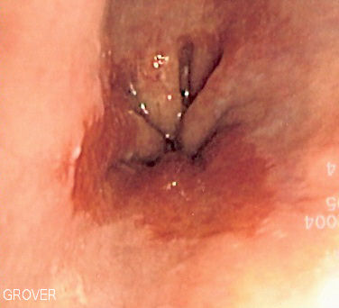
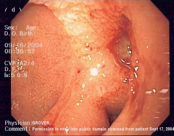
Endoscopic image of Barrett's esophagus, which is the area of red mucosa projecting like a tongue.
-
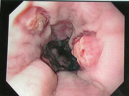
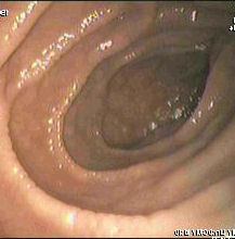
Deep gastric ulcer
-

Endoscopic still of duodenum of patient with celiac disease showing scalloping of folds.
-
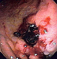
Gastric ulcer in antrum of stomach with overlying clot due to gastric lymphoma.
-

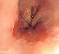
Endoscopic image of a posterior wall duodenal ulcer with a clean base, which is a common cause of upper GI hemorrhage.
References
- ^ "Gastroscopy - examination of oesophagus and stomach by endoscope". BUPA. December 2006. http://hcd2.bupa.co.uk/fact_sheets/html/Gastrointestinal.html. Retrieved 2007-10-07.
- ^ National Digestive Diseases Information Clearinghouse (November 2004). "Upper Endoscopy". National Institutes of Health. http://digestive.niddk.nih.gov/ddiseases/pubs/upperendoscopy/index.htm. Retrieved 2007-10-07.
- ^ "What is Upper GI Endoscopy?". Patient Center -- Procedures. American Gastroenterological Association. Archived from the original on 2007-09-28. http://web.archive.org/web/20070928014913/http://www.gastro.org/wmspage.cfm?parm1=859. Retrieved 2007-10-07.
See also
- Colonscopy
Endoscopy GI tract upper: pharyngoscopy (pharynx) · esophagogastroduodenoscopy (esophagus, stomach, duodenum)
lower: enteroscopy (small intestine) · colonoscopy (colon) · sigmoidoscopy (sigmoid colon, rectum) · rectoscopy (rectum) · proctoscopy (rectum, anus) · anoscopy (anus) · capsule endoscopy
accessory: cholangioscopy (bile duct)Respiratory tract Urinary tract Female reproductive system gynoscopy · colposcopy (cervix) · hysteroscopy (uterus) · falloposcopy (fallopian tubes) · culdoscopyClosed cavity via incision laparoscopy [peritoneoscopy) (abdominal, pelvic cavity) · arthroscopy (joint cavity) · thoracoscopy (pleural cavity) · mediastinoscopy (mediastinum) · coelioscopy (abdominal cavity)During pregnancy Cardiovascular angioscopyOthers Categories:- Endoscopy
- Diagnostic gastroenterology


Wikimedia Foundation. 2010.