- Cataract
-
Cataract Classification and external resources 
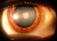
Magnified view of cataract in human eye, seen on examination with a slit lamp using diffuse illuminationICD-10 H25-H26, H28, Q12.0 ICD-9 366 DiseasesDB 2179 MedlinePlus 001001 A cataract is a clouding that develops in the crystalline lens of the eye or in its envelope, varying in degree from slight to complete opacity and obstructing the passage of light. Early in the development of age-related cataract, the power of the lens may be increased, causing near-sightedness (myopia), and the gradual yellowing and opacification of the lens may reduce the perception of blue colours. Cataracts typically progress slowly to cause vision loss, and are potentially blinding if untreated. The condition usually affects both eyes, but almost always one eye is affected earlier than the other.[1]
A senile cataract, occurring in the elderly, is characterized by an initial opacity in the lens, subsequent swelling of the lens and final shrinkage with complete loss of transparency.[2] Moreover, with time the cataract cortex liquefies to form a milky white fluid in a Morgagnian cataract, which can cause severe inflammation if the lens capsule ruptures and leaks. Untreated, the cataract can cause phacomorphic glaucoma. Very advanced cataracts with weak zonules are liable to dislocation anteriorly or posteriorly. Such spontaneous posterior dislocations (akin to the historical surgical procedure of couching) in ancient times were regarded as a blessing from the heavens, because some perception of light was restored in the cataractous patients.
Some children develop cataracts, called congenital cataracts, before or just after birth, but these are usually dealt with in a different way to cataracts in adults.[3]
Cataract derives from the Latin cataracta meaning "waterfall" and that from the Greek καταράκτης (kataraktēs) or καταρράκτης (katarrhaktēs), "down-rushing",[4] from καταράσσω (katarassō) meaning "to dash down"[5] (from kata-, "down"; arassein, "to strike, dash").[6][7] As rapidly running water turns white, the term may later have been used metaphorically to describe the similar appearance of mature ocular opacities. In Latin, cataracta had the alternate meaning "portcullis"[8] and it is possible that the name passed through French to form the English meaning "eye disease" (early 15c.), on the notion of "obstruction".[9] Early Persian physicians called the term nazul-i-ah, or "descent of the water"—vulgarised into waterfall disease or cataract—believing such blindness to be caused by an outpouring of corrupt humour into the eye.[10]
Contents
Epidemiology
 Disability-adjusted life year for cataracts per 100,000 inhabitants in 2004.[11]
 no dataless than 9090-180180-270270-360360-450450-540540-630630-720720-810810-900900-990more than 990
Age-related cataract is responsible for 48% of world blindness, which represents about 18 million people, according to the World Health Organization (WHO).[12] In many countries, surgical services are inadequate, and cataracts remain the leading cause of blindness. As populations age, the number of people with cataracts is growing. Cataracts are also an important cause of low vision in both developed and developing countries. Even where surgical services are available, low vision associated with cataracts may still be prevalent, as a result of long waits for operations and barriers to surgical uptake, such as cost, lack of information and transportation problems.
In the United States, age-related lenticular changes have been reported in 42% of those between the ages of 52 and 64,[13] 60% of those between the ages 65 and 74,[14] and 91% of those between the ages of 75 and 85.[13]
The increase in ultraviolet radiation resulting from depletion of the ozone layer is expected to increase the incidence of cataracts.[15]
History
The earliest records are from the Bible[verification needed], as well as early Hindu records.[16][verification needed] Early cataract surgery was developed by the Indian surgeon, Sushruta (6th century BCE).[17][verification needed] The Indian tradition of cataract surgery was performed with a special tool called the jabamukhi salaka, a curved needle used to loosen the lens and push the cataract out of the field of vision.[17] The eye would later be soaked with warm butter and then bandaged.[17] Though this method was successful, Sushruta cautioned that it should only be used when necessary.[17] Greek physicians and philosophers traveled to India where these surgeries were performed by physicians.[17] The removal of cataract by surgery was also introduced into China from India.[18]
The first references to cataract and its treatment in Ancient Rome are found in 29 CE in De Medicinae, the work of the Latin encyclopedist Aulus Cornelius Celsus.[19] The Romans were pioneers in the health arena—particularly in the area of eye care.[20]
The Muslim ophthalmologist Ammar ibn Ali, in his Choice of Eye Diseases, written circa 1000 CE, wrote of his invention of the hypodermic needle and how he discovered the technique of cataract extraction while experimenting with it on a patient.[21]
Classification
The following is a classification of the various types of cataracts. This is not comprehensive, and other unusual types may be noted.
- Classified by etiology
-
- Age-related cataract
-
- Cortical senile cataract
-
- Immature senile cataract (IMSC): partially opaque lens, disc view hazy
- Mature senile cataract (MSC): completely opaque lens, no disc view
- Hypermature senile cataract (HMSC): liquefied cortical matter: Morgagnian cataract
- Senile nuclear cataract
-
- Cataracta brunescens
- Cataracta nigra
- Cataracta rubra
- Congenital cataract
-
- Sutural cataract
- Lamellar cataract
- Zonular cataract
- Total cataract
- Secondary cataract
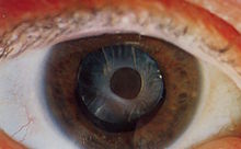
 Slit lamp photo of anterior capsular opacification visible a few months after implantation of intraocular lens in eye, magnified view
-
-
- Drug-induced cataract (e.g. corticosteroids)
- Traumatic cataract
-
- Blunt trauma (capsule usually intact)
- Penetrating trauma (capsular rupture and leakage of lens material—calls for an emergency surgery for extraction of lens and leaked material to minimize further damage)
-
- Classified by opacities, cataract can be classified by using Lens Opacities Classification System III (LOCS III: Nuclear NC1-5, Cortical C1-5 and Posterior P1-5. By application planning in procedures of phacoemulsification, LCOS III can be converted in newer cataract grading systems. Gede Pardianto (2009) introduced Optical Biometry Based Cataract Grading System (OBBCGS) that is helpful in cataract grading due to phacoemulsification planning. LOCS III's NC0, C0 and P0 can be converted as OBBCGS' no cataract (NC), LOCS III's NC1-3, C1-3, P1-4 can be converted to OBBCGS' Optical Biometry Examined Cataract (OBEC) and LOCS III's NC4-5, C4-5, P4-5 can be converted to OBBCGS's Optical Biometry Unexamined Cataract (OBUC); that need examination by Applanation Ultrasound Biometry.[22]
- Classified by location of opacity within lens structure (however, mixed morphology is quite commonly seen, e.g. PSC with nuclear changes and cortical spokes of cataract)
-
- Anterior cortical cataract
- Anterior polar cataract
- Anterior subcapsular cataract
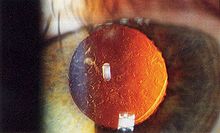
 Slit lamp photo of posterior capsular opacification visible a few months after implantation of intraocular lens in eye, seen on retroillumination
-
- Nuclear cataract—grading correlates with hardness and difficulty of surgical removal
- Posterior cortical cataract
- Posterior polar cataract (importance lies in higher risk of complication—posterior capsular tears during surgery)
- Posterior subcapsular cataract (PSC) (clinically common)
- After-cataract: posterior capsular opacification (PCO) subsequent to a successful extracapsular cataract surgery (usually within three months to two years) with or without IOL implantation. Requires a quick and painless office procedure with Nd:YAG laser capsulotomy to restore optical clarity.
Signs and symptoms
As a cataract becomes more opaque, clear vision is compromised. A loss of visual acuity is noted. Contrast sensitivity is also lost, so that contours, shadows and color vision are less vivid. Veiling glare can be a problem as light is scattered by the cataract into the eye. The affected eye will have an absent red reflex. A contrast sensitivity test should be performed, and if a loss in contrast sensitivity is demonstrated, an eye specialist consultation is recommended.
It may be advisable to seek medical opinion, particularly in high-risk groups such as diabetics, if a "halo" is observed around street lights at night, especially if this phenomenon appears to be confined to one eye only.
The symptoms of cataracts are very similar to the symptoms of ocular citrosis.
Causes
Cataracts develop for a variety of reasons, including long-term exposure to ultraviolet light, exposure to radiation, secondary effects of diseases such as diabetes, hypertension and advanced age, or trauma (possibly much earlier); they are usually a result of denaturation of lens protein. Genetic factors are often a cause of congenital cataracts, and positive family history may also play a role in predisposing someone to cataracts at an earlier age, a phenomenon of "anticipation" in presenile cataracts. Cataracts may also be produced by eye injury or physical trauma. A study among Icelandair pilots showed commercial airline pilots are three times more likely to develop cataracts than people with nonflying jobs. This is thought to be caused by excessive exposure to radiation coming from outer space.[23] Supporting this theory is the report that 33 of the 36 Apollo astronauts involved in the nine Apollo missions to leave Earth orbit have developed early stage cataracts that have been shown to be caused by radiation exposure to cosmic rays during their trip.[24] At least 39 former astronauts have developed cataracts; 36 of those were involved in high-radiation missions such as the Apollo missions.[25] Cataracts are also unusually common in persons exposed to infrared radiation, such as glassblowers, who suffer from "exfoliation syndrome". Exposure to microwave radiation can cause cataracts. Atopic or allergic conditions are also known to quicken the progression of cataracts, especially in children.[26] Cataracts can also be caused by iodine deficiency.[27]
Cataracts may be partial or complete, stationary or progressive, hard or soft.
Some drugs can induce cataract development, such as corticosteroids[28] and Seroquel.
There are various types of cataracts, e.g. nuclear, cortical, mature, and hypermature. Cataracts are also classified by their location, e.g. posterior (classically due to steroid use)[28][29] and anterior (common (senile) cataract related to aging).
Associations with systemic conditions
- Chromosomal disorders
-
- 1q21.1 deletion syndrome
- Cri-du-chat syndrome
- Patau's syndrome
- Schmid-Fraccaro syndrome
- Trisomy 18 (Edward's syndrome)
- Turner's syndrome
- Single-gene disorders
-
- Alport's syndrome
- Conradi's syndrome
- Myotonic dystrophy
- Autosomal dominant severe keratoconus with anterior polar cataract caused by mutation of the miR-184 seed region[30]
- Disease of the skin and mucous membrans
-
- Atopic dermatitis
- Basal-cell nevus syndrome
- Ichthyosis
- Pemphigus
- Metabolic and nutrition diseases
-
- Aminoaciduria (Lowe's syndrome)
- Diabetes mellitus
- Fabry's disease
- Galactosemia / galactosemic cataract
- Homocystinuria
- Hyperparathyroidism
- Hypervitaminosis D
- Hypothyroidism
- Mucopolysaccharidoses
- Wilson's disease
-
- Congenital
-
- Congenital herpes simplex
- Congenital syphilis
- Cytomegalic inclusion disease
- Rubella
- Others
- Toxic substances introduced systemically
-
- Corticosteroids
- Haloperidol
- Miotics
- Triparanol
Prevention
Although cataracts have no scientifically proven prevention, wearing ultraviolet-protecting sunglasses may slow the development of cataracts.[31][32] Regular intake of antioxidants (such as vitamins A, C and E) is theoretically helpful, but taking them as a supplement has not been shown to have a benefit.[33] The less well-known antioxidant N-acetylcarnosine has been shown in randomized controlled clinical trials to treat cataracts, and can be expected to prevent their formation by similar mechanisms.[34] N-acetylcarnosine is a proposed treatment for other ocular disorders that are instigated, or exacerbated, by oxidative stress, including glaucoma, retinal degeneration, corneal disorders, and ocular inflammation.[35]
Treatment
Nonsurgical
See Prevention above.
Surgical
The operation to remove cataracts can be performed at any stage of their development. There is no longer a reason to wait until a cataract is "ripe" before removing it.[36] However, because any surgery involves some risk, it is usually worth waiting until there is some change in vision before removing the cataract.[36]
The most effective and common treatment is to make an incision (capsulotomy) into the capsule of the cloudy lens to surgically remove it. Two types of eye surgery can be used to remove cataracts: extracapsular cataract extraction (ECCE) and intracapsular cataract extraction (ICCE).
ECCE surgery consists of removing the lens, but leaving the majority of the lens capsule intact. High frequency sound waves (phacoemulsification) are sometimes used to break up the lens before extraction.
Intra-capsular (ICCE) surgery involves removing the entire lens of the eye, including the lens capsule, but it is rarely performed in modern practice.
In either extracapsular surgery or intracapsular surgery, the cataractous lens is removed and replaced with a plastic lens (an intraocular lens implant) which stays in the eye permanently.
Cataract operations are usually performed using a local anaesthetic, and the patient is allowed to go home the same day. Recent improvements in intraocular technology now allow cataract patients to choose a multifocal lens to create a visual environment in which they are less dependent on glasses. Such multifocal lenses are flexible and can be controlled using the same eye muscles used to control the natural lens. Under some medical systems, multifocal lenses cost extra. Traditional intraocular lenses are monofocal.
Complications are possible after cataract surgery, including endophthalmitis, posterior capsular opacification and retinal detachment.
Research
Research is scant and mixed, but weakly positive, for the nutrients lutein and zeaxanthin.[37][38][39][40] Bilberry extract shows promise in rat models[41][42] and in clinical studies.[43]
Investigational treatments
In the past few years, eye drops containing acetyl-carnosine have been used by several thousands of cataract patients across the world. The drops are believed to work by reducing oxidation and glycation damage in the lens, particularly reducing crystallin crosslinking.[44][45] Randomized controlled trials indicate the drops may be especially appropriate for seniors, or others where surgery is not advised.[46]
Investigational preventives
Although statins are known for their ability to lower lipids, they are also believed to have antioxidant qualities. Oxidative stress is believed to play a role in the development of nuclear cataracts, which are the most common type of age-related cataracts. To explore the relationship between nuclear cataracts and statin use, a group of researchers treated a group of 1299 patients who were at risk of developing nuclear cataracts with statins. Their results suggest statin use in an at-risk population may be associated with a lower risk of developing nuclear cataract disease.[47][non-primary source needed]
Long term (average five year) observation showed systematic application of azapentacene sodium polysulfonate (Quinax) slows down the progress of the disease.[48]
See also
- Bates method
- List of eye diseases
- List of eye diseases and disorders
- List of systemic diseases with ocular manifestations
References and notes
- Pavan-Langston, Deborah (1990). Manual of Ocular Diagnosis and Therapy. Little, Brown and Company.
- ^ "Common Causes of Vision Loss in Elderly Patients - July 1999 - American Academy of Family Physicians". Aafp.org. 1999-07-01. http://www.aafp.org/afp/990700ap/99.html. Retrieved 2011-11-22.
- ^ eMedicine - Cataract, Senile : Article by Vicente Victor D Ocampo. From eMedicine The Continually Updated Clinical Reference
- ^ [1] Congenital cataracts - RNIB.
- ^ καταρράκτης, Henry George Liddell, Robert Scott, A Greek-English Lexicon, on Perseus
- ^ καταράσσω, Henry George Liddell, Robert Scott, A Greek-English Lexicon, on Perseus
- ^ Dictionary.reference.com, Word of the Day Archive/cataract
- ^ cataract, on Oxford Dictionaries
- ^ cataracta, Charlton T. Lewis, Charles Short, A Latin Dictionary, on Perseus
- ^ Online Etymology Dictionary, etymonline.com
- ^ Mistaken Science - Topic Powered by eve community, Wordcraft Forums, wordcraft.infopop.cc
- ^ "Death and DALY estimates for 2004 by cause for WHO Member States" (xls). World Health Organization. who.int. 2004. http://www.who.int/entity/healthinfo/global_burden_disease/gbddeathdalycountryestimates2004.xls.
- ^ WHO.int, | Priority eye diseases.
- ^ a b Sperduto RD, Seigel D., Seigel D. Sperduto RD (Jul 1980). "Senile lens and senile macular changes in a population-based sample". Am J Ophthalmol 90 (1): 86–91. PMID 7395962.
- ^ Kahn HA, Leibowitz HM, Ganley JP, Kini MM, Colton T, Nickerson RS, Dawber TR (Jul 1977). "The Framingham Eye Study. I. Outline and major prevalence findings". Am J Epidemiol 106 (1): 17–32. PMID 879158.
- ^ Dobson, R. (2005). "Ozone depletion will bring big rise in number of cataracts". BMJ 331 (7528): 1292–1295. doi:10.1136/bmj.331.7528.1292-d. PMC 1298891. PMID 0. http://www.pubmedcentral.nih.gov/articlerender.fcgi?tool=pmcentrez&artid=1298891.
- ^ A short history of cataract surgery, rila.co.uk
- ^ a b c d e Finger, page 66
- ^ Lade & Svoboda, page 85
- ^ Cataract history, mrcophth.com
- ^ "The Romans carried out cataract ops". BBC News. February 9, 2008. http://news.bbc.co.uk/2/hi/health/7194352.stm.
- ^ Finger, Stanley (1994). Origins of Neuroscience: A History of Explorations Into Brain Function. Oxford University Press. p. 70. ISBN 0195146948
- ^ Pardianto G (2009). "Mastering Phacoemulsification in Mimbar Ilmiah". Oftalmologi Indonesia 10: 24–5.
- ^ Rafnsson, V; Olafsdottir E, Hrafnkelsson J, Sasaki H, Arnarsson A, Jonasson F (2005). "Cosmic radiation increases the risk of nuclear cataract in airline pilots: a population-based case-control study". Arch Ophthalmol 123 (8): 1102–1105. doi:10.1001/archopht.123.8.1102. PMID 16087845.
- ^ See Ms. Irene Schneider on the November 20, 2005 episode of The Space Show.
- ^ Barry, Patrick L.. "Blinding Flashes". Science.nasa.gov. http://science.nasa.gov/headlines/y2004/22oct_cataracts.htm. Retrieved November 25, 2008.
- ^ Chen CC, Huang JL, Yang KD, Chen HJ (March 2000). "Atopic cataracts in a child with atopic dermatitis: a case report and review of the literature". Asian Pac. J. Allergy Immunol. 18 (1): 69–71. PMID 12546060.
- ^ Buchberger W, Winkler R, Moser M, Rieger G (1991). "Influence of iodide on cataractogenesis in Emory mice". Ophthalmic Res 23 (6): 303–8. doi:10.1159/000267127. PMID 1803312.
- ^ a b Spencer R., Andelman S. (1965). "Steroid Cataracts. Posterior Subcapsular Cataract Formation In Rheumatoid Arthritis Patients On Long Term Steroid Therapy". Arch Ophthalmol 74: 38–41. PMID 14303339.
- ^ Greiner J, Chylack L (1979). "Posterior subcapsular cataracts: histopathologic study of steroid-associated cataracts". Arch Ophthalmol 97 (1): 135–44. PMID 758890.
- ^ Hughes AE, Bradley DT, Campbell M, Lechner J, Dash DP, Simpson DA, Willoughby CE (2011). "Mutation Altering the miR-184 Seed Region Causes Familial Keratoconus with Cataract". The American Journal of Human Genetics. doi:10.1016/j.ajhg.2011.09.014. http://www.cell.com/AJHG/abstract/S0002-9297(11)00404-6. Retrieved 14/10/2011.
- ^ Neale, RE; JL Purdie, LW Hirst, and AC Green (2003-11). "Sun exposure as a risk factor for nuclear cataract". Epidemiology 14 (6): 707–712. doi:10.1097/01.ede.0000086881.84657.98. PMID 14569187.
- ^ Javitt, JC; Wang, F; West, SK (1996). "Blindness Due to Cataract: Epidemiology and Prevention". Annual review of public health 17: 159–77. doi:10.1146/annurev.pu.17.050196.001111. PMID 8724222. http://www.nei.nih.gov/nehep/pdf/NEHEP_5_year_agenda_2006.pdf. Cited in Five-Year Agenda for the National Eye Health Education Program (NEHEP), p. B-2; National Eye Institute, U.S. National Institutes of Health
- ^ Age-Related Eye Disease Study Research Group (October 2001). "A randomized, placebo-controlled, clinical trial of high-dose supplementation with vitamins C and E and beta carotene for age-related cataract and vision loss: AREDS report no. 9.". Archives of ophthalmology 119 (10): 1439–52. doi:10.1001/archopht.119.10.1439. PMC 1472812. PMID 11594943. http://archopht.ama-assn.org/cgi/content/full/119/10/1439.
- ^ Babizhayev MA, Deyev AI, Yermakova VN, et al. (June 2001). "N-Acetylcarnosine, a natural histidine-containing dipeptide, as a potent ophthalmic drug in treatment of human cataracts.". Peptides 22 (6): 979–94. doi:10.1016/S0196-9781(01)00407-7. PMID 11390029.
- ^ Babizhayev MA, Yermakova VN, Semiletov YA, Deyev AI (May 2000). "The natural histidine-containing dipeptide Nalpha-acetylcarnosine as an antioxidant for ophthalmic use.". Biochemistry. Biokhimiia 65 (5): 588–98. PMID 10851037.
- ^ a b "Cataracts". RNIB. 2011-06-15. http://www.rnib.org.uk/eyehealth/eyeconditions/conditionsac/Pages/cataract.aspx. Retrieved 2011-11-22.
- ^ Olmedilla, B; Granado, F; Blanco, I; Vaquero, M (2003). "Lutein, but not alpha-tocopherol, supplementation improves visual function in patients with age-related cataracts: a 2-y double-blind, placebo-controlled pilot study.". Nutrition (Burbank, Los Angeles County, Calif.) 19 (1): 21–4. PMID 12507634.
- ^ Vu, HT; Robman, L; Hodge, A; McCarty, CA; Taylor, HR (2006). "Lutein and zeaxanthin and the risk of cataract: the Melbourne visual impairment project". Investigative ophthalmology & visual science 47 (9): 3783–6. doi:10.1167/iovs.05-0587. PMID 16936087. http://findarticles.com/p/articles/mi_m0FDN/is_4_11/ai_n27098237/.
- ^ Delcourt, C; Carrière, I; Delage, M; Barberger-Gateau, P; Schalch, W; Pola Study, Group (2006). "Plasma lutein and zeaxanthin and other carotenoids as modifiable risk factors for age-related maculopathy and cataract: the POLA Study.". Investigative ophthalmology & visual science 47 (6): 2329–35. doi:10.1167/iovs.05-1235. PMID 16723441.
- ^ Ribaya-Mercado, JD; Blumberg, JB (2004). "Lutein and zeaxanthin and their potential roles in disease prevention.". Journal of the American College of Nutrition 23 (6 Suppl): 567S–587S. PMID 15640510.
- ^ Fursova AZh, Gesarevich OG, Gonchar AM, Trofimova NA, Kolosova NG (2005). "Dietary supplementation with bilberry extract prevents macular degeneration and cataracts in senesce-accelerated OXYS rats". Advances in gerontology = Uspekhi gerontologii / Rossiiskaia akademiia nauk, Gerontologicheskoe obshchestvo 16: 76–9. PMID 16075680.
- ^ Yamakoshi J et al. (2002). "Procyanidin-rich extract from grape seeds prevents cataract formation in hereditary cataractous (ICR/f) rats.". J. Agric Food Chem. 50 (17): 4983–8. doi:10.1021/jf0201632. PMID 12166994.
- ^ Ann Ottalmol Clin Ocul, 1989
- ^ Williams DL, Munday P (2006). "The effect of a topical antioxidant formulation including N-acetyl carnosine on canine cataract: a preliminary study". Vet Ophthalmol 9 (5): 311–6. doi:10.1111/j.1463-5224.2006.00492.x. PMID 16939459.
- ^ Guo Y, Yan H (2006). "Preventive effect of carnosine on cataract development". Yan Ke Xue Bao. 22 (2): 85–8. PMID 17162883.
- ^ Babizhayev MA, Deyev AI, Yermakova VN, et al. (2002). "Efficacy of N-acetylcarnosine in the treatment of cataracts.". Drugs in R&D 3 (2): 87–103. doi:10.2165/00126839-200203020-00004. PMID 12001824.
- ^ Klein, Barbara; Ronald Klein, Kristine Lee, and Lisa Grady (2006). "Statin Use and Incident Nuclear Cataract". Journal of the American Medical Association 295 (23): 2752–2758. doi:10.1001/jama.295.23.2752. PMID 16788130.
- ^ Stankiewicz A, Poppe E, Stasiewicz B, Gołebiowska-Hrycukowa A (August 1990). "Evaluation of the effectiveness of Quinax in the prevention of the development of senile cataract.". Klinika oczna 92 (3-4): 52–4. PMID 2263035.
External links
- CATRA: Cataract Maps with Snap-on Eyepiece for Mobile Phones, MIT Media Lab
- Cataract Resource Guide from the National Eye Institute (NEI), nei.nih.gov
- eMedicine Health, emedicinehealth.com
- The Childhood Cataract Network, childhoodcataracts.org.uk
- Pediatric Glaucoma and Cataract Family Association, pgcfa.org
- Royal National Institute for Blind People - Cataracts
Categories:- Aging-associated diseases
- Blindness
- Disorders of lens
- Greek loanwords
Wikimedia Foundation. 2010.


