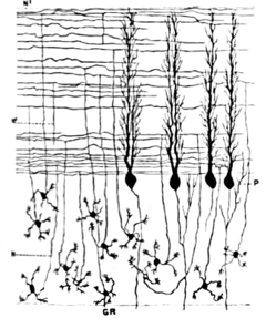
- Cerebellum granule cell
-
Neuron: Cerebellar granule cell 
Granule cells, parallel fibers, and flattened dendritic trees of Purkinje cellsLocation Cerebellum Function excitatory Neurotransmitter glutamate Morphology small cell with few dendrites Presynaptic connections Mossy fibers and Golgi cells Postsynaptic connections Parallel fibers to cerebellar cortex Granule cells of the cerebellum are among the smallest neurons in the brain. (The term granule cell is used for several unrelated types of small neurons in various parts of the brain.) Cerebellar granule cells are also easily the most numerous neurons in the brain: in humans, estimates of their total number average around 50 billion, which means that they constitute about 3/4 of the brain's neurons.[1]
Description
The cell bodies are packed into a thick layer at the bottom of the cerebellar cortex. A granule cell emits only four to five dendrites, each of which ends in an enlargement called a dendritic claw.[1] These enlargements are sites of excitatory input from mossy fibers and inhibitory input from Golgi cells.
The thin, unmyelinated axons of granule cells rise vertically to the upper (molecular) layer of the cortex, where they split in two, with each branch traveling horizontally to form a parallel fiber; the splitting of the vertical branch into two horizontal branches gives rise to a distinctive "T" shape. A parallel fiber runs for an average of 3 mm in each direction from the split, for a total length of about 6 mm (about 1/10 of the total width of the cortical layer).[1] As they run along, the parallel fibers pass through the dendritic trees of Purkinje cells, contacting one of every 3-5 that they pass, making a total of 80-100 synaptic connections with Purkinje cell dendritic spines.[1] Granule cells use glutamate as their neurotransmitter, and therefore exert excitatory effects on their targets.
Function
Granule cells receive all of their input from mossy fibers, but outnumber them 200 to 1 (in humans). Thus, the information in the granule cell population activity state is the same as the information in the mossy fibers, but recoded in a much more expansive way. Because granule cells are so small and so densely packed, it has been very difficult to record their spike activity in behaving animals, so there is little data to use as a basis of theorizing. The most popular concept of their function was proposed by David Marr, who suggested that they could encode combinations of mossy fiber inputs. The idea is that with each granule cell receiving input from only 4–5 mossy fibers, a granule cell would not respond if only a single one of its inputs was active, but would respond if more than one were active. This "combinatorial coding" scheme would potentially allow the cerebellum to make much finer distinctions between input patterns than the mossy fibers alone would permit.[2]
References
- ^ a b c d Llinas RR, Walton KD, Lang EJ (2004). "Ch. 7 Cerebellum". In Shepherd GM. The Synaptic Organization of the Brain. New York: Oxford University Press. ISBN 0-19-515955-1.
- ^ Marr D (1969). "A theory of cerebellar cortex". J. Physiol. Lond. 202: 437–70. PMC 1351491. PMID 5784296. http://www.pubmedcentral.nih.gov/articlerender.fcgi?tool=pmcentrez&artid=1351491.
Histology: nervous tissue (TA A14, GA 9.849, TH H2.00.06, H3.11) CNS GeneralGrey matter · White matter (Projection fibers · Association fiber · Commissural fiber · Lemniscus · Funiculus · Fasciculus · Decussation · Commissure) · meningesOtherPNS GeneralPosterior (Root, Ganglion, Ramus) · Anterior (Root, Ramus) · rami communicantes (Gray, White) · Autonomic ganglion (Preganglionic nerve fibers · Postganglionic nerve fibers)Myelination: Schwann cell (Neurolemma, Myelin incisure, Myelin sheath gap, Internodal segment)
Satellite glial cellNeurons/
nerve fibersPartsPerikaryon (Axon hillock)
Axon (Axon terminals, Axoplasm, Axolemma, Neurofibril/neurofilament)
Dendrite (Nissl body, Dendritic spine, Apical dendrite/Basal dendrite)TypesGSA · GVA · SSA · SVA
fibers (Ia, Ib or Golgi, II or Aβ, III or Aδ or fast pain, IV or C or slow pain)GSE · GVE · SVE
Upper motor neuron · Lower motor neuron (α motorneuron, γ motorneuron, β motorneuron)Termination SynapseHuman brain, rhombencephalon, metencephalon: cerebellum (TA 14.1.07, GA 9.788) Surface anatomy LobesMedial/lateralVermis: anterior (Central lobule, Culmen, Lingula) · posterior (Folium, Tuber, Uvula) · Vallecula of cerebellum
Hemisphere: anterior (Alar central lobule) · posterior (Biventer lobule, Cerebellar tonsil)Grey matter Molecular layer (Stellate cell, Basket cell)
Purkinje cell layer (Purkinje cell, Bergmann glia cell = Golgi epithelial cell)
Granule cell layer (Golgi cell, Granule cell, Unipolar brush cell)
Fibers: Mossy fibers · Climbing fiber · Parallel fiberWhite matter InternalPedunclesInferior (medulla): Dorsal spinocerebellar tract · Olivocerebellar tract · Cuneocerebellar tract · Juxtarestiform body (Vestibulocerebellar tract)
Middle (pons): Pontocerebellar fibers
Superior (midbrain): Ventral spinocerebellar tract · Dentatothalamic tract · Trigeminocerebellar fibersCategories:- Cerebellum
- Human cells
- Neurons
Wikimedia Foundation. 2010.
