- Aneurysm
-
For other uses, see Aneurysm (disambiguation).
Aneurysm Classification and external resources 
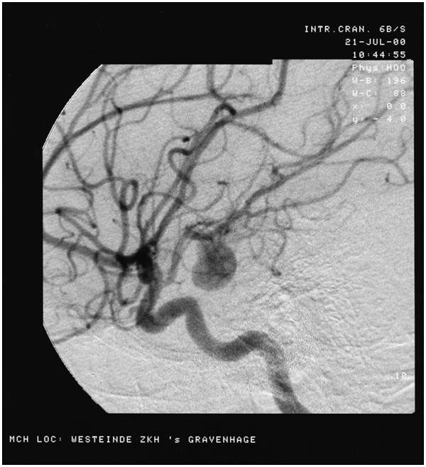
Angiography of an aneurism in a cerebral arteryICD-10 I72 ICD-9 442 DiseasesDB 15088 MedlinePlus 001122 MeSH D000783 An aneurysm or aneurism (from Ancient Greek: ἀνεύρυσμα - aneurusma "dilation", from ἀνευρύνειν - aneurunein "to dilate") is a localized, blood-filled balloon-like bulge in the wall of a blood vessel.[1] Aneurysms can commonly occur in arteries at the base of the brain (the circle of Willis) and an aortic aneurysm occurs in the main artery carrying blood from the left ventricle of the heart. When the size of an aneurysm increases, there is a significant risk of rupture, resulting in severe hemorrhage, other complications or death. Aneurysms can be hereditary or caused by disease, both of which cause the wall of the blood vessel to weaken.
Contents
True and false
A true aneurysm is one that involves all three layers of the wall of an artery (intima, media and adventitia). True aneurysms include atherosclerotic, syphilitic, and congenital aneurysms, as well as ventricular aneurysms that follow transmural myocardial infarctions (aneurysms that involve all layers of the attenuated wall of the heart are also considered true aneurysms).[2]
A false aneurysm or pseudoaneurysm does not primarily involve such distortion of the vessel. It is a collection of blood leaking completely out of an artery or vein, but confined next to the vessel by the surrounding tissue. This blood-filled cavity will eventually either thrombose (clot) enough to seal the leak or rupture out of the tougher tissue enclosing it and flow freely between layers of other tissues or into looser tissues. Pseudoaneurysms can be caused by trauma that punctures the artery and are a known complication of percutaneous arterial procedures, such as arteriography, arterial grafting, or use of an artery for injection, such as by drug abusers unable to find a usable vein. Like true aneurysms, they may be felt as an abnormal pulsatile mass on palpation.
Morphology
Aneurysms are classified by their macroscopic shape and size and are described as either "saccular" or "fusiform." Saccular aneurysms are spherical in shape and involve only a portion of the vessel wall; they vary in size from 5 to 20 cm in diameter, and are often filled, either partially or fully, by thrombus. Fusiform ("spindle-shaped") aneurysms are variable in both their diameter and length; their diameters can extend up to 20 cm. They often involve large portions of the ascending and transverse aortic arch, the abdominal aorta, or less frequently the iliac arteries. The shape of an aneurysm is not pathognomonic for a specific disease.[3]
Location
Cerebral aneurysms occur most commonly in the anterior communicating artery, which is part of the circle of Willis. The next most common sites of cerebral aneurysm occurrence are in the internal carotid artery at the level of the posterior communicating artery, and at the internal carotid artery terminus.
Most (94%) non-intracranial aneurysms arise distal to the origin of the renal arteries at the infrarenal abdominal aorta, a condition mostly caused by atherosclerosis.
The thoracic aorta can also be involved. One common form of thoracic aortic aneurysm involves widening of the proximal aorta and the aortic root, which leads to aortic insufficiency. Aneurysms can also occur in the legs, particularly in the deep vessels (e.g., the popliteal vessels in the knee).
Arterial vs. venous
Arterial aneurysms are much more common, but venous aneurysms do happen (for example, the popliteal venous aneurysm). GEN and VIVO.
Underlying condition
Aneurysms can be classified by the underlying condition.
Many aneurysms are atherosclerotic in nature.
A mycotic aneurysm is an aneurysm that results from an infectious process that involves the arterial wall.[4] The attribute mycotic is a misnomer in implying a fungal cause.[5] In fact, the main pathogens of mycotic aneurysms are gram-positive cocci.[6]
While most aneurysms occur in an isolated form, the occurrence of berry aneurysms of the anterior communicating artery of the circle of Willis is associated with autosomal dominant polycystic kidney disease (ADPKD).
The third stage of syphilis also manifests as aneurysm of the aorta, which is due to loss of the vasa vasorum in the tunica adventitia.
Risk factors
Risk factors for an aneurysm include diabetes, obesity, hypertension, tobacco use, alcoholism, high cholesterol, copper deficiency, and increasing age.[citation needed] Some types are the result of congenital, or inherited, weakness in artery walls.[7]
Copper deficiency
A minority of aneurysms are caused by a copper deficiency. Numerous animal experiments have shown that a copper deficiency can cause diseases affected by elastin[8] tissue strength [Harris]. The lysyl oxidase that cross links connective tissue is secreted normally, but its activity is reduced,[9] due to some of the initial enzyme molecules' (apo-enzyme or enzyme without the copper) lack of copper.[10][11]
Aneurysms of the aorta are the chief cause of death of copper deficient chickens; depleting copper produces aneurysms in turkeys.[12]
Men who die of aneurysms have a liver content (of copper) that can be as little as 26% of normal.[13] In such men the median layer of the blood vessel (where the elastin is) is thinner but its elastin copper content is the same as in the elastin of normal men. The overall thickness is not different.[14] The body must therefore have some way of preventing elastin tissue from growing if there is not enough activated lysyl oxidase for it. Men are more susceptible to aneurysms than young women, probably because estrogen increases the efficiency of absorption of copper. However, women can be affected by some of these problems after pregnancy, probably because women must give the liver of their babies copper in order for them to survive the low levels of copper in milk. A baby’s liver has up to ten times as much copper as an adult liver.[15] Higher alcohol content enhances copper uptake, whilst zinc, iron and copper uptake diminish with increasing sugar and alcohol contents. Handling in iron or galvanized ironware is shown to deplete copper by almost 100% of initial levels and this has potential in health risk reduction applications.[16] Excess intake of zinc can lead to deficiency of copper (hypocupremia). This deficiency happens because excess zinc in the body triggers reduced absorption of copper in the GI tract, resulting in increased fecal loss of copper.[17]
Pathophysiology
The occurrence and expansion of an aneurysm in a given segment of the arterial tree involves local hemodynamic factors and factors intrinsic to the arterial segment itself.
The aorta is a relatively low-resistance circuit for circulating blood. The lower extremities have higher arterial resistance, and the repeated trauma of a reflected arterial wave on the distal aorta may injure a weakened aortic wall and contribute to aneurysmal degeneration. Systemic hypertension compounds the injury, accelerates the expansion of known aneurysms, and may contribute to their formation.
Aneurysm formation is probably the result of multiple factors affecting that arterial segment and its local environment.
Increasing aneurysmal dilatation leads to increasing arterial wall tension or stress. In hemodynamic terms, the coupling of aneurysmal dilatation and increased wall stress is approximated by the law of Laplace. The Law of Laplace applied to a cylinder states that the (arterial) wall tension is equal to the pressure times the radius of the arterial conduit (T = P x R). As diameter increases, wall tension increases, which contributes to more increase in diameter and risk of rupture. Increased blood pressure (systemic hypertension) and increased aneurysm size increase arterial wall tension and therefore increase the risk of rupture. In addition, the vessel wall is supplied by the blood within its lumen in humans (? although aorta has Vasa vasorum). Therefore[citation needed] in a developing aneurysm, the most ischemic portion of the aneurysm is at the farthest end, resulting in weakening of the vessel wall there and aiding further expansion of the aneurysm. Thus eventually all aneurysms will, if left to complete their evolution, rupture without intervention.
Symptoms
Cerebral Aneurysm
Symptoms for a cerebral aneurysm occur when the aneurysm pushes on a structure in the brain. In the case of a cerebral aneurysm, the symptoms for an aneurysm that has ruptured and one that has not ruptured are different.
Symptoms for an aneurysm that has not ruptured:- Fatigue
- Loss of perception
- Loss of balance
- Speech problems
- Double vision
Symptoms for a ruptured aneurysm:
- Severe headaches
- Loss of vision
- Double vision
- Neck pain and/or stiffness
- Pain above and/or behind the eyes
- Death
Diagnosis
Diagnosis of a ruptured cerebral aneurysm is commonly made by finding signs of subarachnoid hemorrhage on a computed tomography (CT) scan. If the CT scan is negative but a ruptured aneurysm is still suspected based on clinical findings, a lumbar puncture can be performed to detect blood in the cerebrospinal fluid. Computed tomography angiography (CTA) is an alternative to traditional angiography and can be performed without the need for arterial catheterization. This test combines a regular CT scan with a contrast dye injected into a vein. Once the dye is injected into a vein, it travels to the cerebral arteries, and images are created using a CT scan. These images show exactly how blood flows into the brain arteries.
Treatment
Historically, the treatment of arterial aneurysms has been limited to either surgical intervention, or watchful waiting in combination with control of blood pressure. In recent years, endovascular or minimally invasive techniques have been developed for many types of aneurysms.
Cranial aneurysms
There are currently two treatment options for brain aneurysms: surgical clipping or endovascular coiling.
Surgical clipping was introduced by Walter Dandy of the Johns Hopkins Hospital in 1937. It consists a craniotomy to expose the aneurysm and closing the base of the aneurysm with a clip. The surgical technique has been modified and improved over the years. Surgical clipping remains the best method to permanently eliminate aneurysms.
Endovascular coiling was introduced by Guido Guglielmi at UCLA in 1991. It consists of passing a catheter into the femoral artery in the groin, through the aorta, into the brain arteries, and finally into the aneurysm itself. Once the catheter is in the aneurysm, platinum coils are pushed into the aneurysm and released. These coils initiate a clotting or thrombotic reaction within the aneurysm that, if successful, will eliminate the aneurysm. In the case of broad-based aneurysms, a stent is passed first into the parent artery to serve as a scaffold for the coils ("stent-assisted coiling").
At this point it appears that the risk of stroke or death associated with surgical clipping and endovascular coiling are the same. The ISAT trials have shown, however, that patients who have experienced aneurysmal rupture have a 7% lower mortality rate when treated by coiling than patients treated by clipping, when all other factors are equal. Despite its lower mortality rate, coiling is associated with a higher rate of recurrence than traditional clipping. For instance, the most recent study by Jacques Moret and colleagues from Paris, France, (a group with one of the largest experiences in endovascular coiling) indicates that 28.6% of aneurysms recurred within one year of coiling, and that the recurrence rate increased with time.[18] These results are similar to those previously reported by other endovascular groups. For instance Jean Raymond and colleagues from Montreal, Canada, (another group with a large experience in endovascular coiling) reported that 33.6% of aneurysms recurred within one year of coiling. (Raymond J et al., Stroke 34(6):1398-1403, June 2003) The long-term coiling results of one of the two prospective, randomized studies comparing surgical clipping versus endovascular coiling, in particular the International Subarachnoid Aneurysm Trial (ISAT) show similar results. However, no studies to date have shown that the higher angiographic recurrence rate equals a higher rate of rebleeding. Thus far, the ISAT trials listed above show no increase in the rate of rebleeding, and show a persistent 7% lower mortality rate in subarachnoid hemorrhage patients who have been treated with coiling.. In ISAT, the need for late retreatment of aneurysms was 6.9 times more likely for endovascular coiling as compared to surgical clipping. (Campi A et al., Stroke 38(5):1538-1544, May 2007)
Patients who undergo endovascular coiling need to have annual studies (such as MRI/MRA, CTA, or angiography) to detect early recurrences, a practice that has not traditionally been performed with clipping. If a recurrence is identified, the aneurysm may need to be retreated with either surgery or further coiling. The risks associated with surgical clipping of previously coiled aneurysms are high. Ultimately, the decision to treat with surgical clipping versus endovascular coiling should be made by a cerebrovascular team with extensive experience in both modalities.
Aortic and peripheral aneurysms
For aneurysms in the aorta, arms, legs, or head, the weakened section of the vessel may be replaced by a bypass graft that is sutured at the vascular stumps. Instead of sewing, the graft tube ends, made rigid and expandable by nitinol wireframe, can be easily inserted in its reduced diameter into the vascular stumps and then expanded up to the most appropriate diameter and permanently fixed there by external ligature.[1] [19] New devices were recently developed to substitute the external ligature by expandable ring allowing use in acute ascending aorta dissection, providing airtight (i.e. not dependent on the coagulation integrity), easy and quick anastomosis extended to the arch concavity [20][2][3] Less invasive endovascular techniques allow covered metallic stent grafts to be inserted through the arteries of the leg and deployed across the aneurysm.
See also
- Abdominal aortic aneurysm
- Aortic aneurysm
- Aortic dissection
- Cerebral aneurysm
- Charcot-Bouchard aneurysms
- Aneurysm of sinus of Valsalva
- Stroke
References
- ^ "aneurysm" at Dorland's Medical Dictionary
- ^ (Kumar, Vinay. Robbins Basic Pathology, 8th Edition. W.B. Saunders Company, 052007. 10.7).
- ^ [Kumar, Vinay. Robbins & Cotran Pathologic Basis of Disease, 7th Edition. Saunders Book tea 082004. 11.2.6]
- ^ emedicine > Cerebral Aneurysm Author: Jonathan L Brisman. Coauthors: Emad Soliman, Abraham Kader, Norvin Perez. Updated: Sep 23, 2010
- ^ Alpagut, U; Ugurlucan, M; Kafali, E; Surmen, B; Sayin, OA; Guven, K; Dayioglu, E; Rozanes, I et al. (2006). "Endoluminal stenting of mycotic saccular aneurysm at the aortic arch". Texas Heart Institute journal / from the Texas Heart Institute of St. Luke's Episcopal Hospital, Texas Children's Hospital 33 (3): 371–5. PMC 1592276. PMID 17041700. http://www.pubmedcentral.nih.gov/articlerender.fcgi?tool=pmcentrez&artid=1592276.
- ^ Table 18-2 in: Trudie A Goers; Washington University School of Medicine Department of Surgery; Klingensmith, Mary E; Li Ern Chen; Sean C Glasgow (2008). The Washington manual of surgery. Philadelphia: Wolters Kluwer Health/Lippincott Williams & Wilkins. ISBN 0-7817-7447-0.
- ^ Understanding Aneurysm -- the Basics, WebMD
- ^ http://herkules.oulu.fi/isbn9514267397/isbn9514267397.pdf elastin review.
- ^ Kosonen T Uriu-Hare JY Clegg MS Keen CL Rucker RB 1996 Copper incorporation into lysyl oxidase. FASEB Journal; 10; A293,
- ^ Rucker RB, Kosonen T, Clegg MS, Mitchell AE, Rucker BR, Uriu-Hare JY, Keen CL 1998 Copper, lysyl oxidase, and extracellular matrix protein cross-linking.; Am J Clin Nutr. May;67(5 Suppl):996S-1002S.
- ^ Smith-Mungo LI, Kagan HM.: Lysyl oxidase 1998 properties, regulation and multiple functions in biology; Matrix Biol. Feb;16(7); 387-98.
- ^ Guenther E Carlson CW Emerick RJ 1978 Copper salts for growth stimulation and alleviation of aortic rupture losses in turkeys., Poult. Sci. Sep;57(5):1313-24.
- ^ Tilson MD 1983 Decreased hepatic copper levels. Arch. Surgery 117; 1212.-1213
- ^ Senapati A Carlsson LK Fletcher CDM Browse NL & Thompson RPH 1995 Is tissue copper deficiency associated with aortic aneuryzms? British Journal of Surgery 72; 352-353.
- ^ Dorea JG, Brito M, Araujo MO. 1987 Concentration of copper and zinc in liver of fetuses and infants. J Am Coll Nutr. Dec;6(6):491-5.
- ^ Food Chemistry Volume 57, Issue 2, October 1996, Pages 205-209 .
- ^ Nations SP, Boyer PJ, Love LA, et al. Denture creams: an unusual source of excess zinc, leading to hypocupremia and neurologic disease. Neurology 2008;71:639-43.
- ^ Piotin M et al., Radiology 243(2):500-508, May 2007. PMID 17293572
- ^ Tex Heart Inst J. 2002;29(1):56-9
- ^ Stefano Nazari: Expandable device type III for easy and reliable approximation of dissection layers in sutureless aortic anastomosis. Ex vivo experimental study.Interact CardioVasc Thorac Surg 2010;10:161-164. doi:10.1510/icvts.2009.216291
External links
- Brain Blood Vessel Disorder Help & Info Site
- @neurIST - Integrated Biomedical Informatics for the Management of Cerebral Aneurysms
- Brain aneurysm and percent packing calculator
Categories:- Gross pathology
- Vascular diseases
Wikimedia Foundation. 2010.