- Lumbar puncture
-
Lumbar puncture Intervention 
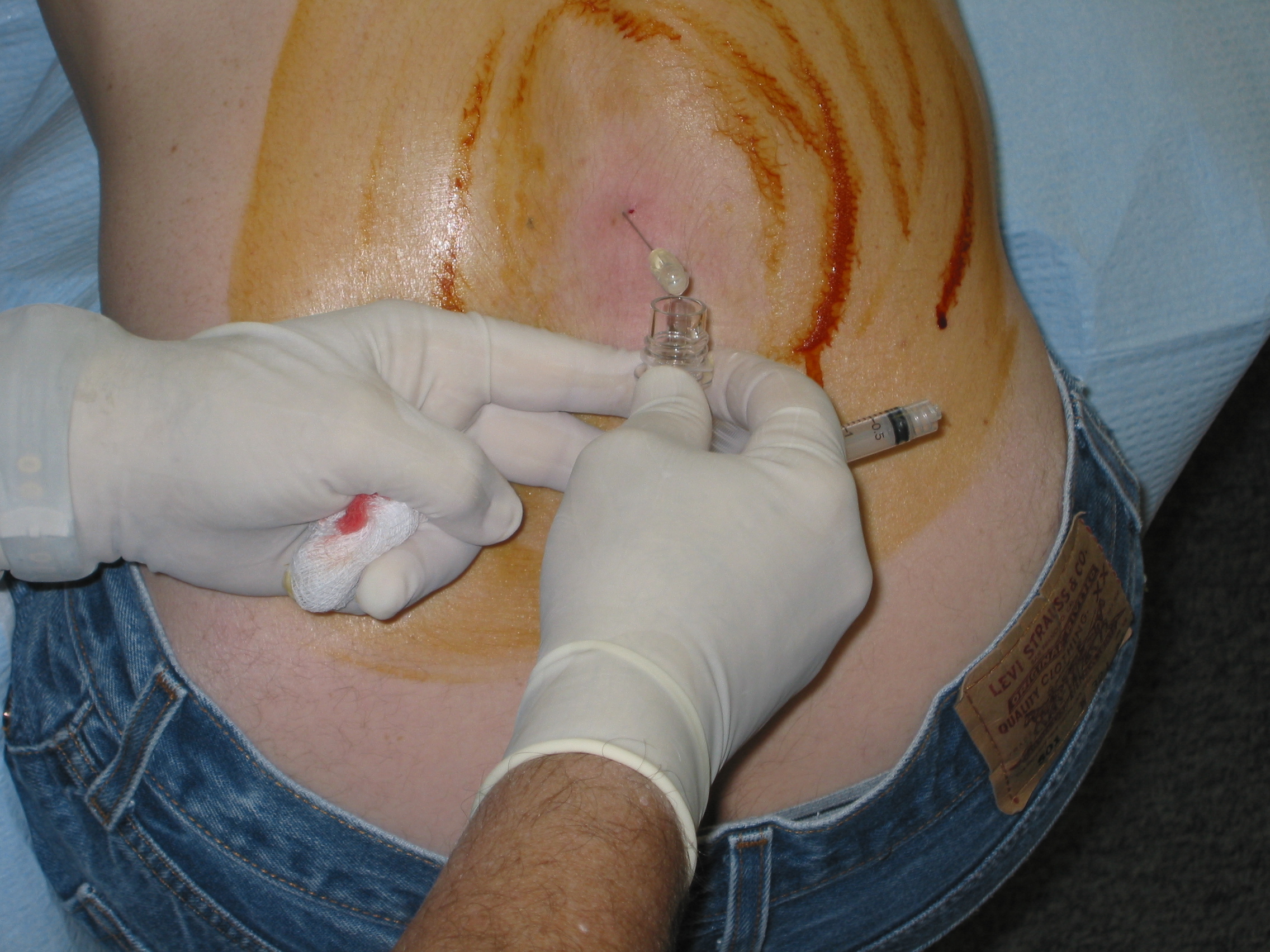
A patient undergoes a lumbar puncture at the hands of a neurologist. The reddish-brown swirls on the patient's back are tincture of iodine (an antiseptic).ICD-9-CM 03.31 MeSH D013129 A lumbar puncture (or LP, and colloquially known as a spinal tap) is a diagnostic and at times therapeutic procedure that is performed in order to collect a sample of cerebrospinal fluid (CSF) for biochemical, microbiological, and cytological analysis, or very rarely as a treatment ("therapeutic lumbar puncture") to relieve increased intracranial pressure.
Contents
Indications
The most common purpose for a lumbar puncture is to collect cerebrospinal fluid in a case of suspected meningitis, since there is no other reliable tool with which meningitis, a life-threatening but highly treatable condition, can be excluded. Young infants commonly require lumbar puncture as a part of the routine workup for fever without a source, as they have a much higher risk of meningitis than older persons and do not reliably show signs of meningeal irritation (meningismus). In any age group, subarachnoid hemorrhage, hydrocephalus, benign intracranial hypertension and many other diagnoses may be supported or excluded with this test.
Lumbar punctures may also be done to inject medications into the cerebrospinal fluid ("intrathecally"), particularly for spinal anesthesia or chemotherapy. It may also be used to detect the presence of malignant cells in the CSF, as in carcinomatous meningitis or medulloblastoma.
Contraindications
Lumbar puncture should not be performed in the following situations
- Idiopathic (unidentified cause) increased intracranial pressure (ICP)
- Rationale: lumbar puncture in the presence of increased ICP may cause uncal herniation
- Exception: therapeutic use of lumbar puncture to reduce ICP
- Precaution
- CT brain is advocated by some, especially in the following situations
- Age >65
- Reduced GCS or conscious state
- Recent history of seizure
- Focal neurological signs
- Ophthalmoscopy for papilledema
- CT brain is advocated by some, especially in the following situations
- Bleeding diathesis
- Coagulopathy
- Decreased platelet count (<50 x 109/L)
- Infections[1]
- Skin infection at puncture site
- Sepsis
- Abnormal respiratory pattern
- Hypertension with bradycardia and deteriorating consciousness
- Vertebral deformities (scoliosis or kyphosis), in hands of an inexperienced physician.[2][3]
Procedure
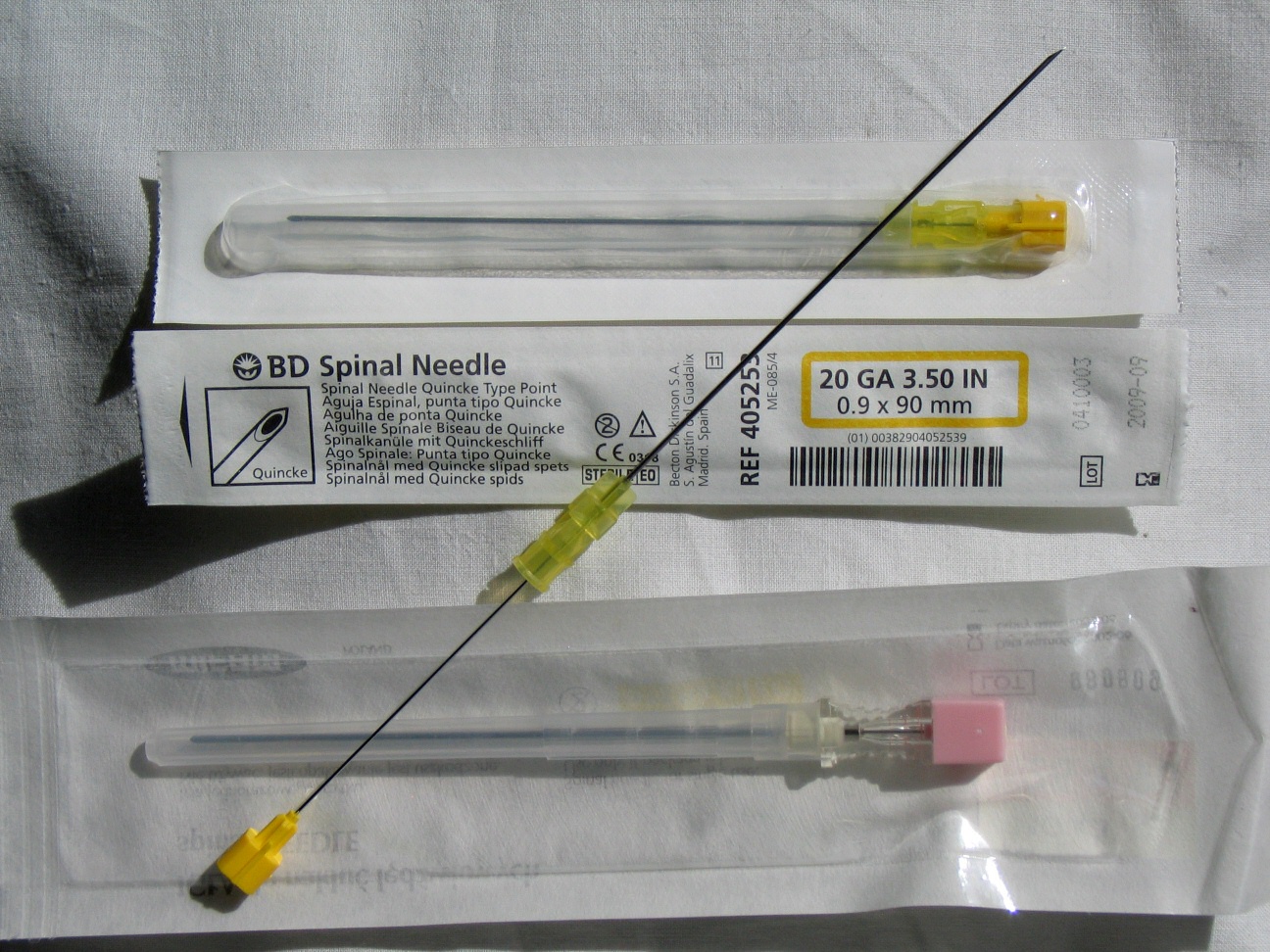
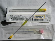 Spinal needles used in lumbar puncture.
In performing a lumbar puncture, first the patient is usually placed in a left (or right) lateral position with his/her neck bent in full flexion and knees bent in full flexion up to his/her chest, approximating a fetal position as much as possible. It is also possible to have the patient sit on a stool and bend his/her head and shoulders forward. The area around the lower back is prepared using aseptic technique. Once the appropriate location is palpated, local anaesthetic is infiltrated under the skin and then injected along the intended path of the spinal needle. A spinal needle is inserted between the lumbar vertebrae L3/L4 or L4/L5 and pushed in until there is a "give" that indicates the needle is past the ligamentum flavum. The needle is again pushed until there is a second 'give' that indicates the needle is now past the dura mater. Since the arachnoid membrane and the dura mater exist in flush contact with one another in the living person's spine (due to fluid pressure from CSF in the subarachnoid space pushing the arachnoid membrane out towards the dura), once the needle has pierced the dura mater it has also traversed the thinner arachnoid membrane and is now in the subarachnoid space. The stylet from the spinal needle is then withdrawn and drops of cerebrospinal fluid are collected. The opening pressure of the cerebrospinal fluid may be taken during this collection by using a simple column manometer. The procedure is ended by withdrawing the needle while placing pressure on the puncture site. In the past, the patient would often be asked to lie on his/her back for at least six hours and be monitored for signs of neurological problems, though there is no scientific evidence that this provides any benefit. The technique described is almost identical to that used in spinal anesthesia, except that spinal anesthesia is more often done with the patient in a seated position.
The upright seated position is advantageous in that there is less distortion of spinal anatomy which allows for easier withdrawal of fluid. It is preferred by some practitioners when a lumbar puncture is performed on an obese patient where having them lie on their side would cause a scoliosis and unreliable anatomical landmarks. On the other hand, opening pressures are notoriously unreliable when measured on a seated patient and therefore the left or right lateral (lying down) position is preferred if an opening pressure needs to be measured.
Patient anxiety during the procedure can lead to increased CSF pressure, especially if the person holds their breath, tenses their muscles or flexes their knees too tightly against their chest. Diagnostic analysis of changes in fluid pressure during lumbar puncture procedures requires attention both to the patient's condition during the procedure and to their medical history.[citation needed]
Reinsertion of the stylet may decrease the rate of post lumbar puncture headaches.[3]
Risks
Post spinal headache with nausea is the most common complication; it often responds to analgesics and infusion of fluids. It was long taught that this complication can often be prevented by strict maintenance of a supine posture for two hours after the successful puncture; this has not been borne out in modern studies involving large numbers of patients. Merritt's Neurology (10th edition), in the section on lumbar puncture, notes that intravenous caffeine injection is often quite effective in aborting these so-called "spinal headaches." Contact between the side of the lumbar puncture needle and a spinal nerve root can result in anomalous sensations (paresthesia) in a leg during the procedure; this is harmless and patients can be warned about it in advance to minimize their anxiety if it should occur. A headache that is persistent despite a long period of bedrest and occurs only when sitting up may be indicative of a CSF leak from the lumbar puncture site. It can be treated by more bedrest, or by an epidural blood patch, where the patient's own blood is injected back into the site of leakage to cause a clot to form and seal off the leak.
Serious complications of a properly performed lumbar puncture are extremely rare.[citation needed] They include spinal or epidural bleeding, and trauma to the spinal cord or spinal nerve roots resulting in weakness or loss of sensation, or even paraplegia. The latter is exceedingly rare, since the level at which the spinal cord ends (normally the inferior border of L1, although it is slightly lower in infants) is several vertebral spaces above the proper location for a lumbar puncture (L3/L4). There are case reports of lumbar puncture resulting in perforation of abnormal dural arterio-venous malformations, resulting in catastrophic epidural hemorrhage; this is exceedingly rare.
The procedure is not recommended when epidural infection is present or suspected, when topical infections or dermatological conditions pose a risk of infection at the puncture site or in patients with severe psychosis or neurosis with back pain. Some authorities believe that withdrawal of fluid when initial pressures are abnormal could result in spinal cord compression or cerebral herniation; others believe that such events are merely coincidental in time, occurring independently as a result of the same pathology that the lumbar puncture was performed to diagnose. In any case, computed tomography of the brain is often performed prior to lumbar puncture if an intracranial mass is suspected.
Removal of cerebrospinal fluid resulting in reduced fluid pressure has been shown to correlate with greater reduction of cerebral blood flow among patients with Alzheimer's disease. Its clinical significance is uncertain.
Diagnostics
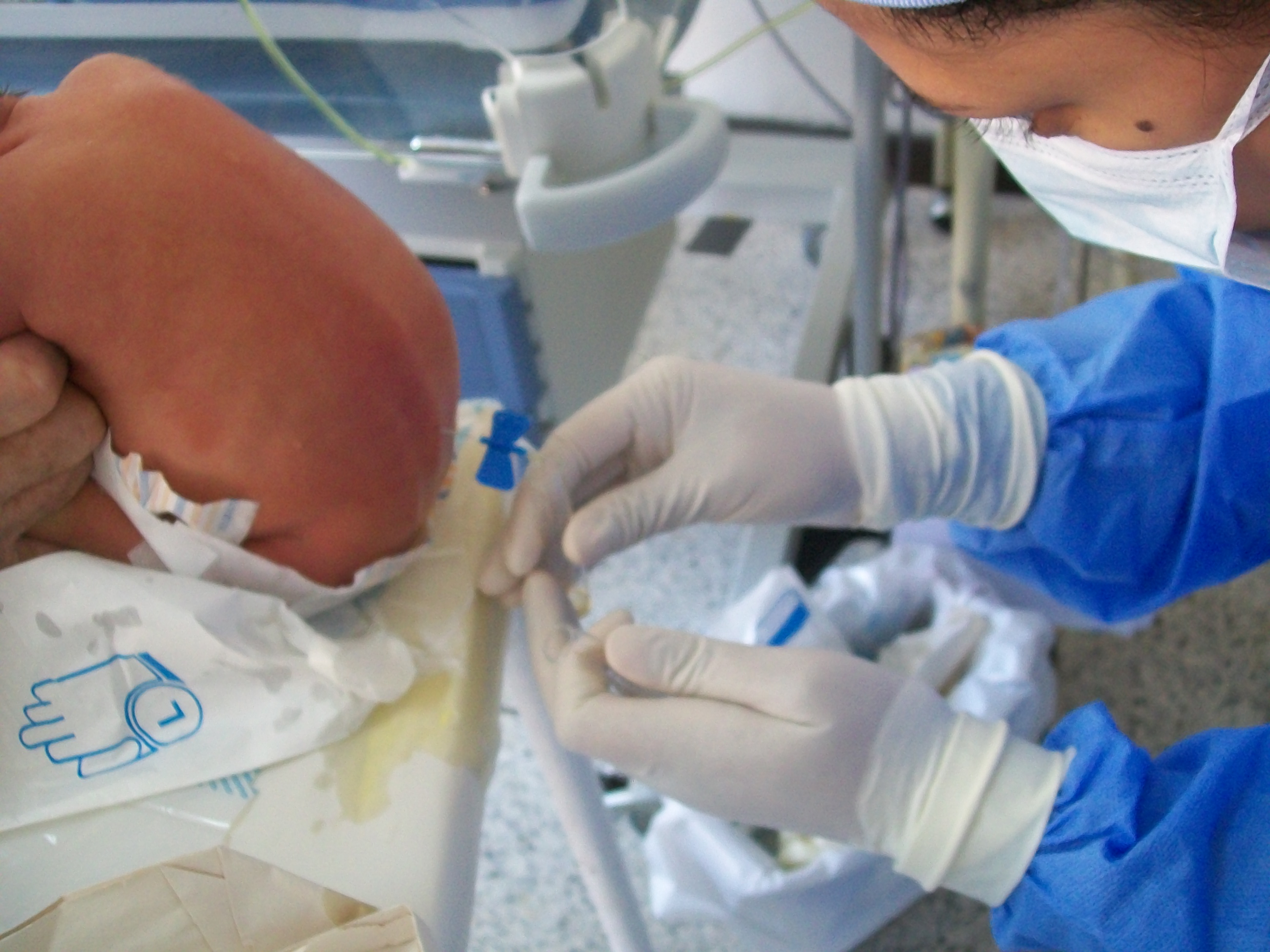
 Lumbar puncture in a child suspected of having meningitis.
Increased CSF pressure can indicate congestive heart failure, cerebral edema, subarachnoid hemorrhage, hypo-osmolality resulting from hemodialysis, meningeal inflammation, purulent meningitis or tuberculous meningitis, hydrocephalus, or pseudotumor cerebri.
Decreased CSF pressure can indicate complete subarachnoid blockage, leakage of spinal fluid, severe dehydration, hyperosmolality, or circulatory collapse. Significant changes in pressure during the procedure can indicate tumors or spinal blockage resulting in a large pool of CSF, or hydrocephalus associated with large volumes of CSF. Lumbar puncture for the purpose of reducing pressure is performed in some patients with idiopathic intracranial hypertension (also called pseudotumor cerebri.)
The presence of white blood cells in cerebrospinal fluid is called pleocytosis. A small number of monocytes can be normal; the presence of granulocytes is always an abnormal finding. A large number of granulocytes often heralds bacterial meningitis. White cells can also indicate reaction to repeated lumbar punctures, reactions to prior injections of medicines or dyes, central nervous system hemorrhage, leukemia, recent epileptic seizure, or a metastatic tumor. When peripheral blood contaminates the withdrawn CSF, a common procedural complication, white blood cells will be present along with erythrocytes, and their ratio will be the same as that in the peripheral blood.
The finding of erythrophagocytosis,[4] where phagocytosed erythrocytes is observed, signifies haemorrhage into the CSF that preceded the lumbar puncture. Therefore, when erythrocytes are detected in the CSF sample, erythrophagocytosis suggests causes other than a traumatic tap, such as intracranial haemorrhage and haemorrhagic herpetic encephalitis. In which case, further investigations are warranted, including imaging and viral culture.
Several substances found in cerebrospinal fluid are available for diagnostic measurement.
- Measurement of chloride levels may aid in detecting the presence of tuberculous meningitis.
- Glucose is usually present in the CSF; the level is usually about 60% that in the peripheral circulation. A fingerstick or venipuncture at the time of lumbar puncture may therefore be performed to assess peripheral glucose levels in order to determine a predicted CSF glucose value. Decreased glucose levels can indicate fungal, tuberculous or pyogenic infections; lymphomas; leukemia spreading to the meninges; meningoencephalitic mumps; or hypoglycemia. A glucose level of less than one third of blood glucose levels in association with low CSF lactate levels is typical in hereditary CSF glucose transporter deficiency also known as De Vivo disease.
- Increased glucose levels in the fluid can indicate diabetes, although the 60% rule still applies.
- Increased levels of glutamine are often involved with hepatic encephalopathies, Reye's syndrome, hepatic coma, cirrhosis and hypercapnia.
- Increased levels of lactate can occur the presence of cancer of the CNS, multiple sclerosis, heritable mitochondrial disease, low blood pressure, low serum phosphorus, respiratory alkalosis, idiopathic seizures, traumatic brain injury, cerebral ischemia, brain abscess, hydrocephalus, hypocapnia or bacterial meningitis.
- The enzyme lactate dehydrogenase can be measured to help distinguish meningitides of bacterial origin, which are often associated with high levels of the enzyme, from those of viral origin in which the enzyme is low or absent.
- Changes in total protein content of cerebrospinal fluid can result from pathologically increased permeability of the blood-cerebrospinal fluid barrier, obstructions of CSF circulation, meningitis, neurosyphilis, brain abscesses, subarachnoid hemorrhage, polio, collagen disease or Guillain-Barré syndrome, leakage of CSF, increases in intracranial pressure or hyperthyroidism. Very high levels of protein may indicate tuberculous meningitis or spinal block.
- IgG synthetic rate is calculated from measured IgG and total protein levels; it is elevated in immune disorders such as multiple sclerosis, transverse myelitis, and neuromyelitis optica of Devic.
- Numerous antibody-mediated tests for CSF are available in some countries: these include rapid tests for antigens of common bacterial pathogens, treponemal titers for the diagnosis of neurosyphilis and Lyme disease, Coccidioides antibody, and others.
- The India ink test is still used for detection of meningitis caused by Cryptococcus neoformans, but the cryptococcal antigen (CrAg) test has a higher sensitivity.
- CSF can be sent to the microbiology lab for various types of smears and cultures to diagnose infections.
- Polymerase chain reaction (PCR) has been a great advance in the diagnosis of some types of meningitis. It has high sensitivity and specificity for many infections of the CNS, is fast, and can be done with small volumes of CSF. Even though testing is expensive, it saves cost of hospitalization.
History
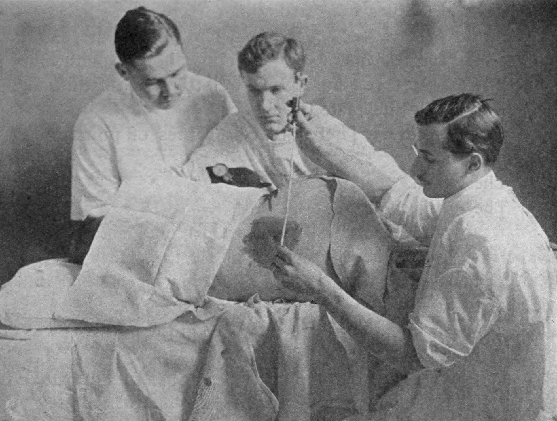
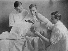 Lumbar puncture, early 20th century.
The first technique for accessing the dural space was described by the London physician Dr Walter Essex Wynter. In 1889, he developed a crude cut down with cannulation in 4 patients with tuberculous meningitis. The main purpose was the treatment of raised intracranial pressure rather than for diagnosis.[5] The technique for needle lumbar puncture was then introduced by the German physician Heinrich Quincke, who credits Wynter with the earlier discovery; he first reported his experiences at an internal medicine conference in Wiesbaden in 1891.[6] He subsequently published a book on the subject.[7][8]
The lumbar puncture procedure was taken to the United States by Arthur H. Wentworth M.D., an assistant professor at the Harvard Medical School, based at Children's Hospital. In 1893, he published a long paper on diagnosing cerebrospinal meningitis by examining spinal fluid. His career took a nosedive, however, when antivivisectionists prosecuted him for having obtained spinal fluid from children. He was acquitted, but he was uninvited from the then forming Johns Hopkins School of Medicine, where he would have been the first professor of pediatrics.[citation needed]
References
- ^ Mary Louise Turgeon (2005). Clinical hematology: theory and procedures. Lippincott Williams & Wilkins. pp. 401–. ISBN 9780781750073. http://books.google.com/books?id=cHAjsUgegpQC&pg=PA401. Retrieved 28 October 2010.
- ^ Roos KL (March 2003). "Lumbar puncture". Semin Neurol 23 (1): 105–14. doi:10.1055/s-2003-40758. PMID 12870112.
- ^ a b Straus SE, Thorpe KE, Holroyd-Leduc J (October 2006). "How do I perform a lumbar puncture and analyze the results to diagnose bacterial meningitis?". JAMA 296 (16): 2012–22. doi:10.1001/jama.296.16.2012. PMID 17062865.
- ^ Harald Kluge (2007). Atlas of CSF cytology. Thieme. pp. 45–46. ISBN 9783131431615. http://books.google.com/books?id=HDLv-LAfqHoC&pg=PA45. Retrieved 28 October 2010.
- ^ Wynter WE (1891). "Four cases of tubercular meningitis in which paracentesis of the theca vertebralis was performed for the relief of fluid pressure". Lancet 1 (3531): 981–2. doi:10.1016/S0140-6736(02)16784-5.
- ^ Quincke HI (1891). Verhandlungen des Congresses für Innere Medizin, Zehnter Congress, Wiesbaden. 10. pp. 321–331.
- ^ Quincke HI (1902). Die Technik der Lumbalpunktion. Berlin & Vienna.
- ^ Heinrich Irenaeus Quincke at Who Named It?
External links
Surgery, Nervous system: neurosurgical and other procedures (ICD-9-CM V3 01–05+89.1, ICD-10-PCS 00-01) Skull CNS thalamus and globus pallidus: Thalamotomy · Thalamic stimulator · Pallidotomy
ventricular system: Ventriculostomy · Suboccipital puncture · Intracranial pressure monitoring
cerebrum: Psychosurgery (Lobotomy, Bilateral cingulotomy) · Hemispherectomy · Anterior temporal lobectomy
pituitary: Hypophysectomy
hippocampus: Amygdalohippocampectomy
Brain biopsyCerebral meningesSpinal cord and roots (Cordotomy, Rhizotomy)
Vertebrae and intervertebral discs: see Template:Bone, cartilage, and joint proceduresCT head · Cerebral angiography · Pneumoencephalography · Echoencephalography/Transcranial doppler · MRI of brain and brain stem · Brain PET · SPECT of brain · MyelographyDiagnosticPNS Sympathetic nerves or gangliaNerves (general)DiagnosticCategories:- CSF tests
- Neurology procedures
- Idiopathic (unidentified cause) increased intracranial pressure (ICP)
Wikimedia Foundation. 2010.