- Blood transfusion
-
Blood transfusion Intervention 
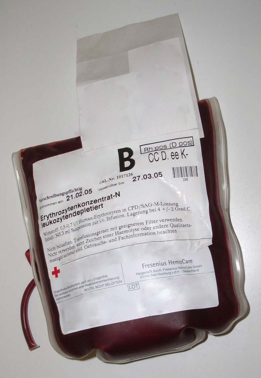
Plastic bag containing packed red blood cells in citrate, phosphate, dextrose, and adenine (CPDA) solutionICD-9-CM 99.0 MeSH D001803 OPS-301 code: 8-80 Blood transfusion is the process of receiving blood products into one's circulation intravenously. Transfusions are used in a variety of medical conditions to replace lost components of the blood. Early transfusions used whole blood, but modern medical practice commonly uses only components of the blood, such as red blood cells, white blood cells, plasma, clotting factors, and platelets.
Contents
History
Early attempts
The first historical attempt at blood transfusion was described by the 17th century chronicler Stefano Infessura. Infessura relates that, in 1492, as Pope Innocent VIII sank into a coma, the blood of three boys was infused into the dying pontiff (through the mouth, as the concept of circulation and methods for intravenous access did not exist at that time) at the suggestion of a physician. The boys were ten years old, and had been promised a ducat each. However, not only did the pope die, but so did the three children. Some authors have discredited Infessura's account, accusing him of anti-papalism.[1]
 World War II syringe for direct inter-human blood transfusion
Beginning with Harvey's experiments with circulation of the blood, more sophisticated research into blood transfusion began in the 17th century, with successful experiments in transfusion between animals. However, successive attempts on humans continued to have fatal results.
The first fully documented human blood transfusion was administered by Dr. Jean-Baptiste Denys, eminent physician to King Louis XIV of France, on June 15, 1667.[2] He transfused the blood of a sheep into a 15-year-old boy, who survived the transfusion.[3] Denys performed another transfusion into a labourer, who also survived. Both instances were likely due to the small amount of blood that was actually transfused into these people. This allowed them to withstand the allergic reaction. Denys' third patient to undergo a blood transfusion was Swedish Baron Gustaf Bonde. He received two transfusions. After the second transfusion Bonde died.[4] In the winter of 1667, Denys performed several transfusions on Antoine Mauroy with calf's blood, who on the third account died.[5] Much controversy surrounded his death. Mauroy's wife asserted Denys was responsible for her husband's death; she was accused as well, though it was later determined that Mauroy actually died from arsenic poisoning, Denys' experiments with animal blood provoked a heated controversy in France.[4] Finally, in 1670 the procedure was banned. In time, the British Parliament and even the pope followed suit. Blood transfusions fell into obscurity for the next 150 years.
First successful transfusion
Richard Lower examined the effects of changes in blood volume on circulatory function and developed methods for cross-circulatory study in animals, obviating clotting by closed arteriovenous connections. His newly devised instruments eventually led to actual transfusion of blood.
"Many of his colleagues were present. Towards the end of February 1665 [when he] selected one dog of medium size, opened its jugular vein, and drew off blood, until ... its strength was nearly gone. Then, to make up for the great loss of this dog by the blood of a second, I introduced blood from the cervical artery of a fairly large mastiff, which had been fastened alongside the first, until this latter animal showed ... it was overfilled ... by the inflowing blood." After he "sewed up the jugular veins," the animal recovered "with no sign of discomfort or of displeasure."
Lower had performed the first blood transfusion between animals. He was then "requested by the Honorable [Robert] Boyle ... to acquaint the Royal Society with the procedure for the whole experiment," which he did in December of 1665 in the Society's Philosophical Transactions. On 15 June 1667 Denys, then a professor in Paris, carried out the first transfusion between humans and claimed credit for the technique, but Lower's priority cannot be challenged.[citation needed]
Six months later in London, Lower performed the first human transfusion in Britain, where he "superintended the introduction in [a patient's] arm at various times of some ounces of sheep's blood at a meeting of the Royal Society, and without any inconvenience to him." The recipient was Arthur Coga, "the subject of a harmless form of insanity." Sheep's blood was used because of speculation about the value of blood exchange between species; it had been suggested that blood from a gentle lamb might quiet the tempestuous spirit of an agitated person and that the shy might be made outgoing by blood from more sociable creatures. Lower wanted to treat Coga several times, but his patient refused. No more transfusions were performed. Shortly before, Lower had moved to London, where his growing practice soon led him to abandon research.[6]
Early successes
The science of blood transfusion dates to the first decade of the 19th century, with the discovery of distinct blood types leading to the practice of mixing some blood from the donor and the receiver before the transfusion (an early form of cross-matching).
In 1818, Dr. James Blundell, a British obstetrician, performed the first successful blood transfusion of human blood, for the treatment of postpartum hemorrhage. He used the patient's husband as a donor, and extracted four ounces of blood from his arm to transfuse into his wife. During the years 1825 and 1830, Dr. Blundell performed 10 transfusions, five of which were beneficial, and published his results. He also invented many instruments for the transfusion of blood. He made a substantial amount of money from this endeavour, roughly $2 million ($50 million real dollars).[citation needed]
In 1840, at St George's Hospital Medical School in London, Samuel Armstrong Lane, aided by Dr. Blundell, performed the first successful whole blood transfusion to treat hemophilia.
In Bram Stoker's novel "Dracula", published in 1897, various incidences of blood transfusion were deliberated upon.
George Washington Crile is credited with performing the first surgery using a direct blood transfusion at the Cleveland Clinic.[when?][7]
Early transfusions were risky and many resulted in the death of the patient. It was not until 1901, when the Austrian Karl Landsteiner discovered human blood groups, that blood transfusions became safer. Mixing blood from two incompatible individuals can lead to an immune response, and the destruction of red blood cells releases free hemoglobin into the bloodstream, which can have fatal consequences. Karl Landsteiner discovered that when incompatible types are mixed, the red blood cells clump, and that this immunological reaction occurs when the receiver of a blood transfusion has antibodies against the donor blood cells. His work made it possible to determine blood type and allowed a way for blood transfusions to be carried out much more safely. For this discovery he was awarded the Nobel Prize in Physiology and Medicine in 1930, and many other blood groups have been discovered since.
Development of blood banking
Main article: Blood bankWhile the first transfusions had to be made directly from donor to receiver before coagulation, in the 1910s it was discovered that by adding anticoagulant and refrigerating the blood it was possible to store it for some days, thus opening the way for blood banks. The first non-direct transfusion was performed on March 27, 1914 by the Belgian doctor Albert Hustin, though this was a diluted solution of blood. The Argentine doctor Luis Agote used a much less diluted solution in November of the same year. Both used sodium citrate as an anticoagulant.[8] The first blood transfusion using blood that had been stored and cooled was performed on January 1, 1916. Oswald Hope Robertson, a medical researcher and U.S. Army officer, is generally credited with establishing the first blood bank while serving in France during World War I.[9]
The first academic institution devoted to the science of blood transfusion was founded by Alexander Bogdanov in Moscow in 1925. Bogdanov was motivated, at least in part, by a search for eternal youth, and remarked with satisfaction on the improvement of his eyesight, suspension of balding, and other positive symptoms after receiving 11 transfusions of whole blood.
In fact, following the death of Vladimir Lenin, Bogdanov was entrusted with the study of Lenin's brain, with a view toward resuscitating the deceased Bolshevik leader. Bogdanov died in 1928 as a result of one of his experiments, when the blood of a student suffering from malaria and tuberculosis was given to him in a transfusion. Some scholars (e.g. Loren Graham) have speculated that his death may have been a suicide, while others attribute it to blood type incompatibility, which was not completely understood at the time.[10]
Today, Red Blood Cells (RBC) can be stored for up to 42 days / 6 weeks from the time of collection, assuming proper storage solutions and conditions. While this particular shelf life has little evidentiary basis and persists primarily for historical reasons, it remains the default metric in the absence of any direct means for measuring actual quality degradation of product units. Likewise, inventory is managed essentially on a "first-in-first-out" basis, due to the need to rely upon storage time as a rough indicator of quality (with many controversies surrounding the extent to which this is reliable).
The modern era
Following Bogdanov's lead, the Soviet Union set up a national system of blood banks in the 1930s. News of the Soviet experience traveled to America, where in 1937 Bernard Fantus, director of therapeutics at the Cook County Hospital in Chicago, established the first hospital blood bank in the United States. In creating a hospital laboratory that preserved and stored donor blood, Fantus originated the term "blood bank". Within a few years, hospital and community blood banks were established across the United States.
In the late 1930s and early 1940s, Dr. Charles R. Drew's research led to the discovery that blood could be separated into blood plasma and red blood cells, and that the plasma could be frozen separately. Blood stored in this way lasted longer and was less likely to become contaminated.
Another important breakthrough came in 1939-40 when Karl Landsteiner, Alex Wiener, Philip Levine, and R.E. Stetson discovered the Rhesus blood group system, which was found to be the cause of the majority of transfusion reactions up to that time. Three years later, the introduction by J.F. Loutit and Patrick L. Mollison of acid-citrate-dextrose (ACD) solution, which reduces the volume of anticoagulant, permitted transfusions of greater volumes of blood and allowed longer term storage.
Carl Walter and W.P. Murphy, Jr. introduced the plastic bag for blood collection in 1950. Replacing breakable glass bottles with durable plastic bags allowed for the evolution of a collection system capable of safe and easy preparation of multiple blood components from a single unit of whole blood.
In the field of cancer surgery massive blood loss became a major problem to replace. The cardiac arrest rate was high. In 1963, C. Paul Boyan and Willam Howland discovered that the temperature of the blood and the rate of infusion greatly affected survival rates, and introduced blood warming to surgery.[11][12]
Further extending the shelf life of stored blood was an anticoagulant preservative, CPDA-1, introduced in 1979, which increased the blood supply and facilitated resource-sharing among blood banks.
As of 2006, there were about 15 million units of blood products transfused per year in the United States.[13]
Pre-transfusion procedures
Before a blood transfusion is given, there are many steps taken to ensure quality of the blood products, compatibility, and safety to the recipient.
Blood donation
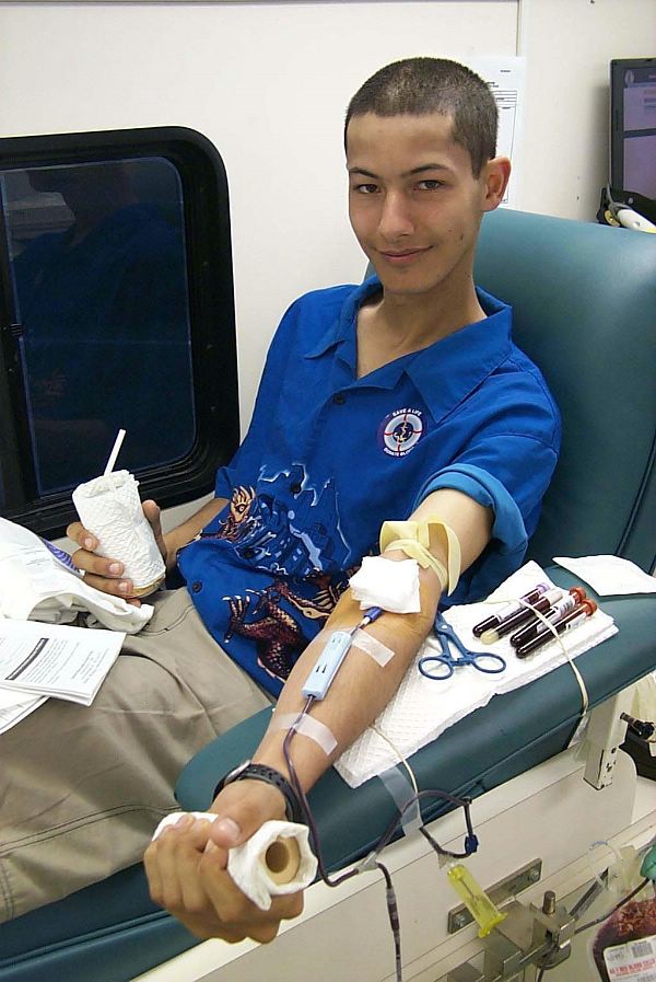
 A United States Navy enlisted man donates blood.Main article: Blood donation
Blood transfusions typically use two sources of blood: one's own (autologous transfusion), or someone else's (allogeneic transfusion). The latter is much more common than the former. Using another's blood must first start with donation of blood. Blood is most commonly donated as whole blood intravenously and collecting it with an anticoagulant. In developed countries, donations are usually anonymous to the recipient, but products in a blood bank are always individually traceable through the whole cycle of donation, testing, separation into components, storage, and administration to the recipient. This enables management and investigation of any suspected transfusion related disease transmission or transfusion reaction. In developing countries the donor is sometimes specifically recruited by or for the recipient, typically a family member, and the donation occurs immediately before the transfusion.
Processing and testing of blood products after donation
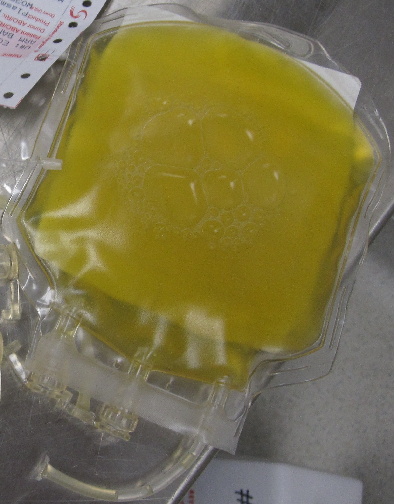
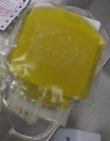 A bag containing one unit of fresh frozen plasma
Donated blood is usually subjected to processing after it is collected, to make it suitable for use in specific patient populations. Collected blood is then separated into blood components by centrifugation: red blood cells, plasma, platelets, albumin protein, clotting factor concentrates, cryoprecipitate, fibrinogen concentrate, and immunoglobulins (antibodies). Red cells, plasma and platelets can also be donated individually via a more complex process called apheresis.
- All donated blood are tested for infections. The current protocol tests donated blood for HIV-1, HIV-2, HTLV-1, HTLV-2, Hepatitis B, Hepatitis C, Syphilis (T pallidum), Chagas disease (T cruzi), and West Nile Virus. In addition, platelet products are also tested for bacterial infections due to its higher inclination for contamination due to storage at room temperature. Presence of Cytomegalovirus (CMV) is also tested because of risk to certain immunocompromised recipients if given, such as those with organ transplant or HIV. However, not all blood is tested for CMV because only a certain amount of CMV-negative blood needs to be available to supply patient needs. Other than positivity for CMV, any products tested positive for infections are not used.
- All donated blood are also tested for ABO and Rh groups, along with the presence of any red blood cell antibodies.
- Leukoreduction is the removal of white blood cells by filtration. Leukoreduced blood products are less likely to cause HLA alloimmunization (development of antibodies against specific blood types), febrile non-hemolytic transfusion reactions, cytomegalovirus infections, and platelet-transfuion refractoriness.
- Pathogen Reduction treatment that involves, for example, the addition of riboflavin with subsequent exposure to UV light has been shown to be effective in inactivating pathogens (viruses, bacteria, parasites and white blood cells) in blood products.[14][15][16] By inactivating white blood cells in donated blood products, riboflavin and UV light treatment can also replace gamma-irradiation as a method to prevent graft-versus-host disease (TA-GVHD).[17][18][19]
Compatibility testing
Main article: ABO blood group systemMain article: Rh blood group systemBefore a recipient receives a transfusion, compatibility testing between donor and recipient blood must be done. The first step before a transfusion is given is to Type and Screen the recipient's blood. Typing of recipient's blood determines the ABO and Rh status. The sample is then Screened for any alloantibodies that may react with donor blood.[20] It takes about 45 minutes to complete (depending on the method used). The blood bank technologist also checks for special requirements of the patient (e.g. need for washed, irradiated or CMV negative blood) and the history of the patient to see if they have a previously identified antibody.
A positive screen warrants an antibody panel/investigation to determine if it is clinically significant. An antibody panel consists of commercially prepared group O red cell suspensions from donors that have been phenotyped for commonly encountered and clinically significant alloantibodies. Donor cells may have homozygous (e.g. K+k-), heterozygous (K+k+) expression or no expression of various antigens (K-k+). The phenotypes of all the donor cells being tested are shown in a chart. The patient's serum is tested against the various donor cells using an enhancement method, e.g. Gel or LISS. Based on the reactions of the patient's serum against the donor cells, a pattern will emerge to confirm the presence of one or more antibodies. Not all antibodies are clinically significant (i.e. cause transfusion reactions, HDN, etc.). Once the patient has developed a clinically significant antibody it is vital that the patient receive antigen negative phenotyped red blood cells to prevent future transfusion reactions. A direct antiglobulin test (Coombs test) is also performed as part of the antibody investigation.[21]
If there is no antibody present, an immediate spin crossmatch or computer assisted crossmatch is performed where the recipient serum and donor serum are incubated. In the immediate spin method, two drops of patient serum are tested against a drop of 3-5% suspension of donor cells in a test tube and spun in a serofuge. Agglutination or hemolysis (i.e., positive Coombs test) in the test tube is a positive reaction and the unit should not be transfused.
If an antibody is suspected, potential donor units must first be screened for the corresponding antigen by phenotyping them. Antigen negative units are then tested against the patient plasma using an antiglobulin/indirect crossmatch technique at 37 degrees Celsius to enhance reactivity and make the test easier to read.
In urgent cases where crossmatching cannot be completed, and the risk of dropping hemoglobin outweighs the risk transfusing uncrossmatched blood, O-negative blood is used, followed by crossmatch as soon as possible. O-negative is also used for children and women of childbearing age. It is preferable for the laboratory to obtain a pre-transfusion sample in these cases so a type and screen can be performed to determine the actual blood group of the patient and to check for alloantibodies.
Neonatal transfusion
To ensure the safety of blood transfusion to pediatric patients, hospitals are taking additional precaution to avoid infection and prefer to use specially tested pediatric blood units that are guaranteed negative for Cytomegalovirus. Most guidelines recommend the provision of CMV-negative blood components and not simply leukoreduced components for newborns or low birthweight infants in whom the immune system is not fully developed.[22] These specific requirements place additional restrictions on blood donors who can donate for neonatal use.
Neonatal transfusions typically fall into one of two categories:
- "Top-up" transfusions, to replace losses due to investigational losses and correction of anemia.
- Exchange (or partial exchange) transfusions are done for removal of bilirubin, removal of antibodies and replacement of red cells (e.g., for anemia secondary to thalassemias and other hemoglobinopathies).[23]
Complications of transfusions
Transfusions of blood products are associated with several complications, many of which can be grouped as immunological or infectious. There is also increasing focus (and controversy) on complications arising directly or indirectly from potential quality degradation during storage.[24] Overall, adverse events from transfusions in the US account for about $17Billion - and in effect add more to the cost of each transfusion than acquisition and procedure costs combined.[25] While some complication risks depend on patient status or specific transfusion quantity involved, a baseline risk of complications simply increases in direct proportion to the frequency and volume of transfusion.
Immunologic complications
- Acute hemolytic reactions occur with transfusion of red blood cells, and occurs in about 0.016 percent of transfusions, with about 0.003 percent being fatal.[citation needed] This is due to destruction of donor erythrocytes by preformed recipient antibodies. Most often this occurs due to clerical errors or improper typing and crossmatching. Symptoms include fever, chills, chest pain, back pain, hemorrhage, increased heart rate, shortness of breath, and rapid drop in blood pressure. When suspected, transfusion should be stopped immediately, and blood sent for tests to evaluate for presence of hemolysis. Treatment is supportive. Kidney injury may occur due to the effects of the hemolytic reaction (pigment nephropathy).
- Delayed hemolytic reactions occur more frequently (about 0.025 percent of transfusions) and are due to the same mechanism as in acute hemolytic reactions. However, the consequences are generally mild and a great proportion of patients may not have symptoms. However, evidence of hemolysis and falling hemoglobin levels may still occur. Treatment is generally not needed, but due to the presence of recipient antibodies, future compatibility may be affected.
- Febrile nonhemolytic reactions are due to recipient antibodies to donor white blood cells, and occurs in about 7% of transfusions. This may occur after exposure from previous transfusions. Fever is generally short lived and is treated with antipyretics, and transfusions may be finished as long as an acute hemolytic reaction is excluded. This is a reason for the now-widespread use of leukoreduction - the filtration of donor white cells from red cell product units.
- Allergic reactions may occur when the recipient has preformed antibodies to certain chemicals in the donor blood, and does not require prior exposure to transfusions. Symptoms include urticaria, pruritus, and may proceed to anaphylactic shock. Treatment is the same as for any other type 1 hypersensitivity reactions. A small population (0.13%) of patients are deficient in the immunoglobin IgA, and upon exposure to IgA-containing blood, may develop an anaphylactic reaction.
- Posttransfusion purpura is a rare complication that occurs after transfusion containing platelets that express a surface protein HPA-1a. Recipients who lack this protein develop sensitization to this protein from prior transfusions, and develop thrombocytopenia about 7–10 days after subsequent transfusions. Treatment is with intravenous immunoglobulin, and recipients should only receive future transfusions with washed cells or HPA-1a negative cells.
- Transfusion-associated acute lung injury (TRALI) is an increasingly recognized adverse event associated with blood transfusion. TRALI is a syndrome of acute respiratory distress, often associated with fever, non-cardiogenic pulmonary edema, and hypotension, which may occur as often as 1 in 2000 transfusions.[26] Symptoms can range from mild to life-threatening, but most patients recover fully within 96 hours, and the mortality rate from this condition is less than 10%.[27] Although the cause of TRALI is not clear, it has been consistently associated with anti-HLA antibodies. Because these types of antibodies are commonly formed during pregnancy, several transfusion organisations have decided to use only plasma from men for transfusion.[28] TRALI is typically associated with plasma components rather than packed red blood cells (RBCs), though there is some residual plasma in RBC units.[28]
Infectious complications
- Blood products can rarely be contaminated with bacteria with possible subsequent life threatening infection, also known as transfusion transmitted bacterial infection. The risk of severe bacterial infection is estimated, as of 2002, at about 1 in 50,000 platelet transfusions, and 1 in 500,000 red blood cell transfusions.[29] It's important to note that blood product contamination, while rare, is still more common than actual infection. The reason platelets are more often contaminated than other blood products is that they are stored at room temperature for short periods of time. Contamination is also more common with longer duration of storage, especially longer than 5 days. Sources of contaminants include the donor's blood, donor's skin, phlebotomist's skin, and from containers. Contaminating organisms varies greatly, and includes skin flora, gut flora, or environmental organisms. There are many strategies in place at blood donation centers and laboratories to reduce such risk. A definite diagnosis of transfusion transmitted bacterial infection includes the identification of a positive culture in the recipient (without an alternative diagnosis) as well as the identification of the same organism in the donor blood.
- Ever since the advent of HIV testing of donor blood starting in the 1980s, the transmission of HIV during transfusion has dropped dramatically. Prior testing of donor blood only included testing for antibodies to HIV. However, due to the existence of a window period (a period of time a person is infectious but has not had time to develop antibodies), many cases of HIV seropositive blood were missed. The development of a nucleic acid test for the HIV-1 RNA has dramatically lowered the rate of donor blood seropositivity to about 1 in 3 million units. Despite this, HIV transmission can still occur but with a rate of even less than this.
- The transmission of hepatitis C via transfusion currently stands at about a rate of 1 in 2 million units. Such low rates has mostly been attributed to the ability to screen for both antibody as well as nucleic acid testing for viral RNA in donor blood.
- Other rare transmissible infections include hepatitis B, syphilis, Chagas disease, cytomegalovirus infections (in immunocompromised recipients), and HTLV.
Other (nonimmunologic/noninfectious) complications
- Transfusion inefficacy, while not itself a "complication," can lead to various complications due in part to the need to repeat transfusions; inefficacy can be especially serious for critical-care patients requiring rapid restoration of oxygen delivery. Insufficient efficacy can result from blood product units damaged by storage lesion - a set of biochemical and biomechanical changes which occur during storage. With red cells, this can decrease viability and ability for tissue oxygenation.[30] (Note that upon transfusion, cells have exhibited some degree of ability to reverse their storage lesion, albeit not entirely - and often too slowly to benefit urgent-care patients.)
- Transfusion-associated volume overload is a common complication simply due to the fact that blood products have a certain amount of volume. This is especially the case in recipients with underlying cardiac or kidney disease. Red cell transfusions can lead to volume overload when they must be repeated due to insufficient efficacy. Plasma transfusion is especially prone to causing volume overload due to its hypertonicity.
- Hypothermia can occur with transfusions with large quantities of blood products which normally are stored at cold temperatures. Core body temperature can go down as low as 32 °C and can produce physiologic disturbances. Prevention should be done with warming the blood to ambient temperature prior to transfusions.
- Transfusions with large amounts of red blood cells, whether due to severe hemorrhaging and/or transfusion inefficacy, can lead to an inclination for bleeding. The mechanism is thought to be due to disseminated intravascular coagulation, along with dilution of recipient platelets and coagulation factors. Close monitoring and transfusions with platelets and plasma is indicated when necessary.
- Metabolic alkalosis can occur with massive blood transfusions due to the breakdown of citrate stored in blood into bicarbonate
- Hypocalcemia can also occur with massive blood transfusions due to the complex of citrate with serum calcium
Ethical issues surrounding transfusion
Objections to blood transfusion
Objections to blood transfusions may arise for personal, medical, or religious reasons. For example, Jehovah's Witnesses object to blood transfusion primarily on religious grounds—they believe that blood is sacred, although they have also highlighted possible complications associated with transfusion.
Nonhuman blood transfusion
Veterinarians also administer transfusions to other animals. Various species require different levels of testing to ensure a compatible match. For example, cats have 3 known blood types, cattle have 11, dogs have 12, pigs 16 and horses have 34. However, in many species (especially horses and dogs), cross matching is not required before the first transfusion, as antibodies against non-self cell surface antigens are not expressed constitutively - i.e. the animal has to be sensitized before it will mount an immune response against the transfused blood.
The rare and experimental practice of inter-species blood transfusions is a form of xenograft.
Blood Transfusion substitutes
Main article: Blood substitutesThus far, there are no available oxygen-carrying blood substitutes, which is the typical objective of a blood (RBC) transfusion; however, there are widely available non-blood volume expanders for cases where only volume restoration is required. These are helping doctors and surgeons avoid the risks of disease transmission and immune suppression, address the chronic blood donor shortage, and address the concerns of Jehovah's Witnesses and others who have religious objections to receiving transfused blood.
A number of blood substitutes have been explored (and still are), but thus far they all suffer from many challenges. Most attempts to find a suitable alternative to blood thus far have concentrated on cell-free hemoglobin solutions. Blood substitutes could make transfusions more readily available in emergency medicine and in pre-hospital EMS care. If successful, such a blood substitute could save many lives, particularly in trauma where massive blood loss results. Hemopure, a hemoglobin-based therapy, is approved for use in South Africa.
See also
- Arnault Tzanck
- Blood transfusion in Sri Lanka
References
- ^ "Vicars of Christ" - Peter de Rossa
- ^ "The First Blood Transfusion?". Heart-valve-surgery.com. 2009-01-03. http://www.heart-valve-surgery.com/heart-surgery-blog/2009/01/03/first-blood-transfusion. Retrieved 2010-02-09.
- ^ "This Month in Anesthesia History". http://www.anesthesia.wisc.edu/AHA/Calendar/June.html. Retrieved 2009-06-15.
- ^ a b "Red Gold . Innovators & Pioneers . Jean-Baptiste Denis". PBS. http://www.pbs.org/wnet/redgold/innovators/bio_denis.html. Retrieved 2010-02-09.
- ^ "Mollison's Blood Transfusion in Clinical Medicine" by H.Klein, D. Anstee (2005), p.406
- ^ http://www.annals.org/cgi/reprint/132/5/420.pdf
- ^ Grunfeld GB, George Crile performs the first direct blood transfusion. In Great Events from History: Science and Technology II edited by Frank N. Magill (Pasadena, CA: Salem Press 1991, pp. 275-9).
- ^ R. Lewisohn. "CITRATE METHOD OF BLOOD TRANSFUSION". Journal of the American Medical Association. http://jama.ama-assn.org/content/114/16/1576.2.
- ^ "Red Gold: the Epic Story of Blood". PBS. http://www.pbs.org/wnet/redgold/history/timeline3.html.
- ^ Bernice Glatzer Rosenthal. New Myth, New World: From Nietzsche to Stalinism, Pennsylvania State University, 2002, ISBN 0-271-02533-6 pp. 161-162.
- ^ Boyan, C. P.; Howland, W. S. (1963). "Cardiac arrest and temperature of bank blood". JAMA : the journal of the American Medical Association 183: 58–60. PMID 14014662.
- ^ Rupreht, J; van Lieburg MJ; Lee JA; Erdman W (1985). Anaesthesia: essays on its history. Springer-Verlag. pp. 99–101. ISBN 3540132554.
- ^ Laura Landro (2007-01-10). "New rules may shrink ranks of blood donors". Wall Street Journal. http://www.post-gazette.com/pg/07010/752655-28.stm.
- ^ Hardwick, CC et al.; Herivel, TR; Hernandez, SC; Ruane, PH; Goodrich, RP (2004). "Separation, Identification and Quantification of Riboflavin and Its Photoproducts in Blood Products Using High-Performance Liquid Chromatography With Fluorescence Detection: A Method to Support Pathogen Reduction Technology". Photochemistry and Photobiology 80 (3): 609–615. doi:10.1562/0031-8655(2004)080<0609:TNSIAQ>2.0.CO;2. ISSN 0031-8655. PMID 15382964.
- ^ The Mirasol Clinical Evaluation Study Group, “A Randomized Controlled Clinical Trial Evaluating the Performance and Safety of Platelets treated with Mirasol Pathogen Reduction Technology.” Transfusion 2010, in press
- ^ Goodrich RP, et al., “The Mirasol PRT System for Pathogen Reduction of Platelets and Plasma: An Overview of Current Status and Future Trends.” Transfusion and ApheresisScience2006a; 35 (1): 5-17.
- ^ Fast, LD et al.; Dileone, G; Cardarelli, G; Li, J; Goodrich, R (2006). "Mirasol PRT Treatment of Donor White Blood Cells Prevents the Development of Xenogeneic Graft-Versus-Host Disease in Rag2-/-γc-/- Double Knockout Mice". Transfusion 46 (9): 1553–1560. doi:10.1111/j.1537-2995.2006.00939.x. PMID 16965583.
- ^ Fast LD, DiLeone G, Marschner S. (2010) Inactivation of human leukocytes in platelet products after pathogen reduction technology treatment in comparison to gamma-irradiation. Transfusion, In press.
- ^ Reddy HL, et.al. , “Toxicity Testing of a Novel Riboflavin-Based Technology for Pathogen Reduction and White Blood Cell Inactivation.” Transfusion Medicine Reviews 2008; 22 (2): 133-153.
- ^ Blood Processing. University of Utah. Available at: http://library.med.utah.edu/WebPath/TUTORIAL/BLDBANK/BBPROC.html. Accessed on: December 15, 2006.
- ^ Harmening, D. (1999). Modern Blood Banking and Transfusion Practices (4th ed.). Philadelphia: F. A. Davis. ISBN 080360419X.
- ^ "Red blood cell transfusions in newborn infants: Revised guidelines". Canadian Paediatric Society (CPS). http://www.cps.ca/english/statements/fn/fn02-02.htm#What%20type%20of%20RBCs%20should%20be%20used. Retrieved 2007-02-02.
- ^ KM Radhakrishnan, Srikumar Chakravarthi, S Pushkala, J Jayaraju (2003 Aug). "Component therapy". Indian J Pediatr 70 (8): 661–6. doi:10.1007/BF02724257. PMID 14510088.
- ^ Wang SS. What's the Shelf Life of Blood? Focus on Whether Older Donations Impair Recovery of Transfusion Recipients. The Wall Street Journal. 2009 Dec. 1.
- ^ Shander A, Hofmann A, Gombotz H, Theusinger OM, Spahn DR: Estimating the cost of blood: past, present, and future directions. Best Pract Res Clin Anaesthesiol; 2007; 21: 271-89.
- ^ Silliman C, Paterson A, Dickey W, Stroneck D, Popovsky M, Caldwell S, Ambruso D (1997). "The association of biologically active lipids with the development of transfusion-related acute lung injury: a retrospective study". Transfusion 37 (7): 719–26. doi:10.1046/j.1537-2995.1997.37797369448.x. PMID 9225936.
- ^ Popovsky M, Chaplin H, Moore S (1992). "Transfusion-related acute lung injury: a neglected, serious complication of hemotherapy". Transfusion 32 (6): 589–92. doi:10.1046/j.1537-2995.1992.32692367207.x. PMID 1502715.
- ^ a b Win N, Chapman CE, Bowles KM, Green A, Bradley S, Edmondson D, Wallis JP (2008). "How much residual plasma may cause TRALI?". Transfusion Medicine 18 (5): 276–280. doi:10.1111/j.1365-3148.2008.00885.x. PMID 18937733.
- ^ Blajchman M (2007). "Incidence and significance of the bacterial contamination of blood components". Dev Biol (Basel) 108 (2): 59–67. doi:10.2478/v10036-007-0007-1. PMID 12220143.
- ^ Zubair AC: Clinical impact of blood storage lesions. Am J Hematol; 2010; 85: 117-22.
Further reading
- Transfusion, ISSN: 1537-2995 (electronic) 0041-1132 (paper)
- Blood Groups and Red Cell Antigens. Free online book at NCBI Bookshelf ID: NBK2261
- Blood Transfusion Indications, information provide by Maharashtra State Blood Transfusion Council.
- Five Myths on Blood Transfusions, an information campaign by the New South Wales Government.
- [1] The Dangers of Illegal Blood Trade
- Cochrane Injuries Group, publishes systematic reviews of interventions for traumatic injury, which include evaluations of blood and blood substitute transfusions
Transfusion medicine General concepts Apheresis (plasmapheresis, plateletpheresis, leukapheresis) · Blood transfusion · Coombs test (direct and indirect) · Cross-matching · Exchange transfusion · International Society of Blood Transfusion · Intraoperative blood salvage · ISBT 128 · Transfusion reactionsBlood group systems/
blood typesBlood products/
blood donationCategories:
Wikimedia Foundation. 2010.