- Coma
-
Coma ICD-10 R40.2 ICD-9 780.01 In medicine, a coma (from the Greek κῶμα koma, meaning deep sleep) is a state of unconsciousness, lasting more than 6 hours[1] in which a person cannot be awakened, fails to respond normally to painful stimuli, light or sound, lacks a normal sleep-wake cycle and does not initiate voluntary actions.[1] A person in a state of coma is described as comatose. According to the Glasgow Coma Scale though, a person with confusion is considered to be in the mildest coma.
Coma may result from a variety of conditions, including intoxication (such as illicit drug abuse, overdose or misuse of over the counter medications, prescribed medication, or controlled substances), metabolic abnormalities, central nervous system diseases, acute neurologic injuries such as strokes or herniations, hypoxia, hypothermia, hypoglycemia or traumatic injuries such as head trauma caused by falls or vehicle collisions. It may also be deliberately induced by pharmaceutical agents in order to preserve higher brain functions following brain trauma, or to save the patient from extreme pain during healing of injuries or diseases.
In order for a patient to maintain consciousness, two important neurological components must function impeccably. The first is the cerebral cortex which is the gray matter covering the outer layer of the brain, and the other is a structure located in the brainstem, called reticular activating system (RAS or ARAS).[2] Injury to either or both of these components is sufficient to cause a patient to experience a coma. The human cortex is a group of tight, dense, "gray matter" composed of the nucleus of the neurons whose axons then form the "white matter", and is responsible for the perception of the universe, relay of the sensory input (sensation) via the thalamic pathway, and most importantly directly or indirectly in charge of all the neurological functions, from simple reflexes to complex thinking. Reticular activating system (RAS) on the other hand is a more primitive structure in the brainstem that is tightly in connection with reticular formation (RF), a critical anatomical structure needed for maintenance of arousal and sleep-wake transitions. Reticular activating system (RAS) takes its name from the effect it has on the reticular formation, which is via its stimulation. It is therefore necessary to investigate in a comatose patient, the integrity of the bilateral cerebral cortices, as well as that of the reticular activating system (RAS).
Contents
Signs and symptoms
Generally, a patient who is unable to voluntarily open the eyes, does not have a sleep-wake cycle, is unresponsive in spite of strong tactile (painful), or verbal stimuli and who generally scores between 3 to 8[7] on the Glasgow Coma Scale is considered to be in coma.[1] Coma may have developed in humans as a response to injury to allow the body to pause bodily actions and heal the most immediate injuries before - if at all - waking. It therefore could be a compensatory state in which the body's expenditure of energy is not superfluous. The severity and mode of onset of coma depends on the underlying cause. For instance, severe hypoglycemia (low blood sugar) or hypercapnia (increased carbon dioxide levels in the blood) initially cause mild agitation and confusion, but progress to obtundation, stupor and finally complete unconsciousness. In contrast, coma resulting from a severe traumatic brain injury or subarachnoid hemorrhage can be instantaneous. The mode of onset may therefore be indicative of the underlying cause. A state of unconsciousness lasting less than 6 hours is not by definition a coma, but a concussion,[1] therefore a patient who loses consciousness for less than this time period should not be immediately labeled as having experienced a coma.
Diagnosis and findings
Diagnosis of coma is simple; however, diagnosing the cause of the underlying disease process often proves to be challenging. The first priority in treatment of a comatose patient is stabilization following the basic ABCs (standing for airway, breathing, and circulation). Once a person in a coma is stable, investigations are performed to assess the underlying cause. Investigative methods are divided into physical examination findings and imaging (such as CAT scan, MRI, etc.) and special studies (EEG, etc.)
Initial assessment and evaluation
In the initial assessment of coma, it is common to gauge the level of consciousness by spontaneously exhibited actions, response to vocal stimuli ("Can you hear me?"), and painful stimuli; this is known as the AVPU (alert, vocal stimuli, painful stimuli, unresponsive) scale. More elaborate scales, such as the Glasgow Coma Scale, quantify an individual's reactions such as eye opening, movement and verbal response on a scale; Glasgow Coma Scale (GCS) is an indication of the extent of brain injury varying from 3 (indicating severe brain injury and death) to a maximum of 15 (indicating mild or no brain injury).
In those with deep unconsciousness, there is a risk of asphyxiation as the control over the muscles in the face and throat is diminished. As a result, those presenting to a hospital with coma are typically assessed for this risk ("airway management"). If the risk of asphyxiation is deemed to be high, doctors may use various devices (such as an oropharyngeal airway, nasopharyngeal airway or endotracheal tube) to safeguard the airway.
Physical examination findings
 Decorticate posturing, indicating a lesion at the red nucleus or above. This positioning is stereotypical for upper brain stem, or cortical damage. The other variant is Decerebrate posturing, not seen in this picture.
Physical examination is critical after stabilization. It should include vital signs, a general portion dedicated to making observations about the patient's respiration (breathing pattern), body movements (if any), and of the patient's body habitus (physique); it should also include assessment of the brainstem and cortical function through special reflex tests such as the oculocephalic reflex test (doll's eyes test), oculovestibular reflex test (cold caloric test), nasal tickle, corneal reflex, and the gag reflex.
Vital signs in medicine are temperature (rectal is most accurate), blood pressure, heart rate (pulse), respiratory rate, and oxygen saturation. It should be easy to evaluate these vitals quickly in order to gain insight into a patient's metabolism, fluid status, heart function, vascular integrity, and tissue oxygenation.
Respiratory pattern (breathing rhythm) is significant and should be noted in a comatose patient. Certain stereotypical patterns of breathing have been identified including Cheyne-Stokes a form of breathing in which the patient's breathing pattern is described as alternating episodes of hyperventilation and apnea. This is a dangerous pattern and is often seen in pending herniations, extensive cortical lesions, or brainstem damage.[2] Another pattern of breathing is apneustic breathing which is characterized by sudden pauses of inspiration and is due to a lesion of the pons.[1][2] Ataxic breathing is irregular and is due to a lesion (damage) of the medulla.
Assessment of posture and body habitus is the next step. It involves general observation about the patient's positioning. There are often two stereotypical postures seen in comatose patients. Decorticate posturing is a stereotypical posturing in which the patient has arms flexed at the elbow, and arms adducted toward the body, with both legs extended. Decerebrate posturing is a stereotypical posturing in which the legs are similarly extended (stretched), but the arms are also stretched (extended at the elbow). The posturing is critical since it indicates where the damage is in the central nervous system. A decorticate posturing indicates a lesion (a point of damage) at or above the red nucleus, whereas a decerebrate posturing indicates a lesion at or below the red nucleus. In other words, a decorticate lesion is closer to the cortex, as opposed to a decerebrate cortex that is closer to the brainstem.
Oculocephalic reflex also known as the doll's eye is performed to assess the integrity of the brainstem. Patient's eye lids are gently elevated and the cornea is visualized. The patient's head is then moved to the patient's left, to observe if the eyes stay or deviate toward the patient's right; same maneuver is attempted on the opposite side. If the patient's eyes move in a direction opposite to the direction of the rotation of the head, then the patient is said to have an intact brainstem. However, failure of both eyes to move to one side, can indicate damage or destruction of the affected side. In special cases, where only one eye deviates and the other does not, this often indicates a lesion (or damage) of the medial longitudinal fasciculus (MLF) which is a brainstem nerve tract. Caloric reflex test also evaluates both cortical and brainstem function; cold water is injected into one ear and the patient is observed for eye movement; if the patient's eyes slowly deviate toward the ear where the water was injected, then the brainstem is intact, however failure to deviate toward the injected ear indicates damage of the brainstem on that side. Cortex is responsible for a rapid nystagmus away from this deviated position and is often seen in patients who are conscious or merely lethargic.
An important part of the physical exam is also assessment of the cranial nerves. Due to the unconscious status of the patient, only a limited number of the nerves can be assessed. These include the cranial nerves number 2 (CN II), number 3 (CN III), number 5 (CN V), number 7 (CN VII), and cranial nerves 9 and 10 (CN IX, CN X). Gag reflex helps assess cranial nerves 9 and 10. Pupil reaction to light is important because it shows an intact retina, and cranial nerve number 2 (CN II); if pupils are reactive to light, then that also indicates that the cranial nerve number 3 (CN III) (or at least its parasympathetic fibers) are intact. Corneal reflex assess the integrity of cranial nerve number 7 (CN VII), and cranial nerve number 5 (CN V). Cranial nerve number 5 (CN V), and its ophthalmic branch (V1) are responsible for the afferent arm of the reflex, and the cranial nerve number 7 (CN VII) also known a facial nerve, is responsible for the efferent arm, causing contraction of the muscle orbicularis oculi resulting in closing of the eyes.
Pupil assessment is often a critical portion of a comatose examination, as it can give information as to the cause of the coma; the following table is a technical, medical guideline for common pupil findings and their possible interpretations:[2]
Pupil sizes (Left eye vs. Right eye) Possible interpretation •ʖ• Normal eye with two pupils equal in size and reactive to light. This means that the patient is probably not in a coma and is probably lethargic, under influence of a drug, or sleeping. •ʖ• "Pinpoint" pupils indicate heroin or opiate overdose, and can be responsible for a patient's coma. The pinpoint pupils are still reactive to light, bilaterally (in both eyes, not just one). Another possibility is the damage of the pons.[2] •ʖ• One pupil is dilated and unreactive, while the other is normal (in this case the L eye is dilated but the R eye is normal in size). This could mean a damage to the oculomotor nerve (cranial nerve number 3, CN III) on the right side, or possibility of vascular involvement. •ʖ• Both pupils are dilated and unreactive to light. This could be due to overdose of certain medications, hypothermia or severe anoxia (lack of oxygen). Imaging and special tests findings
Imaging basically encompasses computed tomography (CAT or CT) scan of the brain, or MRI for example, and is performed to identify specific causes of the coma, such as hemorrhage in the brain or herniation of the brain structures. Special tests such as an EEG can also show a lot about the activity level of the cortex such as semantic processing,[8] presence of seizures, and are important available tools not only for the assessment of the cortical activity but also for predicting the likelihood of the patient's awakening.[9] The autonomous responses such as the Skin Conductance Response may also provide further insight on the patient's emotional processing.[10]
History
When diagnosing any neurological condition, history and examination are fundamental. History is obtained by family, friends or EMS. The Glasgow Coma Scale is a helpful system used to examine and determine the depth of coma, track patients progress and predict outcome as best as possible. In general a correct diagnosis can be achieved by combining findings from physical exam, imaging, and history components and will direct the appropriate therapy.
Severity and classification
Plum and Posner classify coma[2] as either (1) supratentoral (above Tentorium cerebelli), (2) infratentoral (below Tentorium cerebelli), or (3) metabolic or (4) diffuse. This classification is merely dependent on the position of the original damage that caused the coma, and does not correlate with severity or the prognosis. The severity of coma impairment however is categorized into several levels. Patients may or may not progress through these levels. In the first level, the brain responsiveness lessens, normal reflexes are lost, the patient no longer responds to pain and cannot hear.
The Rancho Los Amigos Scale is a complex scale that has eight separate levels, and is often used in the first few weeks or months of coma while the patient is under closer observation, and when shifts between levels are more frequent.
Prognosis
Outcomes range from recovery to death. Comas can last from several days to several weeks. In more severe cases a coma may last for over 5 weeks, while some have lasted as long as several years. After this time, some patients gradually come out of the coma, some progress to a vegetative state, and others die. Some patients who have entered a vegetative state go on to regain a degree of awareness. Others remain in a vegetative state for years or even decades (the longest recorded period being 37 years).[11]
The outcome for coma and vegetative state depends on the cause, location, severity and extent of neurological damage. A deeper coma alone does not necessarily mean a slimmer chance of recovery, because some people in deep coma recover well while others in a so-called milder coma sometimes fail to improve.
People may emerge from a coma with a combination of physical, intellectual and psychological difficulties that need special attention. Recovery usually occurs gradually—patients acquire more and more ability to respond. Some patients never progress beyond very basic responses, but many recover full awareness.[12] Regaining consciousness is not instant: in the first days, patients are only awake for a few minutes, and duration of time awake gradually increases. This is unlike the situation in many movies where people who awake from comas are instantly able to continue their normal lives. In reality, the coma patient awakes sometimes in a profound state of confusion, not knowing how they got there and sometimes suffering from dysarthria, the inability to articulate any speech, and with many other disabilities.
Predicted chances of recovery are variable owing to different techniques used to measure the extent of neurological damage. All the predictions are based on statistical rates with some level of chance for recovery present: a person with a low chance of recovery may still awaken. Time is the best general predictor of a chance of recovery: after 4 months of coma caused by brain damage, the chance of partial recovery is less than 15%, and the chance of full recovery is very low.[13][14]
The most common cause of death for a person in a vegetative state is secondary infection such as pneumonia which can occur in patients who lie still for extended periods.
Occasionally people come out of coma after long periods of time. After 19 years in a minimally conscious state, Terry Wallis spontaneously began speaking and regained awareness of his surroundings.[15] Similarly, Polish railroad worker Jan Grzebski woke up from a 19-year coma in 2007.
A brain-damaged man, trapped in a coma-like state for six years, was brought back to consciousness in 2003 by doctors who planted electrodes deep inside his brain. The method, called deep brain stimulation (DBS) successfully roused communication, complex movement and eating ability in the 38-year-old American man who suffered a traumatic brain injury. His injuries left him in a minimally conscious state (MCS), a condition akin to a coma but characterized by occasional, but brief, evidence of environmental and self-awareness that coma patients lack.[16]
Coma lasting seconds to minutes results in post-traumatic amnesia (PTA) that lasts hours to days; recovery plateau occurs over days to weeks. Coma that lasts hours to days results in PTA lasting days to weeks; recovery plateau occurs over months. Coma lasting weeks results in PTA that lasts months; recovery plateau occurs over months to years.
Treatment and recovery
Medical treatment
Coma is a medical emergency, and attention must first be directed to maintaining the patient's respiration and circulation, using intubation and ventilation, administration of intravenous fluids or blood and other supportive care as needed. Once a patient is stable and no longer in immediate danger, the medical staff may concentrate on maintaining the health of patient’s physical state. The concentration will be directed on preventing infections such as pneumonias, bedsores (decubitus ulcers) and providing a balanced nutrition.[17] These infections may appear from the patient not being able to move around, and being confined to the bed. The nursing staff will move the patient every 2–3 hours from side to side and depending on the state of consciousness sometimes to a chair. The goal is to move the patient as much as possible to try to avoid bedsores, atelectasis and pneumonia. Pneumonia can occur from the person’s inability to swallow leading to aspiration, lack of gag reflex or from feeding tube, (aspiration pneumonia). Physical therapy may also be used to prevent contractures and orthopedic deformities that would limit recovery for those patients who emerge from coma.
A person in a coma may become restless, or seize and need special care to prevent them from hurting themselves. Medicine may be given to calm such individuals. Patients who are restless may also try to pull on tubes or dressings so soft cloth wrist restraints may be put on. Side rails on the bed should be kept up to prevent patient from falling.[17]
Emotional challenges
Coma has a wide variety of emotional reactions from the family members of the affected patients, as well as the primary care givers taking care of the patients. Common reactions, such as desperation, anger, frustration, and denial are possible. The focus of the patient care should be on creating an amicable relationship with the family members or dependents of a comatose patient as well as creating rapport with the medical staff.[18]
Society and culture
Research by Dr. Eelco Wijdicks on the depiction of comas in movies was published in Neurology in May 2006. Dr. Wijdicks studied 30 films (made between 1970 and 2004) that portrayed actors in prolonged comas, and he concluded that only two films accurately depicted the state of a coma victim and the agony of waiting for a patient to awaken: Reversal of Fortune (1990) and The Dreamlife of Angels (1998). The remaining 28 were criticized for portraying miraculous awakenings with no lasting side effects, unrealistic depictions of treatments and equipment required, and comatose patients remaining muscular and tanned.[19]
See also
- Brain death Lack of activity in both cortex, and lack of brainstem function.
- Coma scale, a system to assess the severity of coma
- Locked-in syndrome Paralysis of most muscles, except ocular muscles of the eyes, while patient is conscious.
- Persistent vegetative state (vegetative coma), deep coma without detectable awareness. Damage to the cortex, with an intact brainstem.
- Process Oriented Coma Work, for an approach to working with residual consciousness in comatose patients.
References
- ^ a b c d e Weyhenmyeye, James A.; Eve A. Gallman (2007). Rapid Review Neuroscience 1st Ed. Mosby Elsevier. pp. 177–9. ISBN 0-323-02261-8.
- ^ a b c d e f Hannaman, Robert A. (2005). MedStudy Internal Medicine Review Core Curriculum: Neurology 11th Ed. MedStudy. pp. (11–1)-(11–2). ISBN 1-932703-01-2.
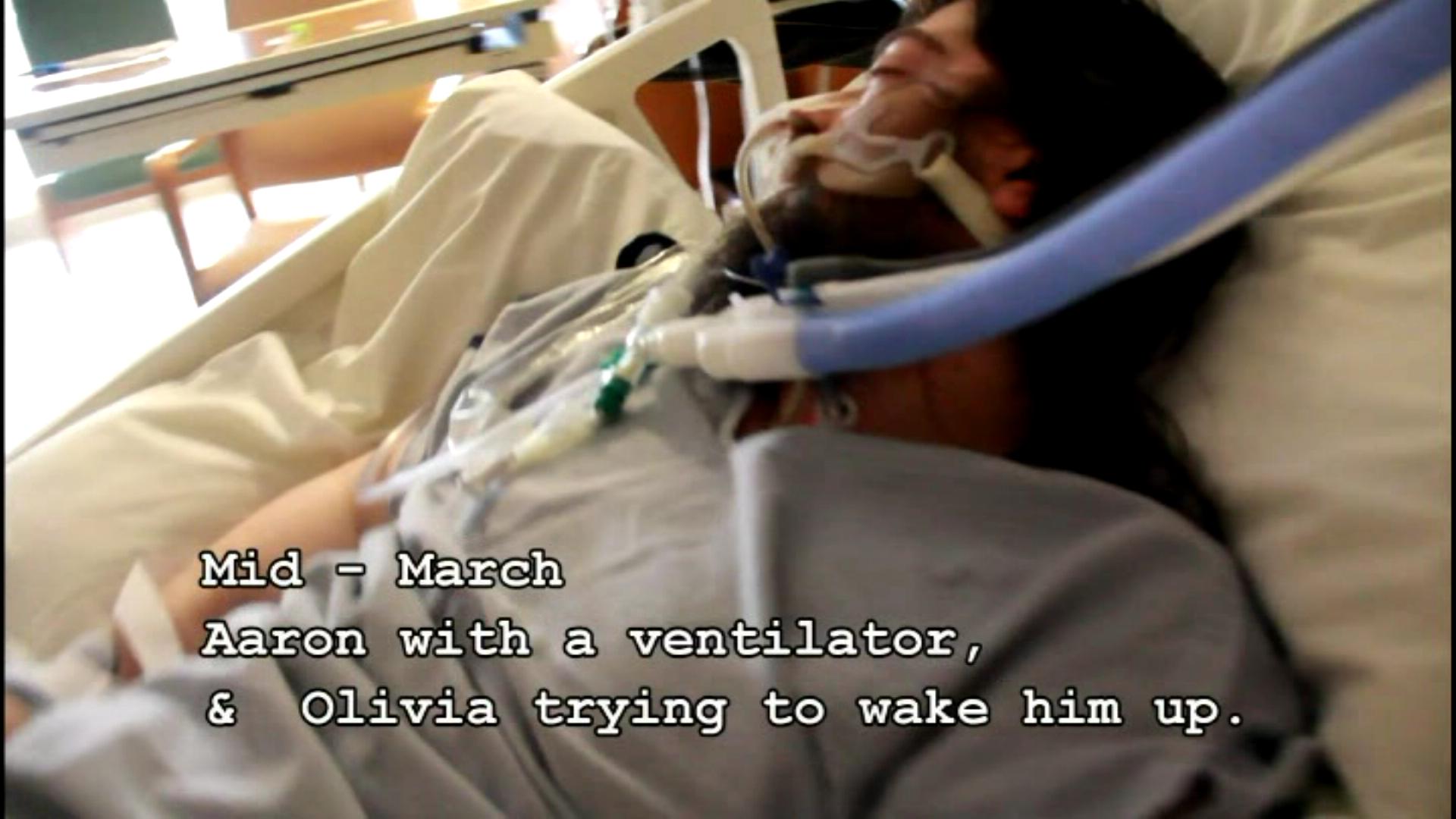
- ^ "Video of Aaron Cohen at beginning of documented 3 month coma.". http://www.youtube.com/watch?v=IW8rBQgiU_c.
- ^ "Video of Aaron Cohen still nonresponsive to stimuli while in coma.". http://www.youtube.com/watch?v=I6t6dDEDVXg.
- ^ "Video of Aaron Cohen nearly out of 3 month coma.". http://www.youtube.com/watch?v=FlwRC7E4TQI.
- ^ "Video of Aaron Cohen walking 2 weeks after 3 month coma.". http://www.youtube.com/watch?v=WcIbULrMBsQ.
- ^ Russ Rowlett. "Glasgow Coma Scale". University of North Carolina at Chapel Hill. http://www.unc.edu/~rowlett/units/scales/glasgow.htm.
- ^ Daltrozzo, J., Wioland, N., Mutschler, V., Lutun, P., Jaeger, A., Calon, B., Meyer, A., Pottecher, T., Lang, S., Kotchoubey, B. (2009c). Cortical Information Processing in Coma, Cognitive & Behavioral Neurology, 22(1), 53-62.[1]
- ^ Daltrozzo, J., Wioland, N., Mutschler, V., Kotchoubey, B. (2007). Predicting Coma and other Low Responsive Patients Outcome using Event-Related Brain Potentials: A Meta-analysis. Clinical Neurophysiology, 118, 606-614.[2]
- ^ Daltrozzo, J., Wioland, N., Mutschler, V., Lutun, P., Calon, B., Meyer, A., Jaeger, A., Pottecher, T., Kotchoubey, B. (2010a). Electrodermal Response in Coma and Other Low Responsive Patients. Neuroscience Letters, 475(1), 44-47.[3]
- ^ According to the Guinness Book of Records, the longest period spent in coma was by Elaine Esposito. She did not wake up after being anaesthetized for an appendectomy on August 6, 1941, at age 6. She died on November 25, 1978 at age 43 years 357 days, having been in a coma for 37 years 111 days.
- ^ NINDS (October 29, 2010). "Coma Information Page: National Institute of Neurological Disorders and Stroke (NINDS)". http://www.ninds.nih.gov/disorders/coma/coma.htm#What_is_the_prognosis. Retrieved 2010-12-08.
- ^ Formisano R, Carlesimo GA, Sabbadini M, et al. (May 2004). "Clinical predictors and neuropleropsychological outcome in severe traumatic brain injury patients". Acta Neurochir (Wien) 146 (5): 457–62. doi:10.1007/s00701-004-0225-4. PMID 15118882.
- ^ brain injury .com | Coma traumatic brain injury - Brain Injury Coma
- ^ "Mother stunned by coma victim's unexpected words". The Sydney Morning Herald. 2003-07-12. http://www.smh.com.au/articles/2003/07/11/1057783356390.html.
- ^ "Electrodes stir man from six-year coma-like state". Cosmos Magazine. 2 August 2007. http://www.cosmosmagazine.com/node/1513.
- ^ a b "Coma". http://medicalcenter.osu.edu/PatientEd/Materials/PDFDocs/dis-cond/general/coma.pdf. Retrieved 2010-12-08.
- ^ Coma Care (2010-03-30). "Caring for Care Giver and Family". http://www.comacare.com/cgi-bin/giga.cgi?. Retrieved 2010-12-08.
- ^ Eelco F.M. Wijdicks, MD and Coen A. Wijdicks, BS (2006). "The portrayal of coma in contemporary motion pictures". Neurology 66 (9): 1300–1303. doi:10.1212/01.wnl.0000210497.62202.e9. PMID 16682658. http://www.neurology.org/cgi/content/abstract/66/9/1300. Retrieved 2009-11-25.
General Related Symptoms and signs: cognition, perception, emotional state and behaviour (R40–R46, 780.0–780.5, 781.1) Cognition Confusion (Delirium) · Somnolence · Obtundation · Stupor · Unconsciousness (Syncope, Coma, Persistent vegetative state)Fainting/SyncopeOtherAmnesia (Anterograde amnesia, Retrograde amnesia) · Dizziness (Vertigo, Presyncope/Lightheadedness, Disequilibrium) · ConvulsionEmotional state Behavior Perception/
sensation
disorderCategories:- Intensive care medicine
- Emergency medicine
- Symptoms and signs: Cognition, perception, emotional state and behaviour


Wikimedia Foundation. 2010.