- Posterior pituitary
-
Posterior pituitary 
Pituitary gland. Posterior pituitary is in blue. Pars nervosa and infundibular stalk are not labeled, but pars nervosa is at bottom and infundibular stalk is at top.) 
Median sagittal through the hypophysis of an adult monkey. (Posterior lobe labeled at bottom right.) Latin glandula pituitaria, pars nervosa Gray's subject #275 1275 Artery inferior hypophyseal artery Vein hypophyseal vein Precursor Neural tube (downward-growth of the diencephalon)[1] MeSH Pituitary+Gland,+Posterior Dorlands/Elsevier Posterior pituitary hormones The posterior pituitary (or neurohypophysis) comprises the posterior lobe of the pituitary gland and is part of the endocrine system. Despite its name, the posterior pituitary gland is not a gland, per se; rather, it is largely a collection of axonal projections from the hypothalamus that terminate behind the anterior pituitary gland.
Contents
Anatomy
The posterior pituitary consists mainly of neuronal projections (axons) extending from the supraoptic and paraventricular nuclei of the hypothalamus. These axons release peptide hormones into the capillaries of the hypophyseal circulation. In addition to axons, the posterior pituitary also contains pituicytes, specialized glial cells resembling astrocytes.
Classification of the posterior pituitary varies, but most sources include the three regions below:
- Pars nervosa
- Also called the neural lobe or posterior lobe, this region constitutes the majority of the posterior pituitary and is the storage site of oxytocin and vasopressin. Sometimes (incorrectly) considered synonymous with the posterior pituitary, the pars nervosa includes Herring bodies and pituicytes.[2]
- Infundibular stalk
- Also known as the infundibulum or pituitary stalk, the infundibular stalk bridges the hypothalamic and hypophyseal systems.
- Median eminence
- This is only occasionally included as part of the posterior pituitary. Other sources specifically exclude it from the pituitary.[3]
A few sources include the pars intermedia as part of the posterior lobe, but this is a minority view. It is based upon the gross anatomical separation of the posterior and anterior pituitary along the cystic remnants of Rathke's pouch, causing the pars intermedia to remain attached to the neurohypophysis.
Hormones secreted
Hormones known classically as posterior pituitary hormones are synthesized by the hypothalamus. They are then stored and secreted by the posterior pituitary into the bloodstream.
Hormone Other names Symbol(s) Target Effect Source Oxytocin - OT Uterus, mammary glands Uterine contractions; lactation supraoptic and paraventricular nuclei Vasopressin (antidiuretic hormone) Arginine vasopressin, argipressin, antidiuretic hormone VP, AVP, ADH Kidneys or Arterioles Stimulates water retention; raises blood pressure by contracting arterioles, induces male aggression supraoptic and paraventricular nuclei Role in disease
Insufficient secretion of vasopressin underlies diabetes insipidus, a condition in which the body loses the capacity to concentrate urine. Affected individuals excrete as much as 20 liters of dilute urine per day. Oversecretion of vasopressin causes the syndrome of inappropriate antidiuretic hormone (SIADH).
See also
References
Additional images
-

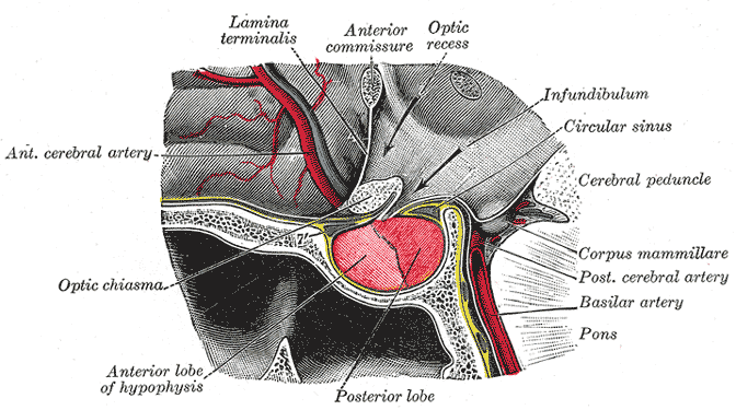
The posterior pituitary comprises the posterior lobe of the pituitary gland.
-

-

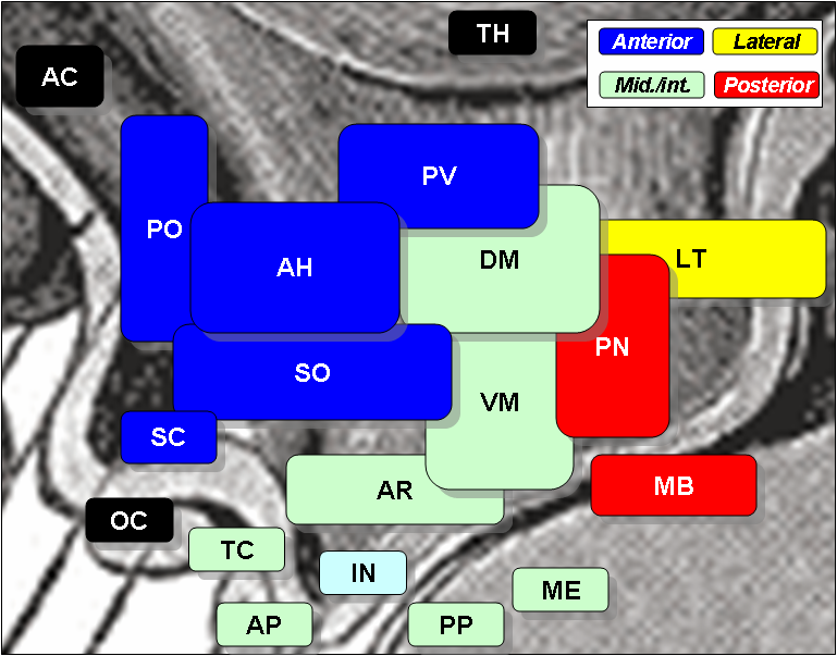
Hypothalamic nuclei
Human anatomy, endocrine system: endocrine glands (TA A11, TH H3.08, GA 11.1269) Islets of pancreas Hypothalamic/
pituitary axes
+parathyroidPituitaryPosterior pituitaryPars intermedia · Pars tuberalis · Pars distalis
Acidophil cell (Somatotropic cell, Prolactin cell) · Basophil cell (Corticotropic cell, Gonadotropic cell, Thyrotropic cell) · Chromophobe cellThyroid isthmus · Lobes of thyroid gland · Pyramidal lobe of thyroid gland
Follicular cell · Parafollicular cellPineal gland Other Categories:


Wikimedia Foundation. 2010.