- Habenula
Infobox Brain
Name = PAGENAME
Latin =
GraySubject =
GrayPage =
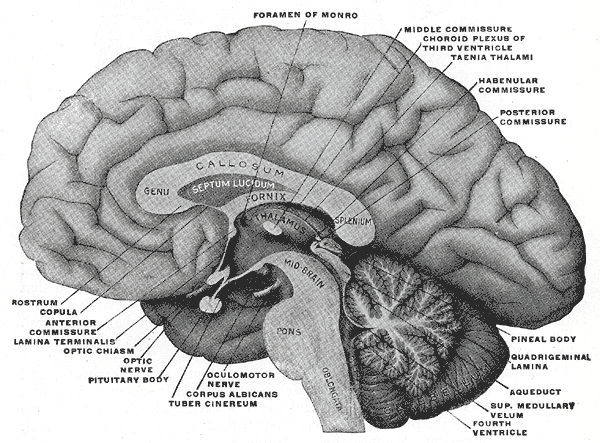
Caption = Mesal aspect of a brain sectioned in the median sagittal plane. Habenula is not labeled directly, but after expanding, look to region with 'habenular commissure', 'pineal body', and 'posterior commissure'

Caption2 = 1.Taenia choroidea (and lateral:Lamina affixa ,Stria terminalis )
2.Thalamus , Pulvinar thalami
3.Third ventricle
4. Stalk ofpineal gland
5.Habenula
6.Stria medullaris
7.Superior colliculus
8.Brachium of superior colliculus
9.Inferior colliculus
10.Brachium of inferior colliculus
11.Medial geniculate nucleus
12.Sulcus medianus
13.Superior cerebellar peduncles
14.Inferior cerebellar peduncle
15.Middle cerebellar peduncles
16.Tuberculum anterius thalami
17.Obex ,Area postrema
IsPartOf =
Components =
Artery =
Vein =
BrainInfoType = hier
BrainInfoNumber = 277
MeshName = habenula
MeshNumber =
DorlandsPre = h_01
DorlandsSuf = 12405682
Inneuroanatomy , habenula (diminutive of Latin "habena" meaning rein) originally denoted the stalk of thepineal gland (pineal habenula; pedunculus of pineal body), but gradually came to refer to a neighboring group of nerve cells with which the pineal gland was believed to be associated, the habenular nucleus. The habenular nucleus is a set of well conserved structures in allvertebrate animals.Andres et al. 1999]Currently, the
Terminologia Anatomica term refers exclusively to this separate cell mass in the caudal and dorsal aspect of the dorsalthalamus (theepithalamus ), embedded in theposterior end of themedullary stria from which it receives most of its afferent fibers. By way of theretroflex fasciculus (habenulointerpeduncular tract) it projects to theinterpeduncular nucleus and other paramedian cell groups of themidbrain tegmentum .The habenula receives input from the brain via the
stria medullaris thalami and outputs to manymidbrain areas involved in releasingneuromodulator s, such asdopamine ,norepinephrine , andserotonin .Anatomy
The habenula was traditionally divided into lateral (limbic) and medial (motor) parts.Iwahori 1977] Detailed examination of the region in the rat, however, suggested that the lateral part should be further divided into ten distinct subnuclei and the medial into five distinct subnuclei.
Lateral habenula
The primary input regions to the lateral habenula are the lateral
preoptic area (bringing input from thehippocampus and lateral steptum), the ventral pallidum (bringing input from thenucleus accumbens and mediodorsal nucleus of thethalamus ), the lateralhypothalamus , and the internal segment of theglobus pallidus (bringing input from otherbasal ganglia structures).Geisler 2008] .The outputs of the lateral habenula target dopaminergic regions (
substantia nigra pars compacta and theventral tegmental area ), serotinergic regions (median raphe and dorsal raphe nuclei), and a cholinergic region (thelaterodorsal tegmental nucleus ).Medial habenula
Input to the medial hebanula comes from a variety of regions and carries a number of different chemicals. Input regions include septal nuclei (the fimbrialis septi and triangularis septi nuclei), dopaminergic inputs from the interfascicular nucleus of the ventral tegmental area, noradrenergic inputs from the
locus ceruleus , and GABAergic inputs from thediagonal band of Broca .Lecourtier and Kelly 2007]The medial habenula sends outputs of
substance P and acetylcholine to the interpeduncular nucleus as well as to the pineal gland.Functions
The habenular nuclei have been shown to be involved in many functions, including pain processing, reproductive behavior, nutrition, sleep-wake cycles, stress responses, and learning. Recent demonstrations using
fMRI Matsumoto and Hikosaka 2007] have closely linked the function of the lateral habenula with reward processing, in particular with regard to encoding negative feedback or negative rewards. For instance, Matsumoto and Hikosaka showed in 2007 that the firing of lateral habenula neurons in rhesus monkeys was complementary to the firing of dopaminergic neurons in the substantia nigra pars compacta: dopaminergic neurons increase in firing rate in response to a stimulus that predicts reward, whereas lateral habenular neurons increase in firing rate in response to a stimulus that predicts a lack of reward.External links
*
References
*cite journal
last =Andres
first =KH
coauthors = During MV, Veh RW
title =Subnuclear organization of the rat habenular complexes.
journal =Journal of Comparative Neurology
volume = 407
pages = 130-150
year = 1999
pmid = 10213193*cite journal
last =Geisler
first =S
title =The lateral habenula: no longer neglected.
journal =CNS Spectrums
volume = 13
pages = 484-489
year =2008*cite journal
last =Iwahori
first =N
title =A Golgi study on the habenular nucleus of the cat.
journal =Journal of Comparative Neurology
volume = 72
pages = 319-344
year =1977*cite journal
last =Lecourtier
first =L
coauthors = Kelly PH
title =A conductor hidden in the orchestra? Role of the habenular complex in monoamine transmission and cognition
journal =Neuroscience & Biobehavioral Reviews
volume = 31
pages = 658-672
year = 2007
pmid = 17379307
doi = 10.1016/j.neubiorev.2007.01.004*cite journal
last =Matsumoto
first =M
coauthors = Hikosaka O
title = Lateral habenula as a source of negative reward signals in dopamine neurons.
journal = Nature
volume = 447
pages = 1111-1115
year = 2007
doi = 10.1038/nature05860*cite journal
last =Ullsperger
first =M
coauthors = von Cramon, DY
title = Error monitoring using external feedback: Specific roles of the habenular complex, the reward system, and the cingulate motor area revealed by functional magnetic resonance imaging.
journal =Journal of Neuroscience
volume = 23
pages = 4308-4314
year = 2003
Wikimedia Foundation. 2010.