- Meiosis
-
For the figure of speech, see meiosis (figure of speech).
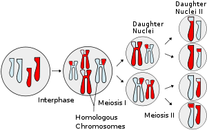 Events involving meiosis, showing chromosomal crossover
Meiosis (pronounced /maɪˈoʊsɨs/ (
 listen)) is a special type of cell division necessary for sexual reproduction. The cells produced by meiosis are gametes or spores. The animals' gametes are called sperm and egg cells.
Whilst the process of meiosis bears a number of similarities with the 'life-cycle' cell division process of mitosis, it differs in two important respects:
- the chromosomes in meiosis undergo a recombination which shuffles the genes producing a different genetic combination in each gamete, compared with the co-existence of each of the two separate pairs of each chromosome (one received from each parent) in each cell which results from mitosis.
- the outcome of meiosis is four (genetically unique) haploid cells, compared with the two (genetically identical) diploid cells produced from mitosis.
Meiosis begins with one diploid cell containing two copies of each chromosome—one from the organism's mother and one from its father—and produces four haploid cells containing one copy of each chromosome. Each of the resulting chromosomes in the gamete cells is a unique mixture of maternal and paternal DNA, ensuring that offspring are genetically distinct from either parent. This gives rise to genetic diversity in sexually reproducing populations, which provides the variation of physical and behavioural attributes (phenotypes) upon which natural selection acts, at a population level, leading to adaptation within the population, resulting in evolution.
Prior to the meiosis process the cell's chromosomes are duplicated by a round of DNA replication, creating a maternal and paternal version of each chromosome (homologs) composed of two exact copies, sister chromatids, attached at the centromere region. In the beginning of meiosis the maternal and paternal homologs pair to each other. Then they typically exchange parts by homologous recombination leading to crossovers of DNA between the maternal and paternal versions of the chromosome. Spindle fibers bind to the centromeres of each pair of homologs and arrange the pairs at the spindle equator. Then the fibers pull the recombined homologs to opposite poles of the cell. As the chromosomes move away from the center the cell divides into two daughter cells, each containing a haploid number of chromosomes composed of two chromatids. After the recombined maternal and paternal homologs have separated into the two daughter cells, a second round of cell division occurs. There meiosis ends as the two sister chromatids making up each homolog are separated and move into one of the four resulting gamete cells. Upon fertilization, for example when a sperm enters an egg cell, two gamete cells produced by meiosis fuse. The gamete from the mother and the gamete from the father each contribute one half of the set of chromosomes that make up the new offsping's genome.
Meiosis uses many of the same mechanisms as mitosis, a type of cell division used by eukaryotes like plants and animals to split one cell into two identical daughter cells. In all plants and in many protists meiosis results in the formation of spores: haploid cells that can divide vegetatively without undergoing fertilization. Some eukaryotes, like Bdelloid rotifers, have lost the ability to carry out meiosis and have acquired the ability to reproduce by parthenogenesis. Meiosis does not occur in archaea or bacteria, which reproduce via asexual processes such as binary fission.
Contents
History
Meiosis was discovered and described for the first time in sea urchin eggs in 1876 by the German biologist Oscar Hertwig. It was described again in 1883, at the level of chromosomes, by the Belgian zoologist Edouard Van Beneden, in Ascaris worms' eggs. The significance of meiosis for reproduction and inheritance, however, was described only in 1890 by German biologist August Weismann, who noted that two cell divisions were necessary to transform one diploid cell into four haploid cells if the number of chromosomes had to be maintained. In 1911 the American geneticist Thomas Hunt Morgan observed crossover in Drosophila melanogaster meiosis and provided the first genetic evidence that genes are transmitted on chromosomes. The term meiosis was coined by J.B Farmer and J.B Moore in 1905.
Occurrence in eukaryotic life cycles
 Gametic life cycle.
 Zygotic life cycle.Main article: Biological life cycle
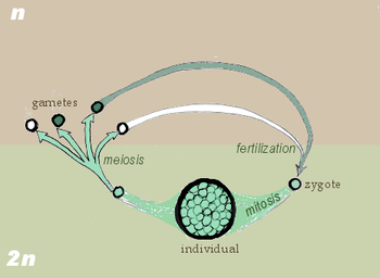
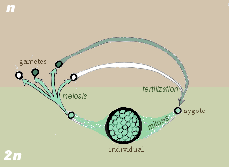
Meiosis occurs in eukaryotic life cycles involving sexual reproduction, consisting of the constant cyclical process of meiosis and fertilization. This takes place alongside normal mitotic cell division. In multicellular organisms, there is an intermediary step between the diploid and haploid transition where the organism grows. The organism will then produce the germ cells that continue in the life cycle. The rest of the cells, called somatic cells, function within the organism and will die with it.
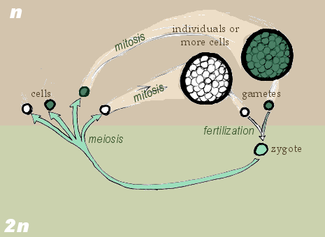
Cycling meiosis and fertilization events produces a series of transitions back and forth between alternating haploid and diploid states. The organism phase of the life cycle can occur either during the diploid state (gametic or diploid life cycle), during the haploid state (zygotic or haploid life cycle), or both (sporic or haplodiploid life cycle, in which there are two distinct organism phases, one during the haploid state and the other during the diploid state). In this sense there are three types of life cycles that utilize sexual reproduction, differentiated by the location of the organisms phase(s).[citation needed]
In the gametic life cycle, of which humans are a part, the species is diploid, grown from a diploid cell called the zygote. The organism's diploid germ-line stem cells undergo meiosis to create haploid gametes (the spermatozoa for males and ova for females), which fertilize to form the zygote. The diploid zygote undergoes repeated cellular division by mitosis to grow into the organism. Mitosis is a related process to meiosis that creates two cells that are genetically identical to the parent cell. The general principle is that mitosis creates somatic cells and meiosis creates germ cells.[citation needed]
In the zygotic life cycle the species is haploid instead, spawned by the proliferation and differentiation of a single haploid cell called the gamete. Two organisms of opposing gender contribute their haploid germ cells to form a diploid zygote. The zygote undergoes meiosis immediately, creating four haploid cells. These cells undergo mitosis to create the organism. Many fungi and many protozoa are members of the zygotic life cycle.[citation needed]
Finally, in the sporic life cycle, the living organism alternates between haploid and diploid states. Consequently, this cycle is also known as the alternation of generations. The diploid organism's germ-line cells undergo meiosis to produce spores. The spores proliferate by mitosis, growing into a haploid organism. The haploid organism's germ cells then combine with another haploid organism's cells, creating the zygote. The zygote undergoes repeated mitosis and differentiation to become the diploid organism again. The sporic life cycle can be considered a fusion of the gametic and zygotic life cycles.[citation needed]
Process
Because meiosis is a "one-way" process, it cannot be said to engage in a cell cycle as mitosis does. However, the preparatory steps that lead up to meiosis are identical in pattern and name to the interphase of the mitotic cell cycle.[citation needed]
Interphase is divided into three phases[citation needed]:
- Growth 1 (G1) phase: This is a very active period, where the cell synthesizes its vast array of proteins, including the enzymes and structural proteins it will need for growth. In G1 stage each of the chromosomes consists of a single (very long) molecule of DNA. In humans, at this point cells are 46 chromosomes, 2N, identical to somatic cells.[citation needed]
- Synthesis (S) phase: The genetic material is replicated: each of its chromosomes duplicates, so that each of the 46 chromosomes becomes a complex of two identical sister chromatids. The cell is still considered diploid because it still contains the same number of centromeres. The identical sister chromatids have not yet condensed into the densely packaged chromosomes visible with the light microscope. This will take place during prophase I in meiosis.[citation needed]
- Growth 2 (G2) phase: G2 phase as seen before mitosis is not present in Meiosis. Actually, the first four stages of prophase I in many respects correspond to the G2 phase of mitotic cell cycle.
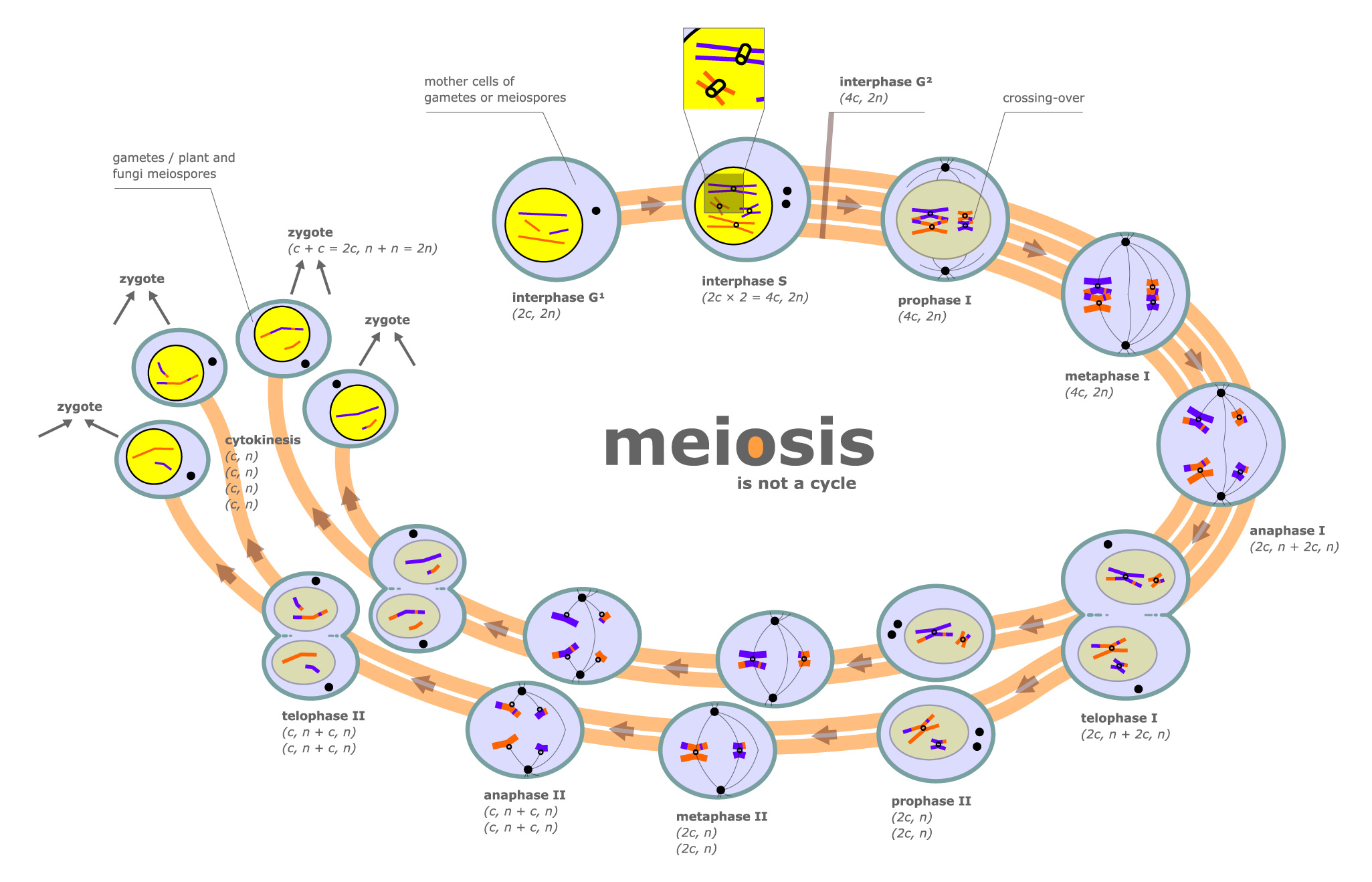
Interphase is followed by meiosis I and then meiosis II. Meiosis I consists of separating the pairs of homologous chromosome, each made up of two sister chromatids, into two cells. One entire haploid content of chromosomes is contained in each of the resulting daughter cells; the first meiotic division therefore reduces the ploidy of the original cell by a factor of 2.[citation needed]
Meiosis II consists of decoupling each chromosome's sister strands (chromatids), and segregating the individual chromatids into haploid daughter cells. The two cells resulting from meiosis I divide during meiosis II, creating 4 haploid daughter cells. Meiosis I and II are each divided into prophase, metaphase, anaphase, and telophase stages, similar in purpose to their analogous subphases in the mitotic cell cycle. Therefore, meiosis includes the stages of meiosis I (prophase I, metaphase I, anaphase I, telophase I), and meiosis II (prophase II, metaphase II, anaphase II, telophase II).[citation needed]
Meiosis generates genetic diversity in two ways: (1) independent alignment and subsequent separation of homologous chromosome pairs during the first meiotic division allows a random and independent selection of each chromosome segregates into each gamete; and (2) physical exchange of homologous chromosomal regions by homologous recombination during prophase I results in new combinations of DNA within chromosomes.[citation needed]
 A diagram of the meiotic phases
Phases
Meiosis is divided into meiosis I and meiosis II which are further divided into Karyokinesis I and Cytokinesis I & Karyokinesis II and Cytokinesis II respectively.
Meiosis I
Meiosis I separates homologous chromosomes, producing two haploid cells (N chromosomes, 23 in humans), so meiosis I is referred to as a reductional division. A regular diploid human cell contains 46 chromosomes and is considered 2N because it contains 23 pairs of homologous chromosomes. However, after meiosis I, although the cell contains 46 chromatids, it is only considered as being N, with 23 chromosomes. This is because later, in Anaphase I, the sister chromatids will remain together as the spindle fibres pull the pair toward the pole of the new cell. In meiosis II, an equational division similar to mitosis will occur whereby the sister chromatids are finally split, creating a total of 4 haploid cells (23 chromosomes, N) - two from each daughter cell from the first division.[citation needed]
Prophase I
During prophase I, DNA is exchanged between homologous chromosomes in a process called homologous recombination. This often results in chromosomal crossover. The new combinations of DNA created during crossover are a significant source of genetic variation, and may result in beneficial new combinations of alleles. The paired and replicated chromosomes are called bivalents or tetrads, which have two chromosomes and four chromatids, with one chromosome coming from each parent. At this stage, non-sister chromatids may cross-over at points called chiasmata (plural; singular chiasma).[citation needed]
Leptotene
The first stage of prophase I is the leptotene stage, also known as leptonema, from Greek words meaning "thin threads".[1]:27In this stage of prophase I, individual chromosomes—each consisting of two sister chromatids—change from the diffuse state they exist in during the cell's period of growth and gene expression, and condense into visible strands within the nucleus.[1]:27[2]:353 However the two sister chromatids are still so tightly bound that they are indistinguishable from one another. During leptotene, lateral elements of the synaptonemal complex assemble. Leptotene is of very short duration and progressive condensation and coiling of chromosome fibers takes place. Chromosome assume a long thread like shape, they contract and become thick. At the beginning chromosomes are present in diploid number as in mitotic prophase. Each chromosome is made up of only one chromatid and half of the total chromosome are paternal and half maternal. For every paternal chromosome there is a corresponding maternal chromosome similar in size, shape and nature of inherited characters and are called homologous chromosome.[citation needed]In animal cells the chromosomes touch the undersurface of nuclear envelope by their telomeres pointing towards the centrioles forming loops. C. D. Darlington called it "bouquet stage".
Zygotene
The zygotene stage, also known as zygonema, from Greek words meaning "paired threads",[1]:27 occurs as the chromosomes approximately line up with each other into homologous chromosome pairs. This is called the bouquet stage because of the way the telomeres cluster at one end of the nucleus. At this stage, the synapsis (pairing/coming together) of homologous chromosomes takes place, facilitated by assembly of central element of the synaptonemal complex. Pairing is brought about by a zipper like fashion and may start at the centromere(procentric), at the chromosome ends(proterminal),or at any other portion(intermediate). Individuals of a pair are equal in length and in position of centromere. Thus pairing is highly specific and exact. The paired chromosomes are called Bivalent or tetrad chromosome.[citation needed]
Pachytene
The pachytene stage, also known as pachynema, from Greek words meaning "thick threads",[1]:27 is the stage when chromosomal crossover (crossing over) occurs. Nonsister chromatids of homologous chromosomes randomly exchange segments over regions of homology. Sex chromosomes, however, are not wholly identical, and only exchange information over a small region of homology. At the sites where exchange happens, chiasmata form. The exchange of information between the non-sister chromatids results in a recombination of information; each chromosome has the complete set of information it had before, and there are no gaps formed as a result of the process. Because the chromosomes cannot be distinguished in the synaptonemal complex, the actual act of crossing over is not perceivable through the microscope, and chiasmata are not visible until the next stage.[citation needed]
Diplotene
During the diplotene stage, also known as diplonema, from Greek words meaning "two threads",[1]:30 the synaptonemal complex degrades and homologous chromosomes separate from one another a little. The chromosomes themselves uncoil a bit, allowing some transcription of DNA. However, the homologous chromosomes of each bivalent remain tightly bound at chiasmata, the regions where crossing-over occurred. The chiasmata remain on the chromosomes until they are severed in anaphase I.[citation needed]
In human fetal oogenesis all developing oocytes develop to this stage and stop before birth. This suspended state is referred to as the dictyotene stage and remains so until puberty.
Diakinesis
Chromosomes condense further during the diakinesis stage, from Greek words meaning "moving through".[1]:30 This is the first point in meiosis where the four parts of the tetrads are actually visible. Sites of crossing over entangle together, effectively overlapping, making chiasmata clearly visible. Other than this observation, the rest of the stage closely resembles prometaphase of mitosis; the nucleoli disappear, the nuclear membrane disintegrates into vesicles, and the meiotic spindle begins to form.[citation needed]
Synchronous processes
During these stages, two centrosomes, containing a pair of centrioles in animal cells, migrate to the two poles of the cell. These centrosomes, which were duplicated during S-phase, function as microtubule organizing centers nucleating microtubules, which are essentially cellular ropes and poles. The microtubules invade the nuclear region after the nuclear envelope disintegrates, attaching to the chromosomes at the kinetochore. The kinetochore functions as a motor, pulling the chromosome along the attached microtubule toward the originating centriole, like a train on a track. There are four kinetochores on each tetrad, but the pair of kinetochores on each sister chromatid fuses and functions as a unit during meiosis I.[3][4]
Microtubules that attach to the kinetochores are known as kinetochore microtubules. Other microtubules will interact with microtubules from the opposite centriole: these are called nonkinetochore microtubules or polar microtubules. A third type of microtubules, the aster microtubules, radiates from the centrosome into the cytoplasm or contacts components of the membrane skeleton.[citation needed]
Metaphase I
Homologous pairs move together along the metaphase plate: As kinetochore microtubules from both centrioles attach to their respective kinetochores, the homologous chromosomes align along an equatorial plane that bisects the spindle, due to continuous counterbalancing forces exerted on the bivalents by the microtubules emanating from the two kinetochores of homologous chromosomes. The physical basis of the independent assortment of chromosomes is the random orientation of each bivalent along the metaphase plate, with respect to the orientation of the other bivalents along the same equatorial line.[citation needed]
Anaphase I
Kinetochore (bipolar spindles) microtubules shorten, severing the recombination nodules and pulling homologous chromosomes apart. Since each chromosome has only one functional unit of a pair of kinetochores,[4] whole chromosomes are pulled toward opposing poles, forming two haploid sets. Each chromosome still contains a pair of sister chromatids. Nonkinetochore microtubules lengthen, pushing the centrioles farther apart. The cell elongates in preparation for division down the center.[citation needed]
Telophase I
The last meiotic division effectively ends when the chromosomes arrive at the poles. Each daughter cell now has half the number of chromosomes but each chromosome consists of a pair of chromatids. The microtubules that make up the spindle network disappear, and a new nuclear membrane surrounds each haploid set. The chromosomes uncoil back into chromatin. Cytokinesis, the pinching of the cell membrane in animal cells or the formation of the cell wall in plant cells, occurs, completing the creation of two daughter cells. Sister chromatids remain attached during telophase I.[citation needed]
Cells may enter a period of rest known as interkinesis or interphase II. No DNA replication occurs during this stage.[citation needed]
Meiosis II
Meiosis II is the second part of the meiotic process. Mechanically, the process is similar to mitosis, though its genetic results are fundamentally different. The end result is production of four haploid cells (23 chromosomes, N in humans) from the two haploid cells (23 chromosomes, N * each of the chromosomes consisting of two sister chromatids) produced in meiosis I. The four main steps of Meiosis II are: Prophase II, Metaphase II, Anaphase II, and Telophase II.[citation needed]
In prophase II we see the disappearance of the nucleoli and the nuclear envelope again as well as the shortening and thickening of the chromatids. Centrioles move to the polar regions and arrange spindle fibers for the second meiotic division.[citation needed]
In metaphase II, the centromeres contain two kinetochores that attach to spindle fibers from the centrosomes (centrioles) at each pole. The new equatorial metaphase plate is rotated by 90 degrees when compared to meiosis I, perpendicular to the previous plate[citation needed].
This is followed by anaphase II, where the centromeres are cleaved, allowing microtubules attached to the kinetochores to pull the sister chromatids apart. The sister chromatids by convention are now called sister chromosomes as they move toward opposing poles.[citation needed]
The process ends with telophase II, which is similar to telophase I, and is marked by uncoiling and lengthening of the chromosomes and the disappearance of the spindle. Nuclear envelopes reform and cleavage or cell wall formation eventually produces a total of four daughter cells, each with a haploid set of chromosomes. Meiosis is now complete and ends up with four new daughter cells.
Significance
Meiosis facilitates stable sexual reproduction. Without the halving of ploidy, or chromosome count, fertilization would result in zygotes that have twice the number of chromosomes as the zygotes from the previous generation. Successive generations would have an exponential increase in chromosome count. In organisms that are normally diploid, polyploidy, the state of having three or more sets of chromosomes, results in extreme developmental abnormalities or lethality.[5] Polyploidy is poorly tolerated in most animal species. Plants, however, regularly produce fertile, viable polyploids. Polyploidy has been implicated as an important mechanism in plant speciation.
Most importantly, recombination and independent assortment of homologous chromosomes allow for a greater diversity of genotypes in the offspring. This produces genetic variation in gametes that promote genetic and phenotypic variation in a population of offspring. Therefore a gene for meiosis will be favoured by natural selection over an allele for mitotic reproduction, because any selection pressure which acts against any clone will act against all clones, whilst inevitably favoring some offspring which are the result of sexual reproduction.
Nondisjunction
Main article: NondisjunctionThe normal separation of chromosomes in meiosis I or sister chromatids in meiosis II is termed disjunction. When the separation is not normal, it is called nondisjunction. This results in the production of gametes which have either too many or too few of a particular chromosome, and is a common mechanism for trisomy or monosomy. Nondisjunction can occur in the meiosis I or meiosis II, phases of cellular reproduction, or during mitosis.
This is a cause of several medical conditions in humans, including but not limited to:
- Down Syndrome - trisomy of chromosome 21
- Patau Syndrome - trisomy of chromosome 13
- Edward Syndrome - trisomy of chromosome 18
- Klinefelter Syndrome - extra X chromosomes in males - i.e. XXY, XXXY, XXXXY, etc.
- Turner Syndrome - lacking of one X chromosome in females - i.e. X0
- Triple X syndrome - an extra X chromosome in females
- XYY Syndrome - an extra Y chromosome in males
Meiosis in mammals
In females, meiosis occurs in cells known as oogonia (singular: oogonium). Each oogonium that initiates meiosis will divide twice to form a single oocyte and three polar bodies.[6] However, before these divisions occur, these cells stop at the diplotene stage of meiosis I and lie dormant within a protective shell of somatic cells called the follicle. Follicles begin growth at a steady pace in a process known as folliculogenesis, and a small number enter the menstrual cycle. Menstruated oocytes continue meiosis I and arrest at meiosis II until fertilization. The process of meiosis in females occurs during oogenesis, and differs from the typical meiosis in that it features a long period of meiotic arrest known as the Dictyate stage and lacks the assistance of centrosomes.
In males, meiosis occurs during spermatogenesis in the seminiferous tubules of the testicles. Meiosis during spermatogenesis is specific to a type of cell called spermatocytes that will later mature to become spermatozoa.
In female mammals, meiosis begins immediately after primordial germ cells migrate to the ovary in the embryo, but in the males, meiosis begins years later at the time of puberty. It is retinoic acid, derived from the primitive kidney (mesonephros) that stimulates meiosis in ovarian oogonia. Tissues of the male testis suppress meiosis by degrading retinoic acid, a stimulator of meiosis. This is overcome at puberty when cells within seminiferous tubules called Sertoli cells start making their own retinoic acid. Sensitivity to retinoic acid is also adjusted by proteins called nanos and DAZL.[7][8]
See also
- Coefficient of coincidence
- Multigene family
- Synizesis (biology)
References
- ^ a b c d e f Snustad, DP; Simmons, MJ (December 2008). Principles of Genetics (5th ed.). Wiley. ISBN 9780470388259.
- ^ Krebs, JE; Goldstein, ES; Kilpatrick, ST (November 2009). Lewin's Genes X (10th ed.). Jones & Barlett Learning. ISBN 9780763766320.
- ^ Raven, Peter H.; Johnson, George B.; Mason, Kenneth A.; Losos, Jonathan & Singer, Susan. Biology, 8th ed. McGraw-Hill 2007.
- ^ a b Petronczki M, Siomos MF, Nasmyth K (February 2003). "Un ménage à quatre: the molecular biology of chromosome segregation in meiosis". Cell 112 (4): 423–40. doi:10.1016/S0092-8674(03)00083-7. PMID 12600308.
- ^ BIL 104 - Lecture 15
- ^ Rosenbusch B (November 2006). "The contradictory information on the distribution of non-disjunction and pre-division in female gametes". Hum. Reprod. 21 (11): 2739–42. doi:10.1093/humrep/del122. PMID 16982661.
- ^ Lin Y, Gill ME, Koubova J, Page DC (December 2008). "Germ cell-intrinsic and -extrinsic factors govern meiotic initiation in mouse embryos". Science 322 (5908): 1685–7. doi:10.1126/science.1166340. PMID 19074348.
- ^ Suzuki A, Saga Y (February 2008). "Nanos2 suppresses meiosis and promotes male germ cell differentiation". Genes Dev. 22 (4): 430–5. doi:10.1101/gad.1612708. PMC 2238665. PMID 18281459. http://www.pubmedcentral.nih.gov/articlerender.fcgi?tool=pmcentrez&artid=2238665.
External links
- Meiosis Flash Animation
- Animations from the U. of Arizona Biology Dept.
- Meiosis at Kimball's Biology Pages
- Khan Academy, video lecture
- CCO The Cell-Cycle Ontology
Cell cycle proteins Cyclin CDK CDK inhibitor P53 p63 p73 family Phases and
checkpointsOther cellular phasesCategories:- Cellular processes
- Cell cycle
- Molecular genetics

Wikimedia Foundation. 2010.