- Gluteus maximus muscle
-
gluteus maximus 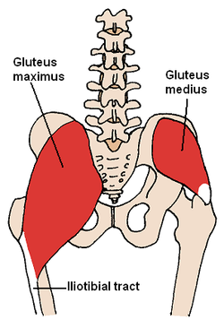
The gluteus medius and nearby muscles 
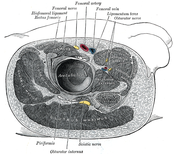
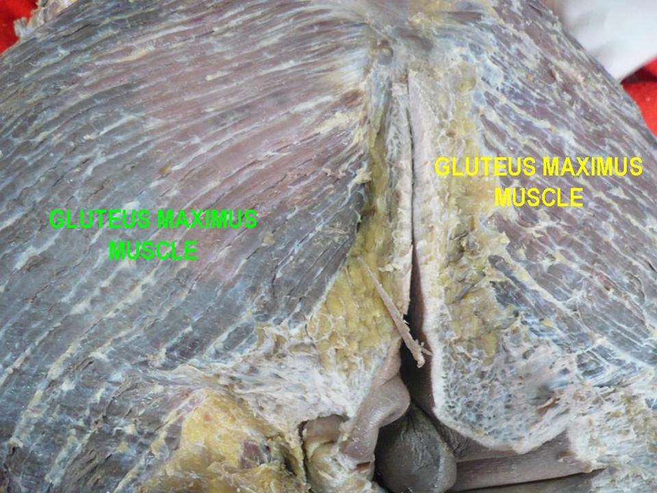
Structures surrounding right hip-joint. (Gluteus maximus visible at bottom.) Latin musculus gluteus maximus Gray's subject #128 474 Origin Gluteal surface of ilium, lumbar fascia, sacrum, sacrotuberous ligament Insertion Gluteal tuberosity of the femur, iliotibial tract Artery superior and inferior gluteal arteries Nerve inferior gluteal nerve (L5, S1, S2 nerve roots) Actions external rotation and extension of the hip joint, supports the extended knee through the iliotibial tract, chief antigravity muscle in sitting Antagonist Iliacus, Psoas major, Psoas minor The gluteus maximus (also known as glutæus maximus or, collectively with the gluteus medius and minimus, the glutes) is the largest and most superficial of the three gluteal muscles. It makes up a large portion of the shape and appearance of the buttocks.
It is a broad and thick fleshy mass of a quadrilateral shape, and forms the prominence of the nates.
Its large size is one of the most characteristic features of the muscular system in humans,[1] connected as it is with the power of maintaining the trunk in the erect posture. Primates have much flatter buttocks.
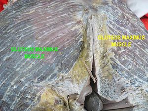
The muscle is remarkably coarse in structure, being made up of fasciculi lying parallel with one another and collected together into large bundles separated by fibrous septa.
Contents
Origin and insertion
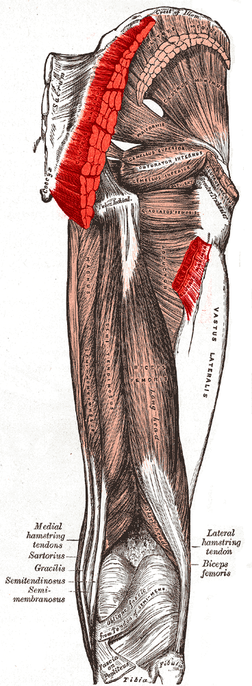
 Muscles of the gluteal and posterior femoral regions, showing origin and insertion of gluteus maximus muscle.
It arises from the posterior gluteal line of the inner upper ilium, and the rough portion of bone including the crest, immediately above and behind it; from the posterior surface of the lower part of the sacrum and the side of the coccyx; from the aponeurosis of the erector spinae (lumbodorsal fascia), the sacrotuberous ligament, and the fascia covering the gluteus medius (gluteal aponeurosis).
The fibers are directed obliquely downward and lateralward;
- those forming the lower and larger portion of the muscle, together with the superficial fibers of the lower portion, end in a thick tendinous lamina, which passes across the greater trochanter, and is inserted into the iliotibial band of the fascia lata;
- the deeper fibers of the lower portion of the muscle are inserted into the gluteal tuberosity between the vastus lateralis and adductor magnus.
Bursae
Three bursae are usually found in relation with the deep surface of this muscle:
- One of these, of large size, and generally multilocular, separates it from the greater trochanter;
- a second, often wanting, is situated on the tuberosity of the ischium;
- a third is found between the tendon of the muscle and that of the vastus lateralis.
Actions
When the gluteus maximus takes its fixed point from the pelvis, it extends the femur and brings the bent thigh into a line with the body.
Taking its fixed point from below, it acts upon the pelvis, supporting it and the trunk upon the head of the femur; this is especially obvious in standing on one leg.
Its most powerful action is to cause the body to regain the erect position after stooping, by drawing the pelvis backward, being assisted in this action by the biceps femoris, semitendinosus, semimembranosus, and adductor magnus.
The gluteus maximus is a tensor of the fascia lata, and by its connection with the iliotibial band steadies the femur on the articular surfaces of the tibia during standing, when the extensor muscles are relaxed.
The lower part of the muscle also acts as an adductor and external rotator of the limb.
Training
- Lunge (exercise)
- Quadruped Hip Extensions
- Step-Ups
- Four-Way Hip Extensions
 Gluteus maximus muscle
See also
References
- ^ Norman Eizenberg et al., General Anatomy: Principles and Applications (2008), p 17.
External links
- -208011185 at GPnotebook
- LUC glmx
- SUNY Labs 13:st-0403
- Cross section at UV pelvis/pelvis-female-17
- Cross section at UV pelvis/pelvis-e12-15
- Cross section at UV pembody/body18b
- Muscles/GluteusMaximus at exrx.net
This article was originally based on an entry from a public domain edition of Gray's Anatomy. As such, some of the information contained within it may be outdated.
List of muscles of lower limbs (TA A04.7, GA 4.465) ILIAC Region
/ ILIOPSOASBUTTOCKS THIGH /
compartmentsLEG/
Crus/
compartmentssuperficial · triceps surae (gastrocnemius, soleus, accessory soleus, Achilles tendon) · plantaris
deep · tarsal tunnel (flexor hallucis longus, flexor digitorum longus, tibialis posterior) · popliteusFOOT DorsalPlantar1st layer (abductor hallucis, flexor digitorum brevis, abductor digiti minimi) · 2nd layer (quadratus plantae, lumbrical muscle) · 3rd layer (flexor hallucis brevis, adductor hallucis, flexor digiti minimi brevis) · 4th layer (dorsal interossei, plantar interossei)Categories:- Hip adductors
- Hip extensors
- Hip lateral rotators
- Muscles of the gluteus

Wikimedia Foundation. 2010.