- Obturator internus muscle
-
Obturator internus muscle 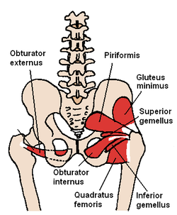
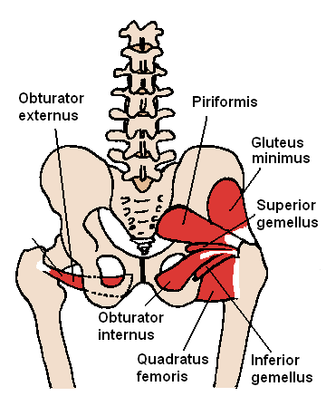
The obturator internus and nearby muscles 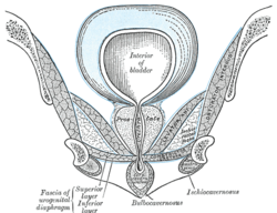
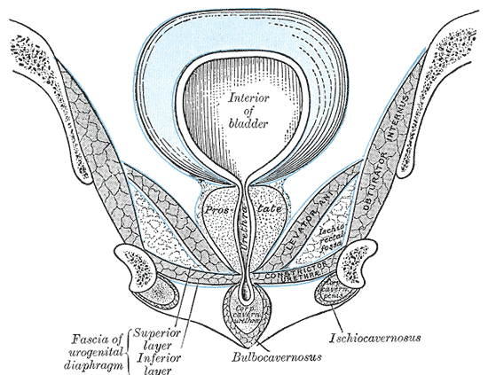
Coronal section of anterior part of pelvis, through the pubic arch. Seen from in front. (Obturator internus labeled at right.) Latin musculus obturatorius internus Gray's subject #128 477 Origin Ischiopubic ramus & obturator membrane Insertion medial aspect of the Greater trochanter Artery Superior gluteal artery Nerve Nerve to obturator internus (L5, S1) Actions Abducts & rotates laterally thigh, and stabiliser of the hip during walking The obturator internus muscle originates on the medial surface of the obturator membrane, the ischium near the membrane, and the rim of the pubis.
It exits the pelvic cavity through the lesser sciatic foramen.
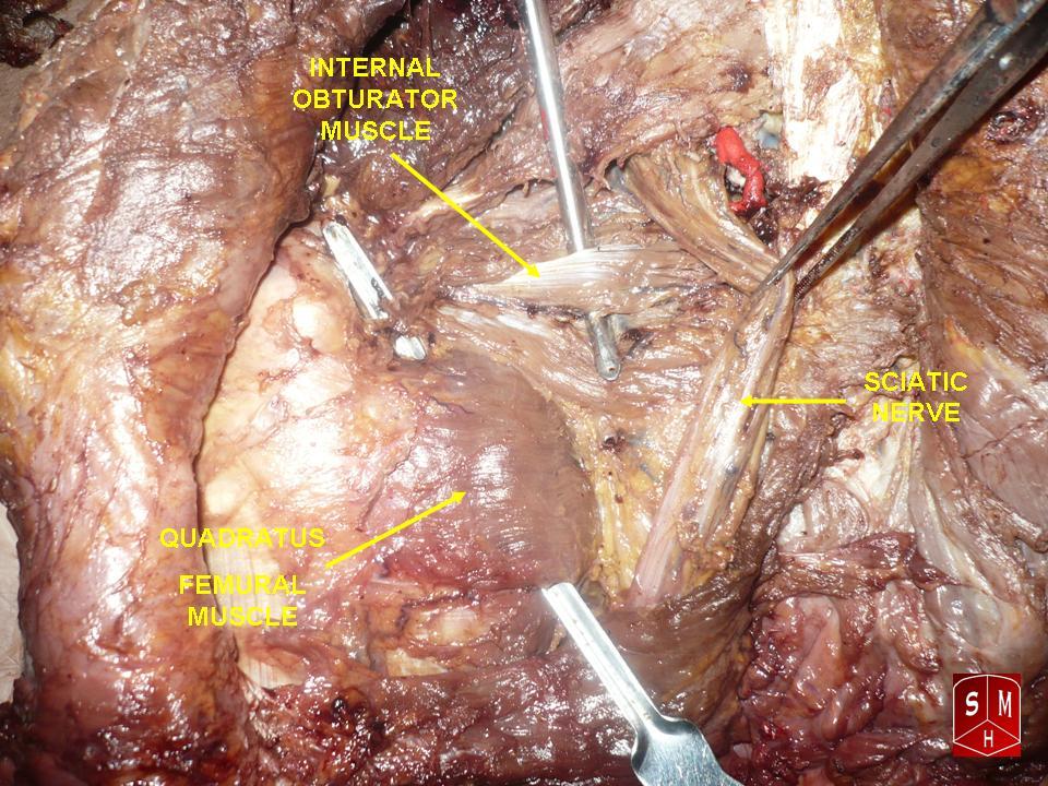
The obturator internus is situated partly within the lesser pelvis, and partly at the back of the hip-joint.
It functions to help laterally rotate extended thigh and abduct flexed thigh, as well as to steady the femoral head in the acetabulum.
Origin and insertion
It arises from the inner surface of the antero-lateral wall of the pelvis, where it surrounds the greater part of the obturator foramen, being attached to the inferior rami of the pubis and ischium, and at the side to the inner surface of the hip bone below and behind the pelvic brim, reaching from the upper part of the greater sciatic foramen above and behind to the obturator foramen below and in front.
It also arises from the pelvic surface of the obturator membrane except in the posterior part, from the tendinous arch which completes the canal for the passage of the obturator vessels and nerve, and to a slight extent from the obturator fascia, which covers the muscle.
The fibers converge rapidly toward the lesser sciatic foramen, and end in four or five tendinous bands, which are found on the deep surface of the muscle; these bands are reflected at a right angle over the grooved surface of the ischium between its spine and tuberosity.
The tendon inserts on the greater trochanter of the proximal femur.
Obturator Internis muscle is also innervated by the nerve to obturator internis (L5, S1).
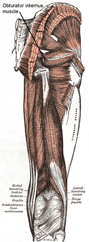
 Muscles of the gluteal and posterior femoral regions.
Bursa/bands
This bony surface is covered by smooth cartilage, which is separated from the tendon by a bursa, and presents one or more ridges corresponding with the furrows between the tendinous bands.
These bands leave the pelvis through the lesser sciatic foramen and unite into a single flattened tendon, which passes horizontally across the capsule of the hip-joint, and, after receiving the attachments of the superior and inferior gemellus muscles, is inserted into the forepart of the medial surface of the greater trochanter above the trochanteric fossa.
A bursa, narrow and elongated in form, is usually found between the tendon and the capsule of the hip-joint; it occasionally communicates with the bursa between the tendon and the ischium.
 Obturator internus muscle
External links
- -1348861872 at GPnotebook
- LUC obi
- SUNY Labs 13:st-0407 - "Gluteal Region: Muscles"
- SUNY Labs 43:st-0603 - "The Female Pelvis: Muscles"
- Cross section at UV pelvis/pelvis-e12-15
- pelvis at The Anatomy Lesson by Wesley Norman (Georgetown University) (femalepelvicdiaphragm, malepelvicdiaphragm)
- perineum at The Anatomy Lesson by Wesley Norman (Georgetown University) (analtriangle3)
- Int. J. Morphol., 25(1):95-98, 2007
This article was originally based on an entry from a public domain edition of Gray's Anatomy. As such, some of the information contained within it may be outdated.
List of muscles of lower limbs (TA A04.7, GA 4.465) ILIAC Region
/ ILIOPSOASBUTTOCKS gluteals: (maximus, medius, minimus) · tensor fasciae latae
lateral rotator group: quadratus femoris · inferior gemellus · obturator internus · superior gemellus · piriformisTHIGH /
compartmentsLEG/
Crus/
compartmentssuperficial · triceps surae (gastrocnemius, soleus, accessory soleus, Achilles tendon) · plantaris
deep · tarsal tunnel (flexor hallucis longus, flexor digitorum longus, tibialis posterior) · popliteusfibularis muscles (longus, brevis)FOOT DorsalPlantar1st layer (abductor hallucis, flexor digitorum brevis, abductor digiti minimi) · 2nd layer (quadratus plantae, lumbrical muscle) · 3rd layer (flexor hallucis brevis, adductor hallucis, flexor digiti minimi brevis) · 4th layer (dorsal interossei, plantar interossei)Categories:- Muscle stubs
- Hip muscles
Wikimedia Foundation. 2010.