- Tensor fasciae latae muscle
-
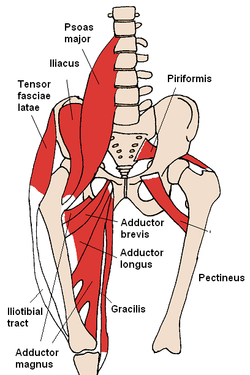
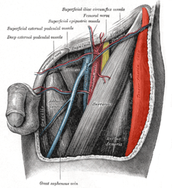
Tensor fasciae latae The tensor fasciae latae and nearby muscles The left femoral triangle. Latin musculus tensor fasciae latae Gray's subject #128 476 Origin iliac crest Insertion iliotibial tract Artery primarily lateral circumflex femoral artery, Superior gluteal artery Nerve Superior gluteal nerve (L4-L5) Actions Thigh - flexion, medial rotation, abduction. Trunk stabilization. The tensor fasciae latae or tensor fasciæ latæ is a muscle of the thigh. The English name for this muscle is the muscle that stretches the band on the side. The word tensor comes from the Latin verb tendere meaning “to stretch." Fascia is the Latin term for “band.” The word latae is the genitive form of the Latin word lata meaning “side.” [1]
Contents
Origin and insertion
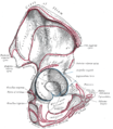
It arises from the posterior part of the outer lip of the iliac crest; from the outer surface of the anterior superior iliac spine, and part of the outer border of the notch below it, between the gluteus medius and sartorius; and from the deep surface of the fascia lata.
It is inserted between the two layers of the iliotibial band of the fascia lata about the junction of the middle and upper thirds of the thigh.
Function
The tensor fasciæ latæ is a tensor of the fascia lata; continuing its action, the oblique direction of its fibers enables it to stabilizes the knee in extension ( Assist Gluteus Maximus in knee extension).
In the erect posture, acting from below, it will serve to steady the pelvis upon the head of the femur; and by means of the iliotibial band it steadies the condyles of the femur on the articular surfaces of the tibia, and assists the Glutæus maximus in supporting the knee in a position of extension.
The basic functional movement of tensor fascia latae is walking. The tensor fascia lata is heavily utilized in horse riding, hurdling and water skiing. Some problems that arise when this muscle is tight or shortened are pelvic imbalances that lead to pain in hips, as well as pain in the lower back and lateral area of knees.[2]
The TFL is a hip abductor muscle. To stretch the tensor fascia latae, the knee may be brought medially across your body (adducted). If one leans a wall with crossed legs (externally/laterally rotated hips) and pushes the pelvis away from the wall (leaning the upper body towards it) sidebending the lumbar spine (ie: curving the spine to the side) should be avoided as it stretches the lumbar region rather than the tensor fascia latae and other muscles which cross the hip rather than the spine.
Nerve
Tensor fascia latae is innervated by the superior gluteal nerve, L4, L5 and S1.[3]
Additional images
The TFL is often involved in lateral meniscus and or knee pain/ problems. Evaluating for strain-sprain and trigger point in the TFl and then correcting with Jones and or Travell technique will significantly aid recovery and reduce pain.
References
- ^ http://www.anatomyexpert.com/structure_detail/5716/1733/
- ^ Jarmey, Chris. The Concise Book of Muscles. North Atlantic Books: Berkeley, CA, 2003. 2nd Ed.
- ^ Saladin. Anatomy and Physiology. 5th ed. Mc-Graw Hill. 2010.
External links
- Template:Http://www.anatomy.tv/interactivehip/release/default.aspx?app=legacyhip flash
- LUC tfl
- 705036366 at GPnotebook
- Cross section at UV pelvis/pelvis-e12-15
- tensor+fasciae+latae+muscle at eMedicine Dictionary
- Muscles/TensorFasciaeLatae at exrx.net
- Coachr
This article was originally based on an entry from a public domain edition of Gray's Anatomy. As such, some of the information contained within it may be outdated.
List of muscles of lower limbs (TA A04.7, GA 4.465) ILIAC Region
/ ILIOPSOASBUTTOCKS gluteals: (maximus, medius, minimus) · tensor fasciae latae
lateral rotator group: quadratus femoris · inferior gemellus · obturator internus · superior gemellus · piriformisTHIGH /
compartmentsLEG/
Crus/
compartmentssuperficial · triceps surae (gastrocnemius, soleus, accessory soleus, Achilles tendon) · plantaris
deep · tarsal tunnel (flexor hallucis longus, flexor digitorum longus, tibialis posterior) · popliteusfibularis muscles (longus, brevis)FOOT DorsalPlantar1st layer (abductor hallucis, flexor digitorum brevis, abductor digiti minimi) · 2nd layer (quadratus plantae, lumbrical muscle) · 3rd layer (flexor hallucis brevis, adductor hallucis, flexor digiti minimi brevis) · 4th layer (dorsal interossei, plantar interossei)Categories:- Hip abductors
- Hip flexors
- Thigh muscles
Wikimedia Foundation. 2010.

