- Fibularis longus
-
Peroneus longus 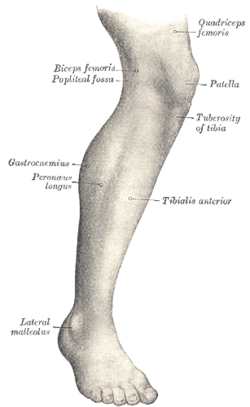
Lateral aspect of right leg. 
Peroneus longus labeled very right Latin musculus peroneus longus Gray's subject #129 486 Origin fibula Insertion first metatarsal, medial cuneiform Artery fibular (peroneal) artery Nerve Superficial fibular (peroneal) nerve Actions plantarflexion, eversion Antagonist Tibialis anterior muscle In human anatomy, the peroneus longus (also known as fibularis longus) is a superficial muscle in the lateral compartment of the leg, and acts to evert and plantar flex the ankle.
It is situated at the upper part of the lateral side of the leg, and is the most superficial of the three peroneus muscles.
It is innervated by the superficial fibular nerve (superficial peroneal nerve).
Blood is supplied by branches of the anterior tibial and peroneal arteries.
Contents
Etymology
The terms Peroneus (i.e., Longus and Brevis) and Peroneal (i.e., Artery, Retinaculum) are derived from the Greek word Perone (pronounced Pair-uh-knee) meaning pin of a brooch or a buckle. In medical terminology, both terms refer to being of or relating to the fibula or to the outer portion of the leg.
Origin and insertion
It is attached proximally to the head of the fibula and its 'belly' runs down most of this bone. It becomes a tendon that goes posteriorly around the lateral malleolus of the ankle, then continues under the foot to attach to the medial cuneiform and first metatarsal.
It arises from the head and upper two-thirds of the lateral surface of the body of the fibula, from the deep surface of the fascia, and from the intermuscular septa between it and the muscles on the front and back of the leg; occasionally also by a few fibers from the lateral condyle of the tibia. Between its attachments to the head and to the body of the fibula there is a gap through which the common peroneal nerve passes to the front of the leg.
It ends in a long tendon, which runs behind the lateral malleolus, in a groove common to it and the tendon of the peroneus brevis; the groove is converted into a canal by the superior peroneal retinaculum, and the tendons in it are contained in a common mucous sheath.
The tendon then extends obliquely forward across the lateral side of the calcaneus, below the trochlear process, and the tendon of the peroneus brevis, and under cover of the inferior peroneal retinaculum.
It crosses the lateral side of the cuboid, and then runs on the under surface of that bone in a groove which is converted into a canal by the long plantar ligament; the tendon then crosses the sole of the foot obliquely, and is inserted into the lateral side of the base of the first metatarsal bone and the lateral side of the medial cuneiform.
Occasionally it sends a slip to the base of the second metatarsal bone.
The tendon changes its direction at two points: first, behind the lateral malleolus; secondly, on the cuboid bone; in both of these situations the tendon is thickened, and, in the latter, a sesamoid fibrocartilage (sometimes a bone), is usually developed in its substance.
Actions
The Peronæi longus and brevis plantar-flex the ankle, in conjunction with the Tibialis posterior, antagonizing the Tibialis anterior and Peronæus tertius, which are dorsi-flexors of the foot.
The Peronæus longus also everts the sole of the foot, and from the oblique direction of the tendon across the sole of the foot is an important agent in the maintenance of the transverse arch.
Taking their fixed points below, the Peronæi serve to steady the leg upon the foot.
This is especially the case in standing upon one leg, when the tendency of the superincumbent weight is to throw the leg medialward; the Peronæus longus overcomes this tendency by drawing on the lateral side of the leg.
Additional images
-
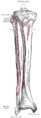
Bones of the right leg. Anterior surface.
-
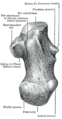
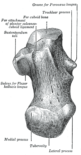
Left calcaneus, inferior surface.
-
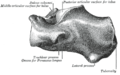
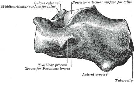
Left calcaneus, lateral surface.
-
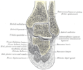
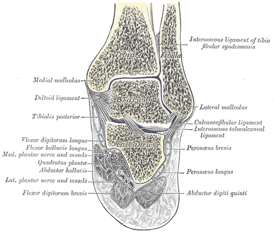
Coronal section through right talocrural and talocalcaneal joints.
-
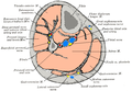
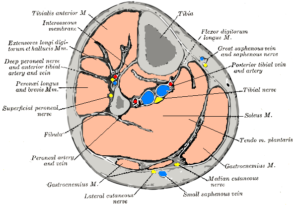
Cross-section through middle of leg.
-
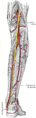
The popliteal, posterior tibial, and peroneal arteries.
-
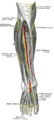
Deep nerves of the front of the leg.
-
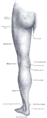
Back of left lower extremity.
See also
- Fibularis brevis
- Fibularis tertius
External links
- Fibularis+longus at eMedicine Dictionary
This article was originally based on an entry from a public domain edition of Gray's Anatomy. As such, some of the information contained within it may be outdated.
List of muscles of lower limbs (TA A04.7, GA 4.465) ILIAC Region
/ ILIOPSOASBUTTOCKS THIGH /
compartmentsLEG/
Crus/
compartmentssuperficial · triceps surae (gastrocnemius, soleus, accessory soleus, Achilles tendon) · plantaris
deep · tarsal tunnel (flexor hallucis longus, flexor digitorum longus, tibialis posterior) · popliteusfibularis muscles (longus, brevis)FOOT DorsalPlantar1st layer (abductor hallucis, flexor digitorum brevis, abductor digiti minimi) · 2nd layer (quadratus plantae, lumbrical muscle) · 3rd layer (flexor hallucis brevis, adductor hallucis, flexor digiti minimi brevis) · 4th layer (dorsal interossei, plantar interossei)Categories:- Muscles of the lower limb
-


Wikimedia Foundation. 2010.