- Piriformis muscle
-
Piriformis muscle 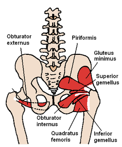
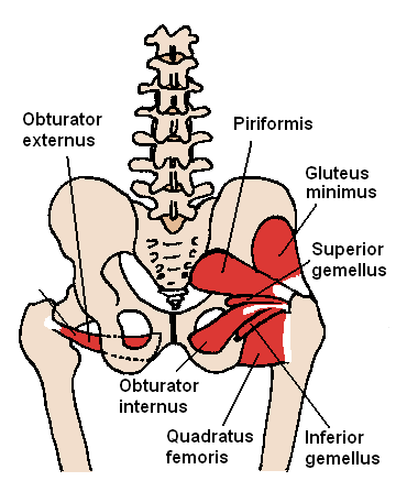
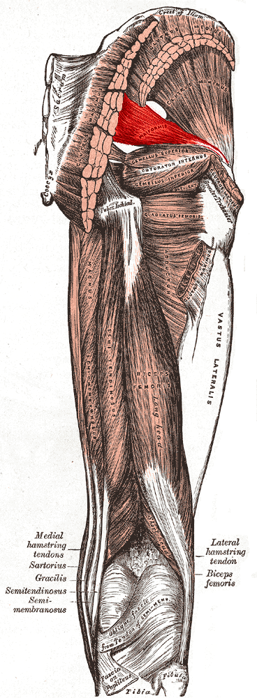
(Posterior view of pelvis) The piriformis and nearby muscles 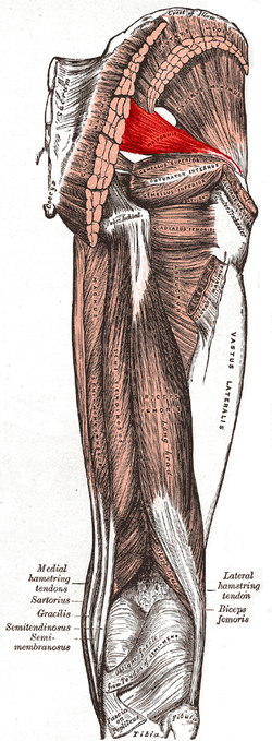
Muscles of the gluteal and posterior femoral regions, piriformis labeled Latin musculus piriformis Gray's subject #128 476 Origin sacrum Insertion greater trochanter Artery Inferior gluteal artery , Lateral sacral artery, Superior gluteal artery, Nerve nerve to the Piriformis (L5, S1, and S2 nerve roots) Actions rotate laterally (outward) the thigh The piriformis (from Latin piriformis = "pear shaped") is a muscle in the gluteal region of the lower limb. It was first named by Spigelius, a professor from the University of Padua in the 16th century.[1]
Contents
Origin and insertion
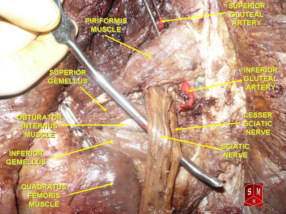
It originates from the anterior (front) part of the sacrum, the part of the spine in the gluteal region, and from the superior margin of the greater sciatic notch (as well as the sacroiliac joint capsule and the sacrotuberous ligament). It exits the pelvis through the greater sciatic foramen to insert on the greater trochanter of the femur. Its tendon often joins with the tendons of the superior gemellus, inferior gemellus, and obturator internus muscles prior to insertion.
Shape and location
The piriformis is a flat muscle, pyramidal in shape, lying almost parallel with the posterior margin of the gluteus medius.
It is situated partly within the pelvis against its posterior wall, and partly at the back of the hip-joint.
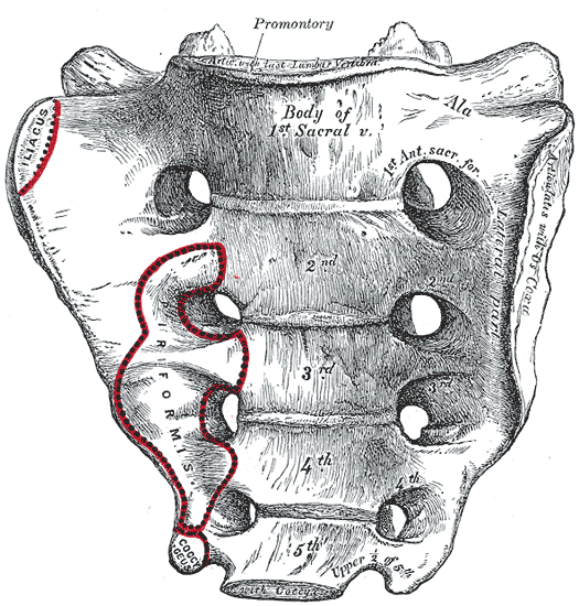
It arises from the front of the sacrum by three fleshy digitations, attached to the portions of bone between the first, second, third, and fourth anterior sacral foramina, and to the grooves leading from the foramina: a few fibers also arise from the margin of the greater sciatic foramen, and from the anterior surface of the sacrotuberous ligament.
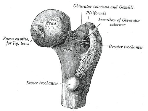
The muscle passes out of the pelvis through the greater sciatic foramen, the upper part of which it fills, and is inserted by a rounded tendon into the upper border of the greater trochanter behind, but often partly blended with, the common tendon of the obturator internus and superior and inferior gemellus muscles.
Action
The piriformis muscle is part of the lateral rotators of the hip, along with the quadratus femoris, gemellus inferior, gemellus superior, obturator externus, and obturator internus. The piriformis laterally rotates the extended thigh and abducts the flexed thigh. Abduction of the flexed thigh is important in the action of walking because it shifts the body weight to the opposite side of the foot being lifted, which keeps us from falling. The action of the lateral rotators can be understood by crossing your legs to rest an ankle on the knee of the other leg. This causes the femur to rotate and point the knee laterally. The lateral rotators also oppose medial rotation by the gluteus medius and gluteus minimus. 'At a range of more than 90 degrees the actions of the piriformis are reversed to adduct and internally rotate (medial).'
Variations
In 17% of limbs, the piriformis muscle is pierced by parts or all of the sciatic nerve. Several variations occur, but the most common type of anomaly (81% of anomalies) is the Beaton's type B which is when the common peroneal nerve pierces the piriformis muscle.[1]
It may be united with the gluteus medius, send fibers to the gluteus minimus, or receive fibers from the superior gemellus.
It may have one or two sacral attachments; or it may be inserted in to the capsule of the hip joint.
Clinicals
Main article: Piriformis syndromeThis syndrome occurs when the piriformis irritates the sciatic nerve, which comes into the gluteal region beneath the muscle, causing pain in the buttocks and referred pain along the sciatic nerve.[2] This referred pain is known as sciatica. Fifteen percent of the population has their sciatic nerve coursing through the piriformis muscle. This subgroup of the population is predisposed to developing sciatica. Sciatica can be described by pain, tingling, or numbness deep in the buttocks and along the sciatic nerve. Sitting down, stretching, climbing stairs, and performing squats usually increases pain. Diagnosing the syndrome is usually based on symptoms and on the physical exam. More testing, including MRIs, X-rays, and nerve conduction tests can be administered to exclude other possible diseases.[2] If diagnosed with piriformis syndrome, the first treatment involves progressive stretching exercises and physical treatment. Corticosteroids can be injected into the piriformis muscle if pain continues. A more invasive, but sometimes necessary treatment involves surgical exploration as a last resort.[2]
 Piriformis muscle
Additional images
-
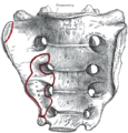
Sacrum, pelvic surface.
-

Upper extremity of right femur viewed from behind and above.
-

Right femur. Anterior surface.
-
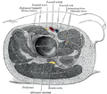
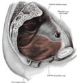
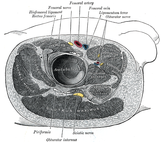
Structures surrounding right hip-joint.
-
Left Levator ani from within.
-
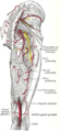
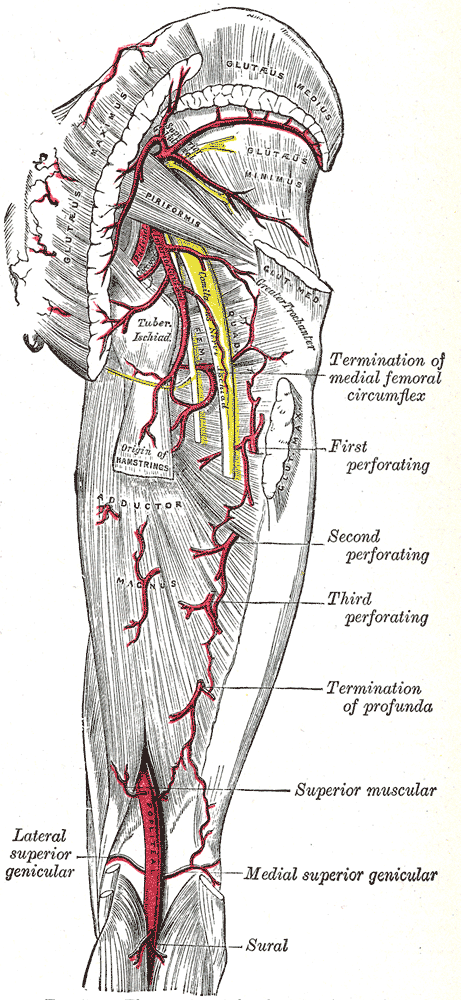
The arteries of the gluteal and posterior femoral regions.
-
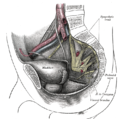
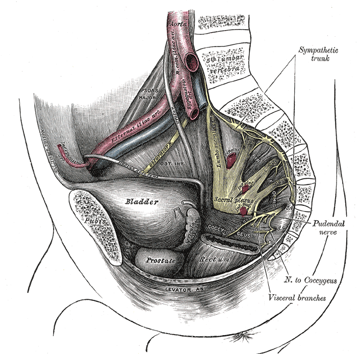
Dissection of side wall of pelvis showing sacral and pudendal plexuses.
-

Nerves of the right lower extremity Posterior view.
External links
- "Piriformis" University of Washington
- SUNY Labs 13:st-0408 - "Gluteal Region: Muscles"
- SUNY Labs 43:15-0101 - "The Female Pelvis: The Posterolateral Pelvic Wall"
- piriformis muscle at GPnotebook
- LUC pirf
- "Piriformis Muscle Stretch, Biomechanics & Piriformis-Sciatic Nerve Relationship Graphics
References
- ^ a b Smoll NR (2010). "Variations of the Piriformis and Sciatic Nerve With Clinical Consequence: A Review". Clinical Anatomy 23: 8–17.
- ^ a b c "The piriformis syndrome". http://www.rice.edu/~jenky/sports/piri.html. Retrieved 2007-11-16.
Saladin, Ken (2007). Anatomy & Physiology: THE UNITY OF FORM AND FUNCTION 4th Edition. The McGraw-Hill Companies, Inc.. ISBN 9780072875065.
This article was originally based on an entry from a public domain edition of Gray's Anatomy. As such, some of the information contained within it may be outdated.
List of muscles of lower limbs (TA A04.7, GA 4.465) ILIAC Region
/ ILIOPSOASBUTTOCKS gluteals: (maximus, medius, minimus) · tensor fasciae latae
lateral rotator group: quadratus femoris · inferior gemellus · obturator internus · superior gemellus · piriformisTHIGH /
compartmentsLEG/
Crus/
compartmentssuperficial · triceps surae (gastrocnemius, soleus, accessory soleus, Achilles tendon) · plantaris
deep · tarsal tunnel (flexor hallucis longus, flexor digitorum longus, tibialis posterior) · popliteusfibularis muscles (longus, brevis)FOOT DorsalPlantar1st layer (abductor hallucis, flexor digitorum brevis, abductor digiti minimi) · 2nd layer (quadratus plantae, lumbrical muscle) · 3rd layer (flexor hallucis brevis, adductor hallucis, flexor digiti minimi brevis) · 4th layer (dorsal interossei, plantar interossei)Categories:- Hip muscles
-

Wikimedia Foundation. 2010.