- Biceps femoris muscle
-
Biceps femoris 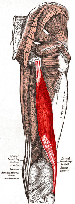
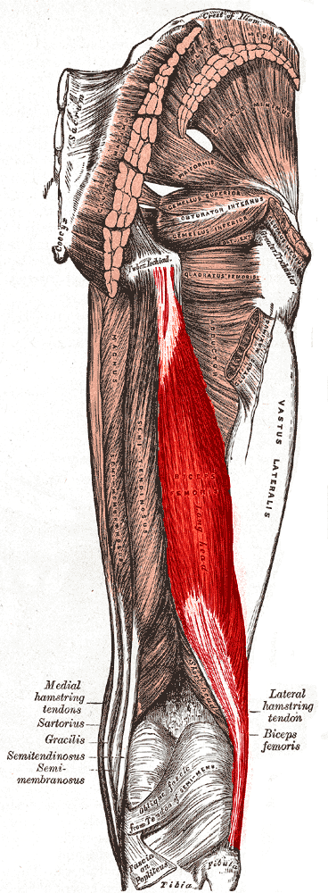
Lateral aspect of right leg. Biceps femoris muscle long head labeled 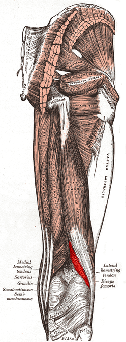
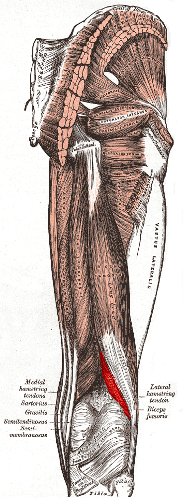
Same picture with the visible part of the short head beneath the long head labeled Latin musculus biceps femoris Gray's subject #128 478 Origin tuberosity of the ischium, linea aspera, femur Insertion the head of the fibula which articulates with the back of the lateral tibial condyle Artery inferior gluteal artery, perforating arteries, popliteal artery Nerve long head: tibial nerve
short head: common peroneal nerveActions flexes knee joint, laterally rotates knee joint (when knee is flexed), extends hip joint (long head only) Antagonist Quadriceps muscle The biceps femoris is a muscle of the posterior (the back) thigh. As its name implies, it has two parts, one of which (the long head) forms part of the hamstrings muscle group.
Contents
Anatomy
Origin and insertion
It has two heads of origin;
- one, the long head, arises from the lower and inner impression on the back part of the tuberosity of the ischium, by a tendon common to it and the semitendinosus, and from the lower part of the sacrotuberous ligament;[1]
- the other, the short head, arises from the lateral lip of the linea aspera, between the adductor magnus and vastus lateralis, extending up almost as high as the insertion of the gluteus maximus; from the lateral prolongation of the linea aspera to within 5 cm. of the lateral condyle; and from the lateral intermuscular septum.[1]
The fibers of the long head form a fusiform belly, which passes obliquely downward and lateralward across the sciatic nerve to end in an aponeurosis which covers the posterior surface of the muscle, and receives the fibers of the short head; this aponeurosis becomes gradually contracted into a tendon, which is inserted into the lateral side of the head of the fibula, and by a small slip into the lateral condyle of the tibia.[1]
At its insertion the tendon divides into two portions, which embrace the fibular collateral ligament of the knee-joint.[1]
From the posterior border of the tendon a thin expansion is given off to the fascia of the leg. The tendon of insertion of this muscle forms the lateral hamstring; the common fibular (peroneal) nerve descends along its medial border.[1]
Variations
The short head may be absent; additional heads may arise from the ischial tuberosity, the linea aspera, the medial supracondylar ridge of the femur, or from various other parts.[1]
A slip may pass to the gastrocnemius.[1]
Action
Both heads of the biceps femoris perform knee flexion.[2]
Since the long head originates in the pelvis it is also involved in hip extension.[2] The long head of the biceps femoris is a weaker knee flexor when the hip is extended (because of active insufficiency). For the same reason the long head is a weaker hip extender when the knee is flexed.
When the knee is semi-flexed, the biceps femoris in consequence of its oblique direction rotates the leg slightly outward.
Innervation
The short head of the biceps femoris develops in the flexor compartment of the thigh and is thus innervated by common peroneal branch of the sciatic nerve (L5, S1), while the long head is innervated by the tibial branch of the sciatic nerve (L5, S1).[3]
Blood supply
The muscle's vascular supply is derived from the anastomoses of a several arteries: the perforating branches of the profunda femoris artery, the inferior gluteal artery, and the popliteal artery.[3]
See also
Additional images
-
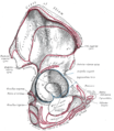
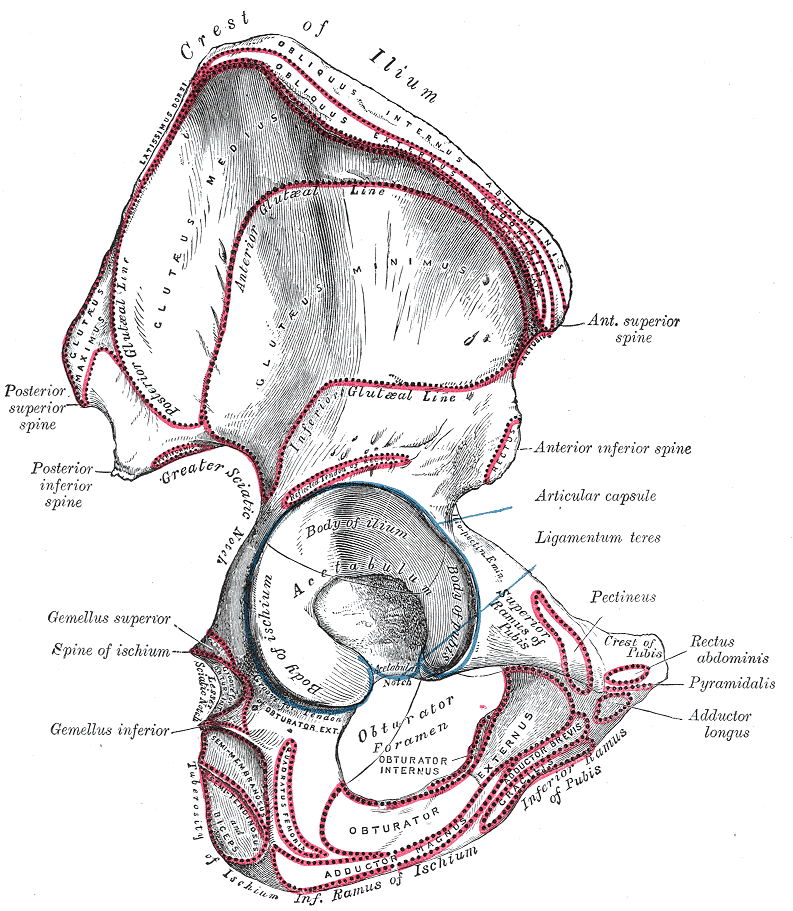
Right hip bone. External surface.
-
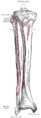
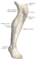
Bones of the right leg. Anterior surface.
-
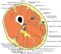
Cross-section through the middle of the thigh.
-
Muscles of the gluteal and posterior femoral regions.
-
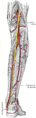
The popliteal, posterior tibial, and peroneal arteries.
-

Nerves of the right lower extremity Posterior view.
-
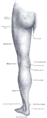
Back of left lower extremity.
-

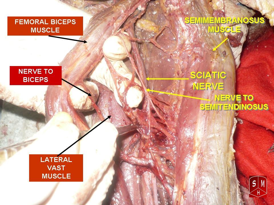
Femoral biceps muscle
References
- ^ a b c d e f g "Gray's Anatomy". 1918. http://education.yahoo.com/reference/gray/subjects/subject/128#p478.
- ^ a b LUC bfem
- ^ a b -865402803 at GPnotebook
This article was originally based on an entry from a public domain edition of Gray's Anatomy. As such, some of the information contained within it may be outdated.
Further reading
- Kumakura, Hiroo (July 1989). "Functional analysis of the biceps femoris muscle during locomotor behavior in some primates". American Journal of Physical Anthropology 79 (3): 379–391. doi:10.1002/ajpa.1330790314. PMID 2504047.
- Marshall, John L.; Girgis, Fakhry G.; Zelko, Russel R. (1972). "The Biceps Femoris Tendon and Its Functional Significance (PDF)". J Bone Joint Surg Am. 54 (54): 1444–1450. http://www.ejbjs.org/cgi/reprint/54/7/1444.
- Sneath, R. S. (1955 October). "The insertion of the biceps femoris". J Anat. 89 (89(Pt 4)): 550–553.. PMC 1244747. PMID 13278305. http://www.pubmedcentral.nih.gov/articlerender.fcgi?tool=pmcentrez&artid=1244747.
External links
- UWash - long head
- UWash - short head
- SUNY Labs 14:06-0100
- SUNY Labs 14:st-0402
List of muscles of lower limbs (TA A04.7, GA 4.465) ILIAC Region
/ ILIOPSOASBUTTOCKS THIGH /
compartmentsLEG/
Crus/
compartmentssuperficial · triceps surae (gastrocnemius, soleus, accessory soleus, Achilles tendon) · plantaris
deep · tarsal tunnel (flexor hallucis longus, flexor digitorum longus, tibialis posterior) · popliteusFOOT DorsalPlantar1st layer (abductor hallucis, flexor digitorum brevis, abductor digiti minimi) · 2nd layer (quadratus plantae, lumbrical muscle) · 3rd layer (flexor hallucis brevis, adductor hallucis, flexor digiti minimi brevis) · 4th layer (dorsal interossei, plantar interossei)Categories:- Knee flexors
- Thigh muscles


Wikimedia Foundation. 2010.