- Common fibular nerve
-
"Peroneal nerve" redirects here. This is not to be confused with Perineal nerve.
Nerve: Common fibular 
Nerves of the right lower extremity Posterior view. (Common fibular labeled at center right as common peroneal.) Latin n. fibularis communis, n. peroneus communis Gray's subject #213 964 Innervates anterior compartment of leg, lateral compartment of leg, extensor digitorum brevis From sacral plexus via sciatic nerve (L4-S3) To Deep fibular nerve and Superficial fibular nerve The common fibular nerve (common peroneal nerve; external popliteal nerve; peroneal nerve; lateral popliteal nerve), about one-half the size of the tibial nerve, is derived from the dorsal branches of the fourth and fifth lumbar and the first and second sacral nerves.
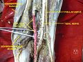
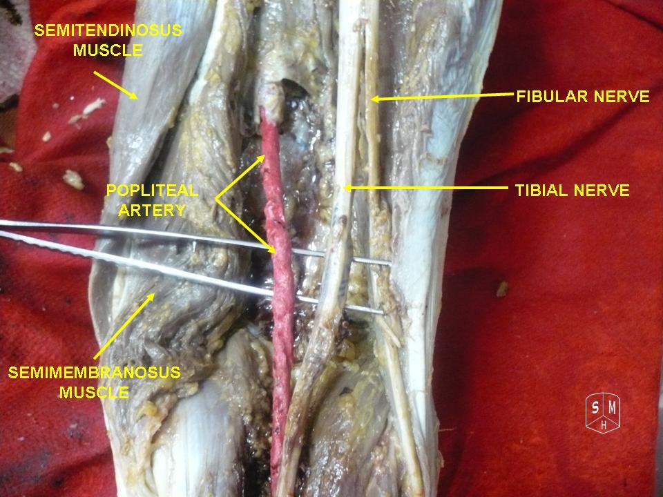
It descends obliquely along the lateral side of the popliteal fossa to the head of the fibula, close to the medial margin of the biceps femoris muscle. Where the common peroneal nerve winds round the head of the fibula, it is palpable[1].
It lies between the tendon of the biceps femoris and lateral head of the gastrocnemius muscle, winds around the neck of the fibula, between the peronæus longus and the bone, and divides beneath the muscle into the superficial fibular nerve (superficial peroneal nerve) and deep fibular nerve (deep peroneal nerve).
It innervates the peroneus longus and peroneus brevis muscles.
Contents
Branches
Previous to its division it gives off articular and lateral sural cutaneous nerves.
- The articular branches (rami articulares) are three in number:
- Two of these accompany the superior and inferior lateral genicular arteries to the knee; the upper one occasionally arises from the trunk of the sciatic nerve.
- The third (recurrent) articular nerve is given off at the point of division of the common fibular nerve; it ascends with the anterior recurrent tibial artery through the tibialis anterior to the front of the knee.
- The lateral sural cutaneous nerve (n. cutaneus suræ lateralis; lateral cutaneous branch) supplies the skin on the posterior and lateral surfaces of the leg.
- The motor branches:
- As the common fibular nerve exits the popliteal fossa, it courses around the lateral aspect of the leg just below the head of the fibula. Here it is apposed with fibula and gives off two branches, the superficial fibular (peroneal) branch and deep fibular (peroneal) branch.
- The superficial peroneal nerve supplies the muscles of the lateral compartment of the leg namely: peroneus longus and peroneus brevis. These two muscle help in eversion and plantar flexion of the foot.
- The deep peroneal nerve innervates the muscles of the anterior compartment of the leg which are: tibialis anterior, extensor hallucis longus, extensor digitorum longus, and the fibularis (peroneus) tertius. Together these muscles are responsible for dorsiflexion of the foot and extension of the toes.
Clinical significance
Chronic peroneal neuropathy can result from, among other conditions, bed rest of long duration, hyperflexion of the knee, peripheral neuropathy, pressure in obstetric stirrups, and conditioning in ballet dancers. The most common cause is habitual leg crossing that compresses the common fibular nerve as it crosses around the head of fibula.[2] Transient trauma to the nerve can result from peroneal strike.
Damage to this nerve typically results in foot drop, where dorsiflexion of the foot is compromised and the foot drags (the toe points) during walking; and in sensory loss to the dorsal surface of the foot and portions of the anterior, lower-lateral leg.
Surgical procedures
- Peroneal nerve decompression:
- In the surgical treatment of fibular nerve compression, an incision is made over the neck of the fibula. Fascia surrounding the nerves to the lateral side of the leg is released.[3]
- Deep peroneal nerve decompression:
- In the surgical treatment of deep fibular nerve entrapment in the foot, a ligament from the extensor digitorum brevis muscle that crosses over the deep peroneal nerve, putting pressure on it and causing pain, is released.[3]
Additional images
-

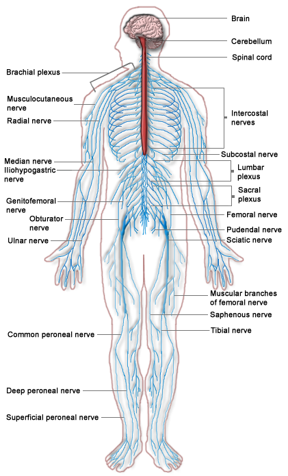
Nervous system
-

Cutaneous nerves of the right lower extremity. Front and posterior views.
-

A schematic of the sacral plexus showing the origin of the common fibular nerve (labeled at the bottom left).
-
Common fibular nerve
See also
- Deep fibular nerve
- Peroneal vein
- Peroneus muscles
- Sacral plexus
References
- ^ Tam, Michael (2006-03-30). "Nerves of the Lower Limb". Medical student's retreat - Michael Tam's anatomy notes for medical students. Archived from the original on 2007-12-13. http://web.archive.org/web/20071213025348/http://download.videohelp.com/vitualis/med/lowrnn.htm#Common_peroneal_nerve. Retrieved 2010-05-08.
- ^ Walter George Bradley (2004). "foot+drop" Neurology in clinical practice (4 ed.). Taylor & Francis. pp. 2545. ISBN 9997625889. http://books.google.com/books?id=vOQqyNhTDl0C&pg=PA454&lpg=PA454&dq="foot+drop". page 453-454
- ^ a b Dellon Institutes Peroneal Nerve Compression Surgical Treatment
External links
- SUNY Labs 14:st-0501
- Duke Orthopedics peroneal_nerve
- Anatomy at MUN nerve/scianerv
- latleg at The Anatomy Lesson by Wesley Norman (Georgetown University)
- Anatomy at Dartmouth arteries-nerves%20LE/nerves4
- Overview at okstate.edu
This article was originally based on an entry from a public domain edition of Gray's Anatomy. As such, some of the information contained within it may be outdated.
Nerves of lower limbs and lower torso: the lumbosacral plexus (L1–Co) (TA A14.2.05–07, GA 9.948) lumbar
plexus
(L1–L4)sacral
plexus
(L4–S4)common
fibularothercoccygeal
plexus
(S4–Co)Categories:- Nerves of the lower limb and lower torso
- The articular branches (rami articulares) are three in number:


Wikimedia Foundation. 2010.