- Gastrocnemius muscle
-
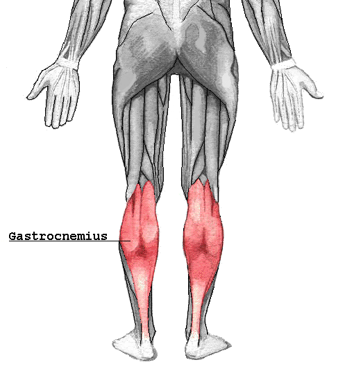
Gastrocnemius muscle 
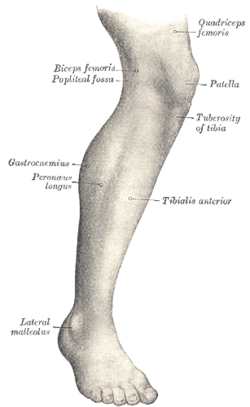
Lateral aspect of right leg. Gray's subject #129 482 Origin superior to articular surfaces of lateral condyle of femur and medial condyle of femur Insertion tendo calcaneus (achilles tendon) into mid-posterior calcaneus Artery sural arteries Nerve tibial nerve from the sciatic, specifically, nerve roots S1–S2 Actions plantar flexes foot, flexes knee Antagonist Tibialis anterior muscle In humans, the gastrocnemius muscle (
 /ˌɡæstrɒkˈniːmiəs/ or /ˌɡæstrəˈniːmiəs/; Latin, from Greek γαστήρ "stomach" and knēmē "leg"; meaning "stomach of leg", referring to the bulging shape of the calf) is a very powerful superficial pennate muscle that is in the back part of the lower leg. It runs from its two heads just above the knee to the heel, and is involved in standing, walking, running and jumping. Along with the soleus muscle it forms the calf muscle. Its function is plantar flexing the foot at the ankle joint and flexing the leg at the knee joint. In a 1967 EMG study, Herman and Bragin concluded that its most important role was plantar flexing in large contractions and in rapid development of tension.
The gastrocnemius is located with the soleus in the posterior (back) compartment of the leg. The lateral head originates from the lateral condyle of the femur, while the medial head originates from the medial condyle of the femur. Its other end forms a common tendon with the soleus muscle; this tendon is known as the calcaneal tendon or Achilles Tendon and inserts onto the posterior surface of the calcaneus, or mountain bone.
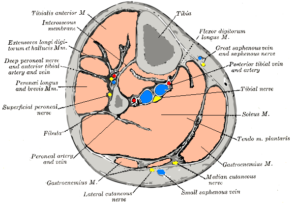
Deep to the gastrocnemius (farther from the skin) is the soleus muscle. Some anatomists consider both to be a single muscle, the triceps surae. The plantaris muscle and a portion of its tendon run between the two muscles, which is involved in "locking" the knee from the standing and posterior tibial vein and the tibial nerve. Since the anterior compartment of the leg is lateral to the tibia, the bulge of muscle medial to the tibia on the anterior side is actually the posterior compartment. The soleus is superficial to the mid-shaft of the tibia. Frequently there is a sesamoid bone called the "fabella" in the lateral head of gastrocnemius muscle.
Clinical significance
The gastrocnemius muscle is very prone to spasms; the painful, involuntary, contraction of the muscle for up to several minutes.[1]
This muscle is prone to injury called torn calf muscle, which is disabling.
The Gastrocnemius muscle may also become inflamed due to overuse. Anti-inflammatory and physical therapy may be necessary.
Anatomical abnormalities involving the medial head of gastrocnemius muscle results in popliteal artery entrapment syndrome.
Additional images
-
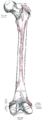
Right femur. Posterior surface.
-
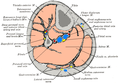
Cross-section through middle of leg.
-
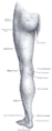
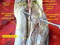
Back of left lower extremity.
-
Gastrocnemius muscle
External links
- WebMD Nighttime Leg Cramps: http://www.webmd.com/a-to-z-guides/nighttime-leg-cramps-topic-overview
- -275120049 at GPnotebook
- LUC gast
- SUNY Labs 14:st-0405
- Anatomy at Dartmouth knee/surface/surface4
- 10th Ed.
Kinesiology: Scientific Basis of Human Motion
Nancy Hamilton, Ph.D. Associate Professor, University of Northern Iowa
Kathryn Luttgens, Ph.D. Professor Emerita, Northeastern University
List of muscles of lower limbs (TA A04.7, GA 4.465) ILIAC Region
/ ILIOPSOASBUTTOCKS THIGH /
compartmentsLEG/
Crus/
compartmentssuperficial · triceps surae (gastrocnemius, soleus, accessory soleus, Achilles tendon) · plantaris
deep · tarsal tunnel (flexor hallucis longus, flexor digitorum longus, tibialis posterior) · popliteusfibularis muscles (longus, brevis)FOOT DorsalPlantar1st layer (abductor hallucis, flexor digitorum brevis, abductor digiti minimi) · 2nd layer (quadratus plantae, lumbrical muscle) · 3rd layer (flexor hallucis brevis, adductor hallucis, flexor digiti minimi brevis) · 4th layer (dorsal interossei, plantar interossei)Categories:- Calf muscles
- Knee flexors
- Plantar flexors
-


Wikimedia Foundation. 2010.