- Semitendinosus muscle
-
Semitendinosus muscle 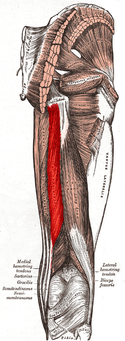
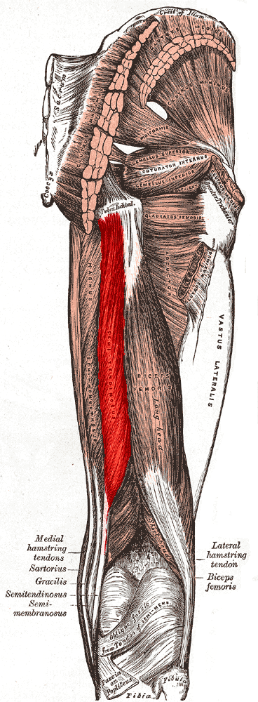
Muscles of the gluteal and posterior femoral regions. Semitendinosus labeled at bottom left. 
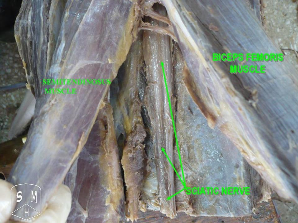
Semitendinosus visible at bottom left. Latin musculus semitendinosus Gray's subject #128 479 Origin tuberosity of the ischium Insertion pes anserinus Artery inferior gluteal artery, perforating arteries Nerve sciatic (tibial, L5, S1, S2) Actions flex knee, extend hip joint Antagonist Quadriceps muscle The semitendinosus is a muscle in the back of the thigh; it is one of the hamstrings.
Contents
Structure
The semitendinosus, remarkable for the great length of its tendon of insertion, is situated at the posterior and medial aspect of the thigh .
It arises from the lower and medial impression on the tuberosity of the ischium, by a tendon common to it and the long head of the biceps femoris; it also arises from an aponeurosis which connects the adjacent surfaces of the two muscles to the extent of about 7.5 cm. from their origin.
The muscle is fusiform and ends a little below the middle of the thigh in a long round tendon which lies along the medial side of the popliteal fossa; it then curves around the medial condyle of the tibia and passes over the tibial collateral ligament of the knee-joint, from which it is separated by a bursa, and is inserted into the upper part of the medial surface of the body of the tibia, nearly as far forward as its anterior crest.
At its insertion it gives off from its lower border a prolongation to the deep fascia of the leg and lies behind the tendon of the sartorius, and below that of the gracilis, to which it is united. These three tendons form what is known as the pes anserinus, so named because it looks like the foot of a goose.
A tendinous intersection is usually observed about the middle of the muscle.
Innervation
The semitendinosus is innervated by the tibial part of the sciatic nerve.
Actions
The semitendinosus helps to extend (straighten) the hip joint and flex (bend) the knee joint. It also helps medially rotate the knee.
Additional images
-
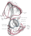
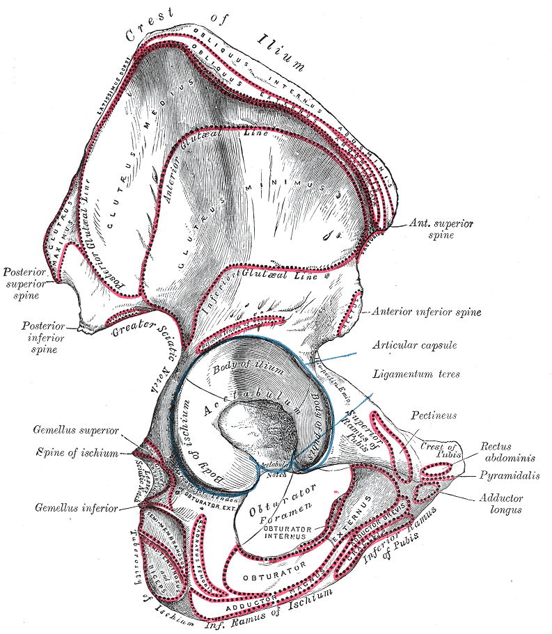
Right hip bone. External surface.
-
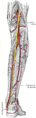
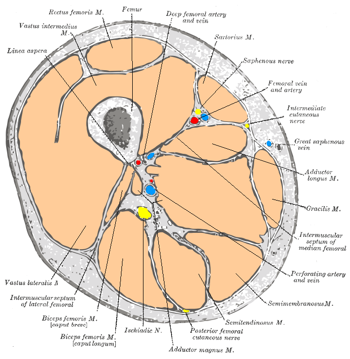
The popliteal, posterior tibial, and peroneal arteries.
-
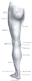
Back of left lower extremity.
-
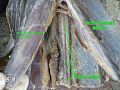
Semitendinosus muscle
See also
External links
- LUC semt
- SUNY Labs 14:st-0410
- Cross section at UV pembody/body18b
- Anatomy at Dartmouth knee/surface/surface4
- PTCentral
This article was originally based on an entry from a public domain edition of Gray's Anatomy. As such, some of the information contained within it may be outdated.
List of muscles of lower limbs (TA A04.7, GA 4.465) ILIAC Region
/ ILIOPSOASBUTTOCKS THIGH /
compartmentsLEG/
Crus/
compartmentssuperficial · triceps surae (gastrocnemius, soleus, accessory soleus, Achilles tendon) · plantaris
deep · tarsal tunnel (flexor hallucis longus, flexor digitorum longus, tibialis posterior) · popliteusfibularis muscles (longus, brevis)FOOT DorsalPlantar1st layer (abductor hallucis, flexor digitorum brevis, abductor digiti minimi) · 2nd layer (quadratus plantae, lumbrical muscle) · 3rd layer (flexor hallucis brevis, adductor hallucis, flexor digiti minimi brevis) · 4th layer (dorsal interossei, plantar interossei)Categories:- Muscle stubs
- Hip extensors
- Knee flexors
- Knee medial rotators
- Thigh muscles
-
Wikimedia Foundation. 2010.