- Medial cuneiform bone
-
Bone: First cuneiform bone 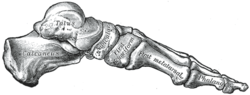
Skeleton of foot. Medial aspect. Latin os cuneiforme mediale, os cuneiform primum Gray's subject #63 270 The medial cuneiform (also known as first cuneiform) is the largest of the cuneiforms.
It is situated at the medial side of the foot, anterior to the navicular and posterior to the base of the first metatarsal.
It articulates with four bones: the navicular, second cuneiform, and first and second metatarsals.
Additional images
-
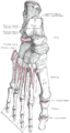
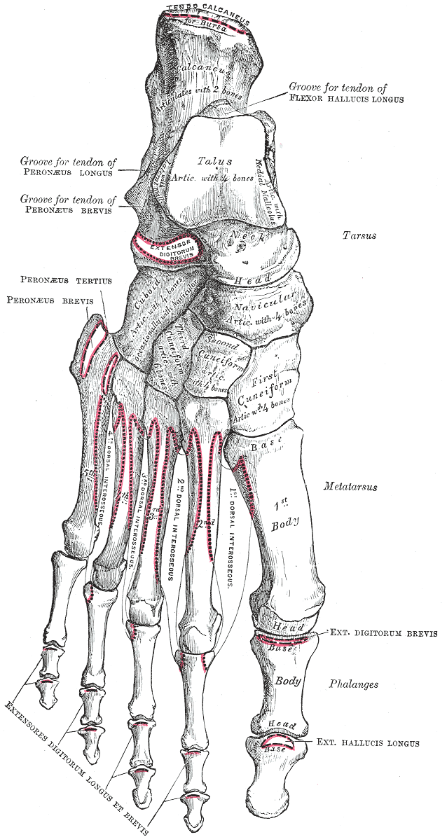
Bones of the right foot. Dorsal surface.
-
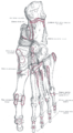
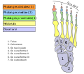
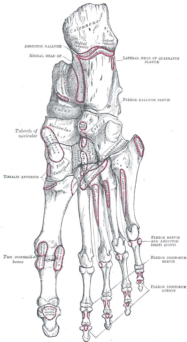
Bones of the right foot. Plantar surface.
-
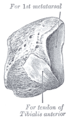
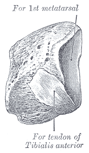
The left first cuneiform. Antero-medial view.
-
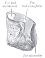
The left first cuneiform. Postero-lateral view.
-

Skeleton of foot. Lateral aspect.
-
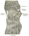
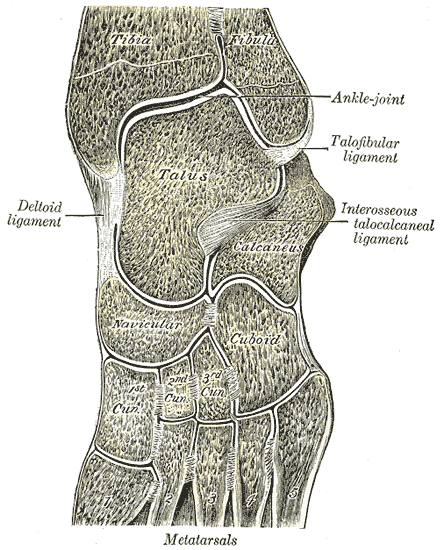
Oblique section of left intertarsal and tarsometatarsal articulations, showing the synovial cavities.
-
Bones of foot
External links
- medial+cuneiform+bone at eMedicine Dictionary
This article was originally based on an entry from a public domain edition of Gray's Anatomy. As such, some of the information contained within it may be outdated.
Bones of lower limbs (TA A02.5.04–18, GA 2.242–277) Femur head (fovea) · neck · greater trochanter (trochanteric fossa) · lesser trochanter · intertrochanteric line · intertrochanteric crest · quadrate tubercleadductor tubercle · patellar surface · epicondyles (lateral, medial) · condyles (lateral, medial) · intercondylar fossaCrus Otherpatella (apex of patella)Foot calcaneus (sustentaculum tali, trochlear process) · talus (body, neck, head) · navicular · cuboid · cuneiform (medial, intermediate, lateral)OtherCategories:- Skeletal system
-


Wikimedia Foundation. 2010.