- Body of femur
Infobox Bone
Name = Body of femur
Latin = corpus femoris
GraySubject = 59
GrayPage = 243
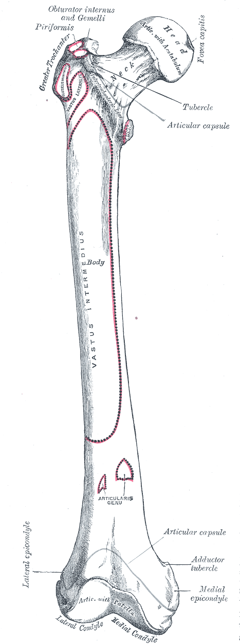
Caption = Right femur. Anterior surface.
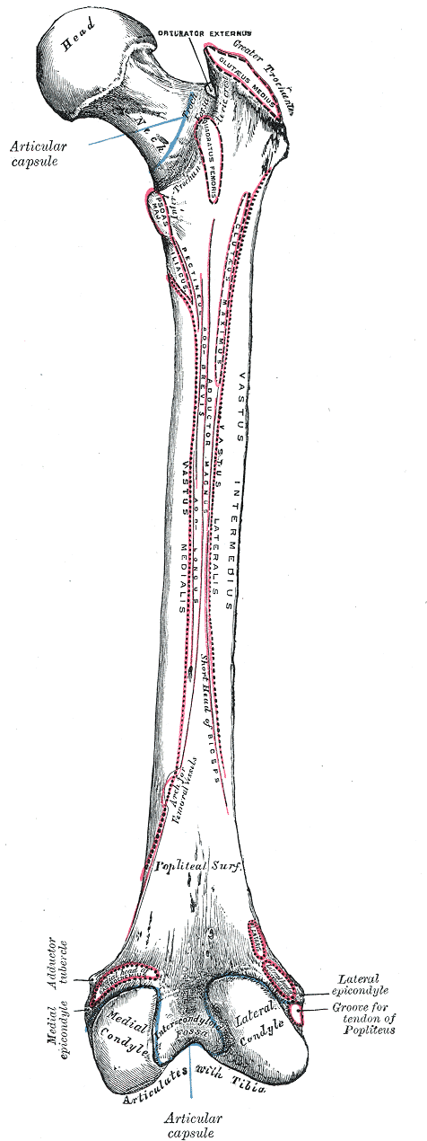
Caption2 = Right femur. Posterior surface.
System =
MeshName =
MeshNumber =
DorlandsPre = s_09
DorlandsSuf = 12732595
The body of thefemur (or shaft), almost cylindrical in form, is a little broader above than in the center, broadest and somewhat flattened from before backward below. It is slightly arched, so as to be convex in front, and concave behind, where it is strengthened by a prominent longitudinal ridge, thelinea aspera .It presents for examination three borders, separating three surfaces.
Of the borders, one, the linea aspera, is posterior, one is medial, and the other, lateral.
Linea aspera border
The linea aspera is a prominent longitudinal ridge or crest, on the middle third of the bone, presenting a medial and a lateral lip, and a narrow rough, intermediate line.
Above, the linea aspera is prolonged by three ridges.
The lateral ridge is very rough, and runs almost vertically upward to the base of the greater trochanter.
It is termed the
gluteal tuberosity , and gives attachment to part of theglutæus maximus : its upper part is often elongated into a roughened crest, on which a more or less well-marked, rounded tubercle, thethird trochanter , is occasionally developed.The intermediate ridge or pectineal line is continued to the base of the lesser trochanter and gives attachment to the
pectineus ; the medial ridge is lost in the intertrochanteric line; between these two a portion of theiliacus is inserted.Below, the linea aspera is prolonged into two ridges, enclosing between them a triangular area, the popliteal surface, upon which the
popliteal artery rests.Of these two ridges, the lateral is the more prominent, and descends to the summit of the
lateral condyle .The medial is less marked, especially at its upper part, where it is crossed by the
femoral artery .It ends below at the summit of the medial condyle, in a small tubercle, the adductor tubercle, which affords insertion to the tendon of the
adductor magnus .From the medial lip of the linea aspera and its prolongations above and below, the
vastus medialis arises; and from the lateral lip and its upward prolongation, thevastus lateralis takes origin.The adductor magnus is inserted into the linea aspera, and to its lateral prolongation above, and its medial prolongation below.
Between the vastus lateralis and the adductor magnus two muscles are attached—viz., the glutæus maximus inserted above, and the short head of the
biceps femoris arising below.Between the adductor magnus and the vastus medialis four muscles are inserted: the iliacus and pectineus above; the adductor brevis and adductor longus below.
The linea aspera is perforated a little below its center by the
nutrient canal , which is directed obliquely upward.Lateral border and medial border
The other two borders of the femur are only slightly marked: the "lateral border" extends from the antero-inferior angle of the greater trochanter to the anterior extremity of the lateral condyle; the "medial border" from the
intertrochanteric line , at a point opposite the lesser trochanter, to the anterior extremity of the medial condyle.Anterior surface
The anterior surface includes that portion of the shaft which is situated between the lateral and medial borders.
It is smooth, convex, broader above and below than in the center.
From the upper three-fourths of this surface the
Vastus intermedius arises; the lower fourth is separated from the muscle by the intervention of thesynovial membrane of theknee-joint and a bursa; from the upper part of it theArticularis genu takes origin.The lateral surface includes the portion between the lateral border and the linea aspera; it is continuous above with the corresponding surface of the greater trochanter, below with that of the lateral condyle: from its upper three-fourths the Vastus intermedius takes origin.
The medial surface includes the portion between the medial border and the linea aspera; it is continuous above with the lower border of the neck, below with the medial side of the medial condyle: it is covered by the Vastus medialis.
Wikimedia Foundation. 2010.