- Body of talus
-
Body of talus 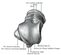
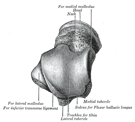
Left talus, from above. Latin corpus tali Gray's subject #63 266 The body of the talus comprises most of the volume of the talus bone. It presents with five surfaces.
Contents
Superior surface
The superior surface of the body presents, behind, a smooth trochlear surface, the trochlea, for articulation with the tibia.
The trochlea is broader in front than behind, convex from before backward, slightly concave from side to side: in front it is continuous with the upper surface of the neck of the bone.
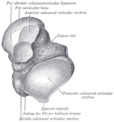
Inferior surface
The inferior surface presents two articular areas, the posterior and middle calcaneal surfaces, separated from one another by a deep groove, the sulcus tali.
The groove runs obliquely forward and lateralward, becoming gradually broader and deeper in front: in the articulated foot it lies above a similar groove upon the upper surface of the calcaneus, and forms, with it, a canal (sinus tarsi) filled up in the fresh state by the interosseous talocalcaneal ligament.
The posterior calcaneal articular surface is large and of an oval or oblong form.
It articulates with the corresponding facet on the upper surface of the calcaneus, and is deeply concave in the direction of its long axis which runs forward and lateralward at an angle of about 45° with the median plane of the body.
The middle calcaneal articular surface is small, oval in form and slightly convex; it articulates with the upper surface of the sustentaculum tali of the calcaneus.
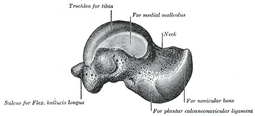
Medial surface
The medial surface presents at its upper part a pear-shaped articular facet for the medial malleolus, continuous above with the trochlea; below the articular surface is a rough depression for the attachment of the deep portion of the deltoid ligament of the ankle-joint.
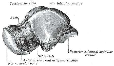
Lateral surface
The lateral surface carries a large triangular facet, concave from above downward, for articulation with the lateral malleolus; its anterior half is continuous above with the trochlea; and in front of it is a rough depression for the attachment of the anterior talofibular ligament.
Between the posterior half of the lateral border of the trochlea and the posterior part of the base of the fibular articular surface is a triangular facet which comes into contact with the transverse inferior tibiofibular ligament during flexion of the ankle-joint; below the base of this facet is a groove which affords attachment to the posterior talofibular ligament.
Posterior surface
The posterior surface is narrow, and traversed by a groove running obliquely downward and medialward, and transmitting the tendon of the Flexor hallucis longus.
Lateral to the groove is a prominent tubercle, the posterior process, to which the posterior talofibular ligament is attached; this process is sometimes separated from the rest of the talus, and is then known as the os trigonum.
Medial to the groove is a second smaller tubercle.
Additional images
-
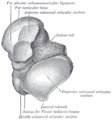
Left talus, from below.
-
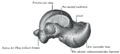
Left talus, medial surface.
-
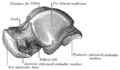
Left talus, lateral surface.
This article was originally based on an entry from a public domain edition of Gray's Anatomy. As such, some of the information contained within it may be outdated.
Bones of lower limbs (TA A02.5.04–18, GA 2.242–277) Femur head (fovea) · neck · greater trochanter (trochanteric fossa) · lesser trochanter · intertrochanteric line · intertrochanteric crest · quadrate tubercleadductor tubercle · patellar surface · epicondyles (lateral, medial) · condyles (lateral, medial) · intercondylar fossaCrus Otherpatella (apex of patella)Foot calcaneus (sustentaculum tali, trochlear process) · talus (body, neck, head) · navicular · cuboid · cuneiform (medial, intermediate, lateral)OtherCategories: -
Wikimedia Foundation. 2010.