- Lower extremity of tibia
-
Bone: Lower extremity of tibia 
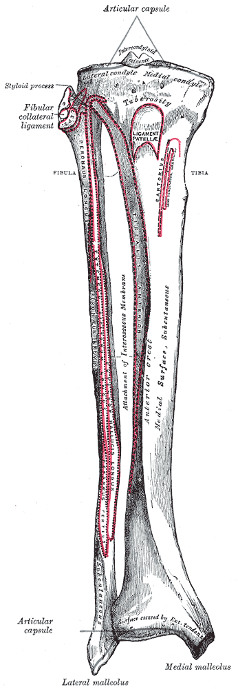
Bones of the right leg. Anterior surface. 
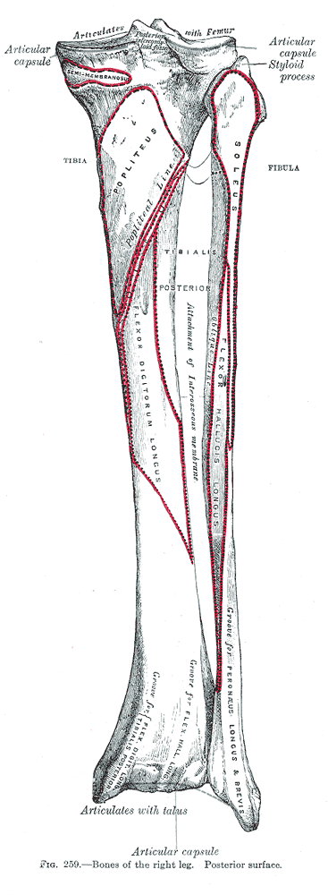
Bones of the right leg. Posterior surface. Gray's subject #61 259 The lower extremity of the tibia, much smaller than the upper extremity of tibia, presents five surfaces; it is prolonged downward on its medial side as a strong process, the medial malleolus.
Surfaces
The inferior articular surface is quadrilateral, and smooth for articulation with the talus. It is concave from before backward, broader in front than behind, and traversed from before backward by a slight elevation, separating two depressions. It is continuous with that on the medial malleolus.
The anterior surface of the lower extremity is smooth and rounded above, and covered by the tendons of the Extensor muscles; its lower margin presents a rough transverse depression for the attachment of the articular capsule of the ankle-joint.
The posterior surface is traversed by a shallow groove directed obliquely downward and medialward, continuous with a similar groove on the posterior surface of the talus and serving for the passage of the tendon of the Flexor hallucis longus.
The lateral surface presents a triangular rough depression for the attachment of the inferior interosseous ligament connecting it with the fibula; the lower part of this depression is smooth, covered with cartilage in the fresh state, and articulates with the fibula. The surface is bounded by two prominent borders, continuous above with the interosseous crest; they afford attachment to the anterior and posterior ligaments of the lateral malleolus.
The medial surface -- see medial malleolus for details.
This article was originally based on an entry from a public domain edition of Gray's Anatomy. As such, some of the information contained within it may be outdated.
Bones of lower limbs (TA A02.5.04–18, GA 2.242–277) Femur head (fovea) · neck · greater trochanter (trochanteric fossa) · lesser trochanter · intertrochanteric line · intertrochanteric crest · quadrate tubercleadductor tubercle · patellar surface · epicondyles (lateral, medial) · condyles (lateral, medial) · intercondylar fossaCrus lower extremityOtherFoot calcaneus (sustentaculum tali, trochlear process) · talus (body, neck, head) · navicular · cuboid · cuneiform (medial, intermediate, lateral)OtherCategories:- Bones of the lower limb
- Musculoskeletal system stubs
Wikimedia Foundation. 2010.