- Navicular bone
-
- For the bone in the wrist/hand formerly called navicular, see scaphoid bone
Bone: Navicular bone 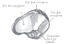
The left navicular. Antero-lateral view 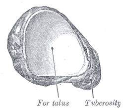
The left navicular. Postero-medial view. Latin os naviculare Gray's subject #63 270 The navicular bone is a small bone found in the feet of both humans and horses.
Contents
Human anatomy
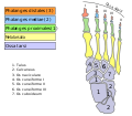
The navicular bone is one of the tarsal bones, found in the foot. Its name derives from the bone's resemblance to a small boat, caused by the strongly concave proximal articular surface. The term navicular bone or hand navicular bone was formerly used[1] for the scaphoid bone, one of the carpal bones of the wrist.
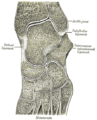
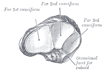
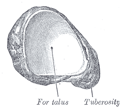
It is located on the medial side of the foot, and articulates proximally with the talus, distally with the three cuneiform bones, and occasionally laterally with the cuboid.
As the bones in the foot develop, a particular process is important for the navicular structure. In the early stage, the navicular bone is actually cartilaginous and has to progress and calcify in order to maintain a strong form. There is a condition, however, in which the three cuneiforms that compose the bone do not completely calcify as a unit. In other words, they do not fuse properly, which leads to the most medial third creating a protrusion along the medial arch. This extension of the foot has a tendency to put stress on two tendons and the ligament that run along its side. The tendon of the peroneus brevis muscle which is the most distal of the two tendons, the tendon of the peroneus longus muscle which extends to the posterior of the ankle, and the posterior talofibular ligament which extends upward partway along the calf muscle, can all be potentially affected by this protrusion. Sometimes referred to as an extra navicular bone, this bump can cause wear and tear on the tendons causing sharp pain with increased activity. This condition can be corrected with surgery to file down or remove the protrusion and repair the tendons that were affected.[2]
Horse anatomy
The navicular bone of the horse, also known as the distal sesamoid bone, lies on the palmar aspect of the coffin joint between the second phalanx and third phalanx, known as the coffin or pedal bone. It is an important structure in relation to lameness and is involved with a significant disease process called Navicular Disease. Recently much of the original literature concerning navicular disease has been called into question, particularly the significance of some radiographic changes.[3]
Notes
- ^ "Gray's Anatomy, 6b. The Hand. 1. The Carpus. 4". 1918. http://www.bartleby.com/107/54.html. Retrieved December 2009.
- ^ Wexler, Randell K., M.D. “The Injured Ankle.” American Academy of Family Physicians. 1998 Vol. 57 No. 3. <http://www.aafp.org/afp/980201ap/wexler.html>
- ^ Citing Clinical Anatomy and Physiology Laboratory Manual for Veterinary Technicians, Colville, Thomas and Bassert, Joanna M. 2008 Publ. Mosby/Elsevier, Canada. "Navicular Bone - The distal sesamoid bone of the horse. The navicular bone is located deep in the hoof behind the joint between the middle and distal phalanges."
See also
- Bone terminology
- Terms for anatomical location
- Equine forelimb anatomy
Additional images
-
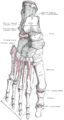
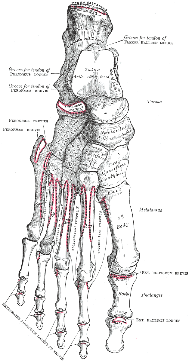
Bones of the right foot. Dorsal surface.
-
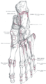
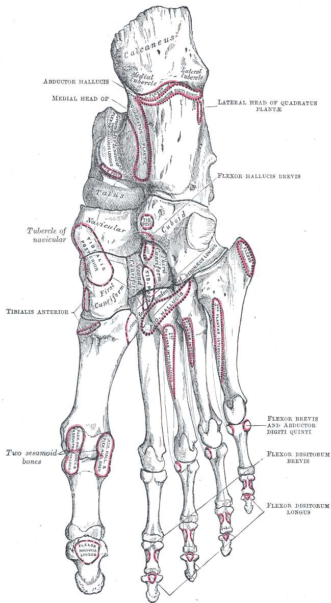
Bones of the right foot. Plantar surface.
-

Skeleton of foot. Medial aspect.
-

Skeleton of foot. Lateral aspect.
-

Talocalcaneal and talocalcaneonavicular articulations exposed from above by removing the talus.
-
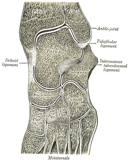
Oblique section of left intertarsal and tarsometatarsal articulations, showing the synovial cavities.
-
Bones of foot
Bones (TA A02, GA 2) Axial Thoracic skeletonFacial bonesnasal · maxilla · lacrimal · zygomatic · palatine · inferior nasal conchae · vomer · mandible · THROAT: hyoid (greater cornu, lesser cornu, body)Appendicular LowerCategories:- Skeletal system
- Horse anatomy


Wikimedia Foundation. 2010.