- Parietal bone
-
Bone: Parietal bone 
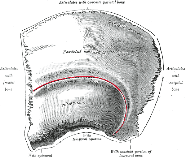
Figure 1 : Left parietal bone. Outer surface. 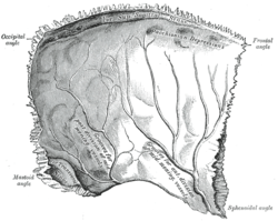
Figure 2 : Left parietal bone. Inner surface. Latin os parietale Gray's subject #32 133 MeSH Parietal+bone The parietal bones are bones in the human skull which, when joined together, form the sides and roof of the cranium. Each bone is roughly quadrilateral in form, and has two surfaces, four borders, and four angles. It is named from the Latin pariet-, wall.
Contents
Surfaces
External
The external surface [Fig. 1] is convex, smooth, and marked near the center by an eminence, the parietal eminence (tuber parietale), which indicates the point where ossification commenced.
Crossing the middle of the bone in an arched direction are two curved lines, the superior and inferior temporal lines; the former gives attachment to the temporal fascia, and the latter indicates the upper limit of the muscular origin of the temporalis.
Above these lines the bone is covered by the galea aponeurotica (epicranial aponeurosis); below them it forms part of the temporal fossa, and affords attachment to the temporalis muscle.
At the back part and close to the upper or sagittal border is the parietal foramen, which transmits a vein to the superior sagittal sinus, and sometimes a small branch of the occipital artery; it is not constantly present, and its size varies considerably.
Internal
The internal surface [Fig. 2] is concave; it presents depressions corresponding to the cerebral convolutions, and numerous furrows (grooves) for the ramifications of the middle meningeal artery; the latter run upward and backward from the sphenoidal angle, and from the central and posterior part of the squamous border.
Along the upper margin is a shallow groove, which, together with that on the opposite parietal, forms a channel, the sagittal sulcus, for the superior sagittal sinus; the edges of the sulcus afford attachment to the falx cerebri.
Near the groove are several depressions, best marked in the skulls of old persons, for the arachnoid granulations (Pacchionian bodies).
In the groove is the internal opening of the parietal foramen when that aperture exists.
Borders
- The sagittal border, the longest and thickest, is dentated (has toothlike projections) and articulates with its fellow of the opposite side, forming the sagittal suture.
- The squamous border is divided into three parts: of these:
- the anterior is thin and pointed, bevelled at the expense of the outer surface, and overlapped by the tip of the great wing of the sphenoid;
- the middle portion is arched, bevelled at the expense of the outer surface, and overlapped by the squama of the temporal;
- the posterior part is thick and serrated for articulation with the mastoid portion of the temporal.
- The frontal border is deeply serrated, and bevelled at the expense of the outer surface above and of the inner below; it articulates with the frontal bone, forming half of the coronal suture. The point where the coronal suture intersects with the sagittal suture forms a T-shape and is called the bregma.
- The occipital border, deeply denticulated (finely toothed), articulates with the occipital bone, forming half of the lambdoid suture. That point where the sagittal suture intersects the lambdoid suture is called the lambda, because of its resemblance to the Greek letter.
Angles
- The frontal angle is practically a right angle, and corresponds with the point of meeting of the sagittal and coronal sutures; this point is named the bregma; in the fetal skull and for about a year and a half after birth this region is membranous, and is called the anterior fontanelle.
- The sphenoidal angle, thin and acute, is received into the interval between the frontal bone and the great wing of the sphenoid. Its inner surface is marked by a deep groove, sometimes a canal, for the anterior divisions of the middle meningeal artery.
- The occipital angle is rounded and corresponds with the point of meeting of the sagittal and lambdoidal sutures—a point which is termed the lambda; in the fetus this part of the skull is membranous, and is called the posterior fontanelle.
- The mastoid angle is truncated; it articulates with the occipital bone and with the mastoid portion of the temporal, and presents on its inner surface a broad, shallow groove which lodges part of the transverse sinus. The point of meeting of this angle with the occipital and the mastoid part of the temporal is named the asterion.
Ossification
The parietal bone is ossified in membrane from a single center, which appears at the parietal eminence about the eighth week of fetal life.
Ossification gradually extends in a radial manner from the center toward the margins of the bone; the angles are consequently the parts last formed, and it is here that the fontanelles exist.
Occasionally the parietal bone is divided into two parts, upper and lower, by an antero-posterior suture.
In other animals
In non-human vertebrates, the parietal bones typically form the rear or central part of the skull roof, lying behind the frontal bones. In many non-mammalian tetrapods, they are bordered to the rear by a pair of postparietal bones that may be solely in the roof of the skull, or slope downwards to contribute to the back of the skull, depending on the species. In the living tuatara, and many fossil species, a small opening, the parietal foramen, lies between the two parietal bones. This opening is the location of a third eye in the midline of the skull, which is much smaller than the two main eyes.[1]
Additional images
-
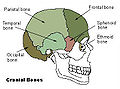
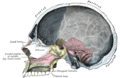
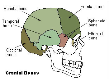
Cranial bones
-
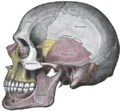
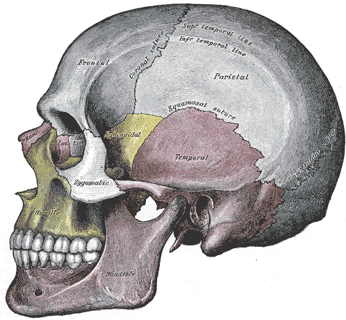
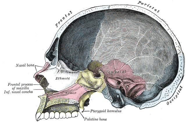
Side view of the skull.
-
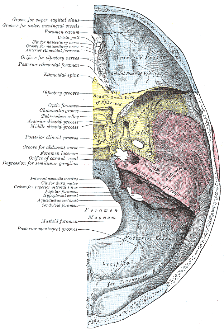
Base of the skull. Upper surface.
-
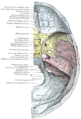
Sagittal section of skull.
See also
- Bone terminology
- Terms for anatomical location
References
External links
This article was originally based on an entry from a public domain edition of Gray's Anatomy. As such, some of the information contained within it may be outdated.
Categories:- Bones of the head and neck

Wikimedia Foundation. 2010.