- Squama temporalis
Infobox Bone
Name = Squama temporalis
Latin = squama temporalis
GraySubject = 34
GrayPage = 139
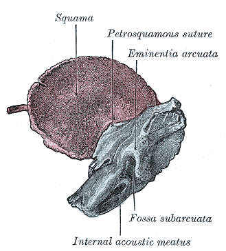
Caption =Temporal bone at birth. Inner aspect.
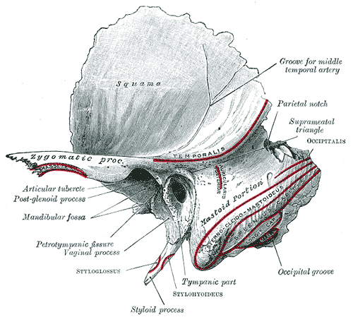
Caption2 = Lefttemporal bone . Outer surface. (Squama is visible at top left.)
System =
MeshName =
MeshNumber =
The squama of thetemporal bone forms the anterior and upper part of the bone, and is scale-like, thin, and translucent.Surfaces
Its outer surface is smooth and ; it affords attachment to the
temporalis muscle , and forms part of thetemporal fossa ; on its hinder part is a vertical groove for the middletemporal artery . A curved line, the "temporal line", or "supramastoid crest", runs backward and upward across its posterior part; it serves for the attachment of the temporalfascia , and limits the origin of the temporalis muscle. The boundary between the squama and the mastoid portion of the bone, as indicated by traces of the original suture, lies about 1 cm. below this line.Projecting from the lower part of the squama is a long, arched process, the "zygomatic process". This process is at first directed lateralward, its two surfaces looking upward and downward; it then appears as if twisted inward upon itself, and runs forward, its surfaces now looking medialward and lateralward. The superior border is long, thin, and sharp, and serves for the attachment of the temporal fascia; the inferior, short, thick, and arched, has attached to it some fibers of the
masseter . The lateral surface is convex andsubcutaneous ; the medial is , and affords attachment to the masseter. The anterior end is deeply serrated and articulates with thezygomatic bone . The posterior end is connected to the squama by two roots, the anterior and posterior roots. The posterior root, a prolongation of the upper border, is strongly marked; it runs backward above theexternal auditory meatus , and is continuous with the temporal line. The anterior root, continuous with the lower border, is short but broad and strong; it is directed medialward and ends in a rounded eminence, thearticular tubercle (eminentia articularis).This tubercle forms the front boundary of the
mandibular fossa , and in the fresh state is covered with cartilage. In front of the articular tubercle is a small triangular area which assists in forming theinfratemporal fossa ; this area is separated from the outer surface of the squama by a ridge which is continuous behind with the anterior root of the zygomatic process, and in front, in the articulated skull, with theinfratemporal crest on the great wing of the sphenoid. Between the posterior wall of theexternal acoustic meatus and the posterior root of the zygomatic process is the area called the suprameatal triangle (Macewen), ormastoid fossa , through which an instrument may be pushed into thetympanic antrum .At the junction of the anterior root with the zygomatic process is a projection for the attachment of the
temporomandibular ligament ; and behind the anterior root is an oval depression, forming part of the mandibular fossa, for the reception of the condyle of the mandible. The mandibular fossa (glenoid fossa) is bounded, in front, by the articular tubercle; behind, by the tympanic part of the bone, which separates it from the external acoustic meatus; it is divided into two parts by a narrow slit, the petrotympanic fissure (Glaserian fissure ). The anterior part, formed by the squama, is smooth, covered in the fresh state withcartilage , and articulates with the condyle of the mandible. Behind this part of the fossa is a small conical eminence; this is the representative of a prominent tubercle which, in some mammals, descends behind the condyle of the mandible, and prevents its backward displacement. The posterior part of the mandibular fossa, formed by thetympanic part of the bone, is non-articular, and sometimes lodges a portion of theparotid gland .The
petrotympanic fissure leads into themiddle ear ortympanic cavity ; it lodges the anterior process of themalleus , and transmits the tympanic branch of theinternal maxillary artery . Thechorda tympani nerve passes through a canal (canal ofHuguier ), separated from the anterior edge of the petrotympanic fissure by a thin scale of bone and situated on the lateral side of theauditory tube , in the retiring angle between the squama and the petrous portion of the temporal.The internal surface of the squama is concave; it presents depressions corresponding to the convolutions of the temporal lobe of the brain, and grooves for the branches of the middle
meningeal vessel s.Borders
The superior border is thin, and bevelled at the expense of the internal table, so as to overlap the squamous border of the
parietal bone , forming with it the squamosal suture. Posteriorly, the superior border forms an angle, the parietal notch, with the mastoid portion of the bone.The antero-inferior border is thick, serrated, and bevelled at the expense of the inner table above and of the outer below, for articulation with the great wing of the
sphenoid .External links
*
Wikimedia Foundation. 2010.