- Squama frontalis
Infobox Bone
Name = Squama frontalis
Latin =
GraySubject = 33
GrayPage = 135
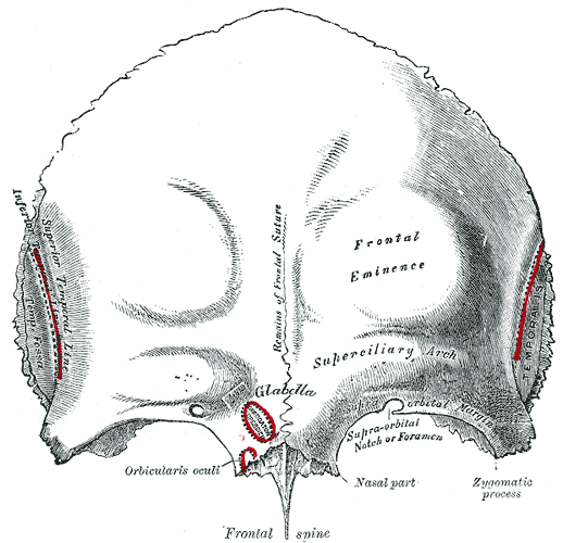
Caption =Frontal bone . Outer surface. (The Squama frontalis is the upper two thirds.)
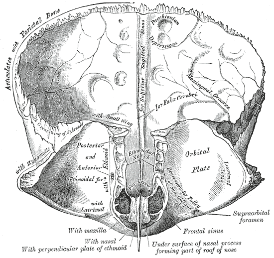
Caption2 = Frontal bone. Inner surface. (The Squama frontalis is the upper two thirds.)
System =
MeshName =
MeshNumber =
There are two surfaces of thesquama of thefrontal bone : the external surface, and the internal surface.External surface
The external surface is convex and usually exhibits, in the lower part of the middle line, the remains of the frontal or
metopic suture; in infancy this suture divides the bone into two, a condition which may persist throughout life.On either side of this suture, about 3 cm. above the supraorbital margin, is a rounded elevation, the
frontal eminence (tuber frontale).These eminences vary in size in different individuals, are occasionally unsymmetrical, and are especially prominent in young skulls; the surface of the bone above them is smooth, and covered by the
galea aponeurotica .Below the frontal eminences, and separated from them by a shallow groove, are two arched elevations, the
superciliary arches ; these are prominentmedially , and are joined to one another by a smooth elevation named theglabella . They are larger in the male than in the female, and their degree of prominence depends to some extent on the size of thefrontal air sinuses ; prominent ridges are, however, occasionally associated with small air sinuses.Beneath each superciliary arch is a curved and prominent margin, the
supraorbital margin , which forms the upper boundary of the base of theorbit , and separates the squama from the orbital portion of the bone.The lateral part of this margin is sharp and prominent, affording to the eye, in that situation, considerable protection from injury; the medial part is rounded.
At the junction of its medial and intermediate thirds is a notch, sometimes converted into a
foramen , thesupraorbital notch or foramen, which transmits the supraorbital vessels and nerve.A small aperture in the upper part of the notch transmits a vein from the
diploë to join thesupraorbital vein .The supraorbital margin ends laterally in the zygomatic process, which is strong and prominent, and articulates with the
zygomatic bone .Running upward and backward from this process is a well-marked line, the
temporal line , which divides into the upper and lower temporal lines, continuous, in the articulated skull, with the corresponding lines on the parietal bone.The area below and behind the temporal line forms the anterior part of the
temporal fossa , and gives origin to theTemporalis muscle .Between the supraorbital margins the squama projects downward to a level below that of the zygomatic processes; this portion is known as the nasal part and presents a rough, uneven interval, the nasal notch, which articulates on either side of the middle line with the nasal bone, and laterally with the frontal process of the
maxilla and with thelacrimal .The term
nasion is applied to the middle of thefrontonasal suture . From the center of the notch the nasal process projects downward and forward beneath the nasal bones and frontal processes of themaxillæ , and supports the bridge of the nose.The nasal process ends below in a sharp spine, and on either side of this is a small grooved surface which enters into the formation of the roof of the corresponding nasal cavity.
The spine forms part of the
septum of the nose, articulating in front with the crest of the nasal bones and behind with the perpendicular plate of theethmoid .Internal surface
The internal surface of the
squama is concave and presents in the upper part of the middle line a vertical groove, thesagittal sulcus , the edges of which unite below to form a ridge, the frontal crest; the sulcus lodges the superior sagittal sinus, while its margins and the crest afford attachment to thefalx cerebri .The crest ends below in a small notch which is converted into a foramen, the
foramen cecum , by articulation with theethmoid .This foramen varies in size in different subjects, and is frequently impervious; when open, it transmits a vein from the nose to the
superior sagittal sinus .On either side of the middle line the bone presents depressions for the convolutions of the brain, and numerous small furrows for the anterior branches of the middle meningeal vessels.
Several small, irregular fossæ may also be seen on either side of the
sagittal sulcus , for the reception of thearachnoid granulations .External links
*
Wikimedia Foundation. 2010.