Posterior clinoid processes
- Posterior clinoid processes
Infobox Bone
Name = Posterior clinoid processes
Latin = p. clinoideus posterior
GraySubject = 35
GrayPage = 147

Caption = Sphenoid bone. Upper surface. (Posterior clinoid process labeled at upper right.)
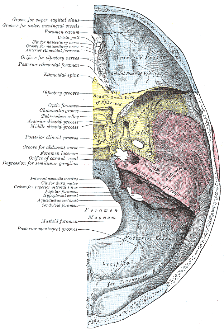
Caption2 = Base of the skull. Upper surface. (Caption for posterior clinoid process visible at center left. Sphenoid bone is yellow.))
System =
MeshName =
MeshNumber =
DorlandsPre = p_34
DorlandsSuf = 12667375
In the sphenoid bone, the anterior boundary of the sella turcica is completed by two small eminences, one on either side, called the anterior clinoid processes, while the posterior boundary is formed by a square-shaped plate of bone, the dorsum sellæ, ending at its superior angles in two tubercles, the posterior clinoid processes, the size and form of which vary considerably in different individuals. The posterior clinoid processes deepen the sella turcica, and give attachment to the tentorium cerebelli.
Etymology
Clinoid likely comes from the Greek root "klinein" or the Latin "clinare", both meaning "sloped" as in "inclined."
External links
*
*
Wikimedia Foundation.
2010.
Look at other dictionaries:
Clinoid process — can refer to: Anterior clinoid process Posterior clinoid processes This disambiguation page lists articles associated with the same title. If an internal link led you here, you may wish to change t … Wikipedia
Middle clinoid process — Bone: Middle clinoid process Sphenoid bone. Upper surface. (Middle clinoid process labeled at upper left.) … Wikipedia
Bone — This article is about the skeletal organ. For other uses, see Bone (disambiguation) and Bones (disambiguation). For the tissue, see Osseous tissue. Drawing of a human femur Bones are rigid organs that constitute part of the endoskeleton of… … Wikipedia
Middle cranial fossa — Base of the skull. Upper surface. (Middle cranial fossa is the centermost of the three indentations, in pink and yellow.) Latin fossa cranii media Gray s … Wikipedia
Dorsum sellae — Bone: Dorsum sellae Sphenoid bone. Upper surface. (Dorsum sellae is labeled in the white portion in the center.) … Wikipedia
Ligament — A ligament is a tough band of connective tissue that connects various structures such as two bones. Ligament is a fitting term; it comes from the Latin ligare meaning to bind or tie. * * * 1. A band or sheet of fibrous tissue connecting two or… … Medical dictionary
Body of sphenoid bone — Infobox Bone Name = Body of sphenoid bone Latin = corpus ossis sphenoidalis GraySubject = 35 GrayPage = 147 Caption = Figure 2: Sphenoid bone, anterior and inferior surfaces. Caption2 = Figure 3: Sphenoid bone, upper and posterior surfaces.… … Wikipedia
Sphenoid bone — Bone: Sphenoid bone Cranial Bones. Only the end of the wing of the sphenoid bone is visible … Wikipedia
Medial pterygoid plate — Bone: Medial pterygoid plate Sphenoid bone. Anterior and inferior surfaces. (Medial pterygoid plate labeled at bottom left.) … Wikipedia
Occipital bone — Bone: Occipital bone Sagittal section of skull. (Occipital bone is at right, in blue.) … Wikipedia