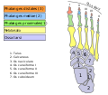
- Talus bone
-
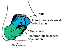
Bone: Talus bone Subtalar Joint, viewed from an angle between lateral and frontal. Latin Os trigonum, Astralagus Gray's subject #63 266 MeSH Talus The talus bone (Latin for ankle[1]) or astragalus is a bone in the collection of bones in the foot called the tarsus. The tarsus forms the lower part of the ankle joint through its articulations with the lateral and medial malleoli of the two bones of the lower leg, the tibia and fibula. Within the tarsus, it articulates with the calcaneus below and navicular in front. Through these articulations, it transmits the entire weight of the body to the foot.[2]
The second largest of the tarsal bones, it is also one of the bones in the human body with the highest percentage of its surface area covered by articular cartilage. Additionally, it is also unusual in that it has a retrograde blood supply, i.e. arterial blood enters the bone at the distal end.[citation needed]
Contents
Structure
Though irregular in shape, the talus can be subdivided into three parts.
Facing anteriorly, the head carries the articulate surface of the navicular bone, and the neck, the roughened area between the body and the head, has small vascular channels.[2]
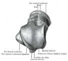
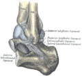
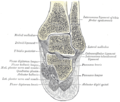
The body features several prominent articulate surfaces: On its superior side is the trochlea tali flanked by the articulate facets for the two malleoli.[2] The ankle mortise, the fork-like structure of the malleoli, holds these three articulate surfaces in a steady grip, which guarantees the stability of the ankle joint. However, because the trochlea is wider in front than at the back (approximately 5-6 mm) the stability in the joint vary with the position of the foot: with the foot dorsiflexed (toes pulled upward) the ligaments of the joint are kept stretched, which guarantees the stability of the joint; but with the foot plantarflexed (as when standing on the toes) the narrower width of the trochlea causes the stability to decrease.[3] Behind the trochlea is a posterior process with a medial and a lateral tubercle separated by a groove for the tendon of the flexor hallucis longus. Exceptionally, the lateral of these tubercles forms an independent bone called os trigonum or "accessory talus". On the bone's inferior side, three articular surfaces serve for the articulation with the calcaneus, and several variously developed articular surfaces exist for the articulation with ligaments.[2]
During the 7-8th intrauterine month an ossification center is formed in the talus.[2]
The Talus bone lacks a good blood supply. Because of this, healing a broken talus can take longer than most other bones. The bone does not get an ample supply of oxygen through the blood due to the low supply, which prolongs the healing process. One with a broken talus may not be able to walk for many months without crutches and will further wear a walking cast or boot of some kind after that.
Evolution
The talus is apparently derived from the fusion of three separate bones in the feet of primitive amphibians; the tibiale, articulating with tibia, the intermedium, between the bases of the tibia and fibula, and the fourth centrale, lying in the mid-part of the tarsus. These bones are still partially separate in modern amphibians, which therefore do not have a true talus. The talus forms a considerably more flexible joint in mammals than it does in reptiles. This reaches its greatest extent in artiodactyls, where the distal surface of the bone has a smooth keel to allow greater freedom of movement of the foot, and thus increase running speed.[4]
Additional images
See also
- Squatting facets
- Knucklebones, a dice game using astragali
Notes
- ^ Mosby’s Medical, Nursing and Allied Health Dictionary, Fourth Edition, Mosby-Year Book Inc., 1994, p. 1526
- ^ a b c d e Platzer (2004), p 216
- ^ Thieme Atlas of Anatomy (2006), p 406
- ^ Romer, Alfred Sherwood; Parsons, Thomas S. (1977). The Vertebrate Body. Philadelphia, PA: Holt-Saunders International. p. 207. ISBN 0-03-910284-X.
References
- Platzer, Werner (2004). Color Atlas of Human Anatomy, Vol. 1: Locomotor System (5th ed.). Thieme. ISBN 3-13-533305-1.
- Thieme Atlas of Anatomy: General Anatomy and Musculoskeletal System. Thieme. 2006. ISBN 1-58890-419-9.
External links
- Illustration at orthoinfo.aaos.org
- Fractures of the Talus at mdmercy.com
- lljoints at The Anatomy Lesson by Wesley Norman (Georgetown University) (posterioranklejoint)
Bones of lower limbs (TA A02.5.04–18, GA 2.242–277) Femur head (fovea) · neck · greater trochanter (trochanteric fossa) · lesser trochanter · intertrochanteric line · intertrochanteric crest · quadrate tubercleadductor tubercle · patellar surface · epicondyles (lateral, medial) · condyles (lateral, medial) · intercondylar fossaCrus Otherpatella (apex of patella)Foot calcaneus (sustentaculum tali, trochlear process) · talus (body, neck, head) · navicular · cuboid · cuneiform (medial, intermediate, lateral)OtherCategories:- Skeletal system
- Bones of the lower limb
Wikimedia Foundation. 2010.

