- Body of fibula
Infobox Bone
Name = Body of fibula
Latin = corpus fibulae
GraySubject = 62
GrayPage = 260
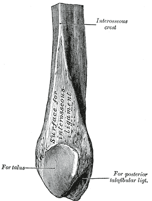
Caption = Lower extremity of right fibula. Medial aspect.
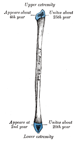
Caption2 = Plan of ossification of the fibula. From three centers.
System =
MeshName =
MeshNumber =
DorlandsPre = c_56
DorlandsSuf = 12260483
The body offibula presents four borders - the antero-lateral, the antero-medial, the postero-lateral, and the postero-medial; and four surfaces - anterior, posterior, medial, and lateral.Borders
The antero-lateral border begins above in front of the head, runs vertically downward to a little below the middle of the bone, and then curving somewhat lateralward, bifurcates so as to embrace a triangular subcutaneous surface immediately above the lateral malleolus. This border gives attachment to an intermuscular septum, which separates the Extensor muscles on the anterior surface of the leg from the Peronæi longus and brevis on the lateral surface.
The antero-medial border, or interosseous crest, is situated close to the medial side of the preceding, and runs nearly parallel with it in the upper third of its extent, but diverges from it in the lower two-thirds. It begins above just beneath the head of the bone (sometimes it is quite indistinct for about 2.5 cm. below the head), and ends at the apex of a rough triangular surface immediately above the articular facet of the lateral malleolus. It serves for the attachment of the
interosseous membrane , which separates the Extensor muscles in front from the Flexor muscles behind.The postero-lateral border is prominent; it begins above at the apex, and ends below in the posterior border of the lateral malleolus. It is directed lateralward above, backward in the middle of its course, backward, and a little medialward below, and gives attachment to an aponeurosis which separates the Peronæi on the lateral surface from the Flexor muscles on the posterior surface.
The postero-medial border, sometimes called the oblique line, begins above at the medial side of the head, and ends by becoming continuous with the interosseous crest at the lower fourth of the bone. It is well-marked and prominent at the upper and middle parts of the bone. It gives attachment to an aponeurosis which separates the Tibialis posterior from the Soleus and Flexor hallucis longus.
Surfaces
The anterior surface is the interval between the antero-lateral and antero-medial borders. It is extremely narrow and flat in the upper third of its extent; broader and grooved longitudinally in its lower third; it serves for the origin of three muscles: the
Extensor digitorum longus ,Extensor hallucis longus , andPeronæus tertius .The posterior surface is the space included between the postero-lateral and the postero-medial borders; it is continuous below with the triangular area above the articular surface of the lateral malleolus; it is directed backward above, backward and medialward at its middle, directly medialward below. Its upper third is rough, for the origin of the Soleus; its lower part presents a triangular surface, connected to the tibia by a strong interosseous ligament; the intervening part of the surface is covered by the fibers of origin of the Flexor hallucis longus. Near the middle of this surface is the nutrient foramen, which is directed downward.
The medial surface is the interval included between the antero-medial and the postero-medial borders. It is grooved for the origin of the Tibialis posterior.The lateral surface is the space between the antero-lateral and postero-lateral borders. It is broad, and often deeply grooved; it is directed lateralward in the upper two-thirds of its course, backward in the lower third, where it is continuous with the posterior border of the lateral malleolus. This surface gives origin to the Peronæi longus and brevis.
Wikimedia Foundation. 2010.