- Lateral malleolus
Infobox Bone
Name = Lateral malleolus
Latin = malleolus lateralis
GraySubject = 62
GrayPage = 260

Caption = Lateral aspect of right leg. (Lateral malleolus labeled at lower left.)
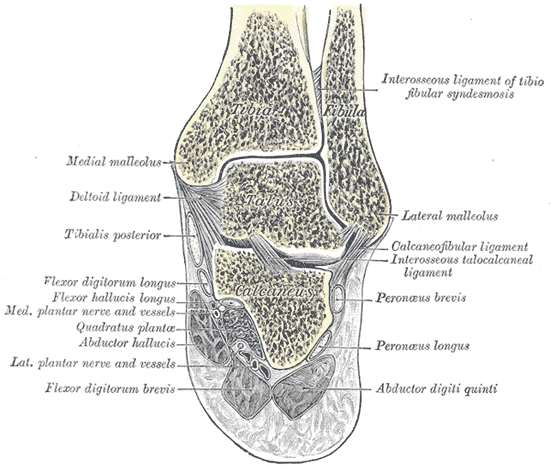
Caption2 = Coronal section through right talocrural and talocalcaneal joints. (Lateral malleolus labeled at center right.)
System =
MeshName =
MeshNumber =
DorlandsPre = m_02
DorlandsSuf = 12511177The lower extremity (distal extremity; external malleolus) of the
fibula is of a pyramidal form, and somewhat flattened from side to side; it descends to a lower level than themedial malleolus .The lateral surface is convex, subcutaneous, and continuous with the triangular, subcutaneous surface on the lateral side of the body.
The medial surface presents in front a smooth triangular surface, convex from above downward, which articulates with a corresponding surface on the lateral side of the talus. Behind and beneath the articular surface is a rough depression, which gives attachment to the
posterior talofibular ligament .The anterior border is thick and rough, and marked below by a depression for the attachment of the
anterior talofibular ligament .The posterior border is broad and presents the
shallow malleolar sulcus , for the passage of the tendons of thePeronæi longus and brevis.The summit is rounded, and gives attachment to the
calcaneofibular ligament .Often when a patient fractures their ankle, this is the bone that suffers a chip or other significant damage.
=Additionalee also
*
Ankle
*Medial malleolus
*Posterior malleolus
*Trimalleolar fracture External links
* [http://academic.wsc.edu/faculty/jatodd1/351/tibia_fibula.jpgDiagram at wsc.edu]
* [http://footandankle.mdmercy.com/conditions/ankle_injury/instability.html Diagnostic imaging]
Wikimedia Foundation. 2010.