Pulmonary consolidation

Pulmonary consolidation
Classification and external resources

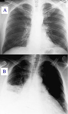
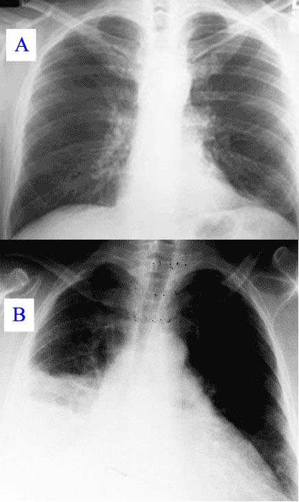
Pneumonia as seen on chest x-ray. A: Normal chest x-ray. B: Abnormal chest x-ray with consolidation from pneumonia in the right lung, middle or inferior lobe (white area, left side of image).
DiseasesDB 10949

Consolidation is a clinical term for solidification into a firm, dense mass. It is more specifically used in reference to a region of lung tissue that, normally compressible, has filled with liquid,[1] a condition marked by induration[2] (swelling or hardening of normally soft tissue) of a normally aerated lung. Consolidation occurs through accumulation of inflammatory cellular exudate in the alveoli and adjoining ducts. Simply, it is defined as alveolar space that contains liquid instead of gas. The fluid can be pulmonary edema, inflammatory exudate, pus, inhaled water, or blood (from bronchial tree or haemorrhage from a pulmonary artery). It is clinically important in pneumonia: the signs of lobar pneumonia are characteristic and clinically referred to as consolidation.[3]

Signs

Signs that consolidation may have occurred include:

  • Expansion of the thorax on inspiration is reduced on the affected side
  • Vocal fremitus is increased on the side with consolidation
  • Percussion is dull in affected area
  • Breath sounds are bronchial
  • Possible medium, late, or pan-inspiratory crackles
  • Vocal resonance is increased. Vocal resonance testing can be done with a stethoscope. Here, the patient's voice (or whisper, as in whispered pectoriloquy) can be heard more clearly when there is consolidation, as opposed to in the healthy lung where speech sounds muffled.
  • A pleural rub may be present[4]

Radiology

  • Typically, an area of white lung is seen on a standard X-ray. [1] Consolidated tissue is radio-opaque, so that it is clearly demonstrable in X-rays and CT (computerized tomography) scans. Consolidation is often a middle-to-late stage feature/complication in pulmonary infections.

References

  1. ^ Chest X-Ray Made Easy. Churchill Livingstone. 2002. ISBN 0443070083. 
  1. ^ "Consolidation - Definition". Merriam-Webster. http://www2.merriam-webster.com/cgi-bin/mwmednlm?book=Medical&va=consolidation. Retrieved 2009-01-16. 
  2. ^ "Induration- Definition". Merriam-Webster. http://www2.merriam-webster.com/cgi-bin/mwmednlm?book=Medical&va=induration. Retrieved 2009-01-16. 
  3. ^ Metlay, JP, Kapoor, WN, Fine, MJ. Does this patient have community-acquired pneumonia? Diagnosing pneumonia by history and physical examination. JAMA 1997; 278:1440. doi:10.1001/jama.278.17.1440 PMID 9356004
  4. ^ Talley and O'Connor (2001). Clinical Examination, a Clinical Guide to Physical Diagnosis. Elsevier 4:121

Wikimedia Foundation. 2010.

Игры ⚽ Поможем сделать НИР

Look at other dictionaries:

  • Consolidation — may refer to: Consolidation (business), the mergers or acquisitions of many smaller companies into much larger ones Consolidation (soil), a geological process whereby a soil decreases in volume Consolidation (media), consolidation of United… …   Wikipedia

  • Pulmonary contusion — Classification and external resources A CT scan showing a pulmonary contusion (red arrow) accompanied by a rib fracture (blue arrow) ICD 10 S …   Wikipedia

  • Consolidation (medicine) — Pneumonia as seen on chest x ray. A : Normal chest x ray. B : Abnormal chest x ray with consolidation from pneumonia in the right lung, middle or inferior lobe (white area, left side of image).Consolidation is a clinical term for solidification… …   Wikipedia

  • consolidation — Solidification into a firm dense mass; applied especially to inflammatory induration of a normally aerated lung due to the presence of cellular exudate in the pulmonary alveoli as commonly seen in pneumonia. [L. consolido, to make thick, condense …   Medical dictionary

  • Pneumonia — For other uses, see Pneumonia (disambiguation). Pneumonia Classification and external resources A chest X ray showing a very prominent wedge shaped bacterial pneumonia in the right lung …   Wikipedia

  • Sign — Any abnormality that indicates a disease process, such as a change in appearance, sensation, or function, that is observed by a physician when evaluating a patient. * * * 1. Any abnormality indicative of disease, discoverable on examination of… …   Medical dictionary

  • Atelectasis — DiseaseDisorder infobox Name = Atelectasis ICD10 = ICD10|J|98|1|j|95 ICD9 = ICD9|518.0 ICDO = Caption = OMIM = OMIM mult = MedlinePlus = 000065 eMedicineSubj = med eMedicineTopic = 180 DiseasesDB = 10940 MeshID = D001261 Atelectasis is a collapse …   Wikipedia

  • bronchopneumonia — Acute inflammation of the walls of the smaller bronchial tubes, with varying amounts of pulmonary consolidation due to spread of the inflammation into peribronchiolar alveoli and the alveolar ducts; may become confluent or may be hemorrhagic …   Medical dictionary

  • pneumocystis pneumonia — (PCP) a pulmonary disease caused by Pneumocystis jiroveci, occurring in premature or malnourished infants and immunocompromised persons. It is characterized by dyspnea, tachypnea, fever, cough, and cyanosis, with cellular detritus containing… …   Medical dictionary

  • Plague — The plague is an infectious disease due to a bacteria called Yersinia pestis. Y. pestis mainly infects rats and other rodents. Rodents are the prime reservoir for the bacteria. Fleas function as the prime vectors carrying the bacteria from one… …   Medical dictionary

Share the article and excerpts

Direct link
Do a right-click on the link above
and select “Copy Link”