- Myocardial perfusion imaging
-
Myocardial perfusion imaging Intervention 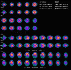
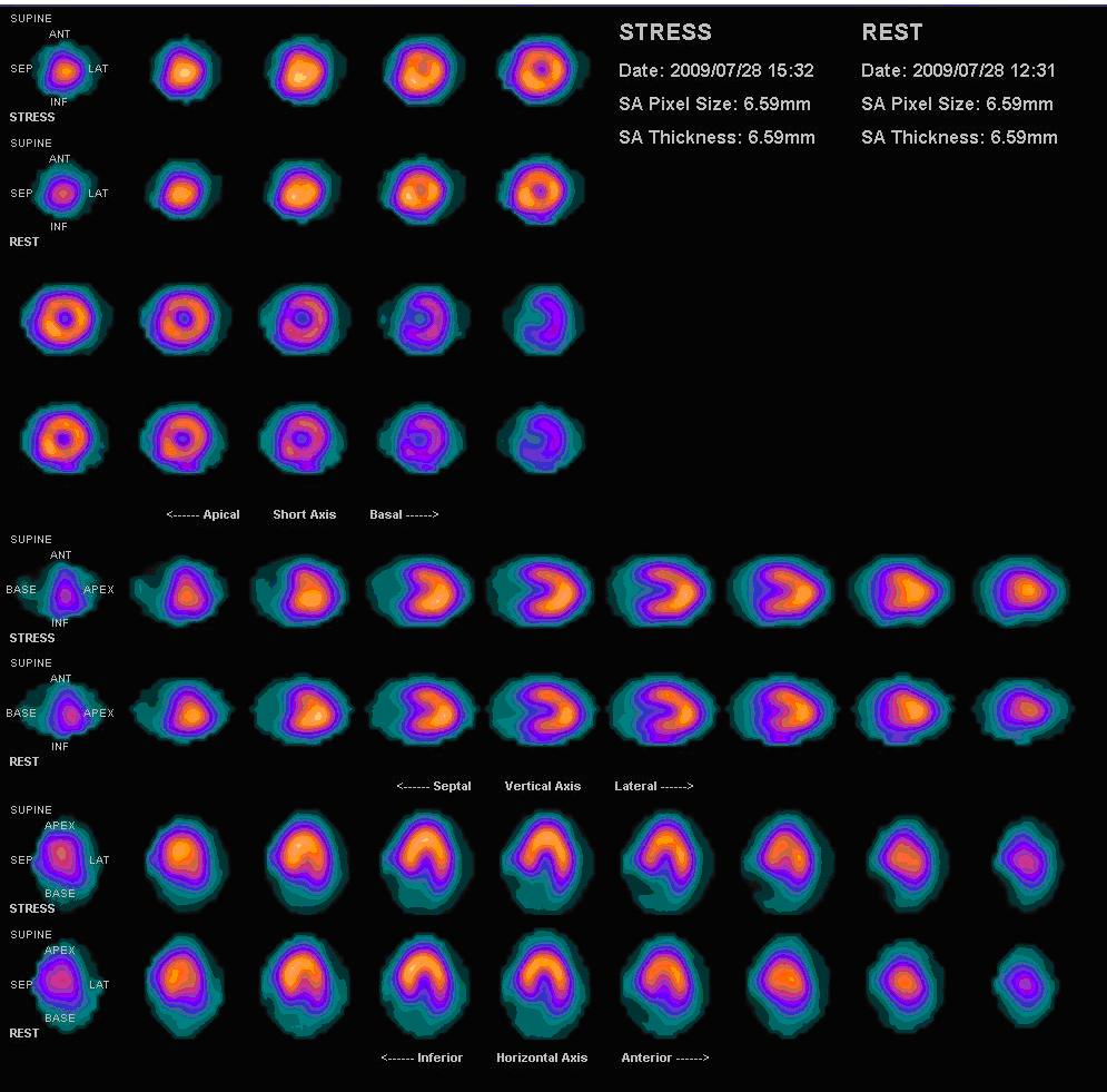
Nuclear medicine myocardial perfusion scan with Thallium-201 for the rest images (bottom rows) and Tc-Sestamibi for the stress images (top rows). The nuclear medicine myocardial perfusion scan plays a pivotal role in the noninvasive evaluation of coronary artery disease. The study not only identifies patients with coronary artery disease, it also provides overall prognostic information or overall risk of adverse cardiac events for the patient.ICD-10-PCS C22G MeSH D055414 OPS-301 code: 3-704, 3-721 Myocardial perfusion scan is a nuclear medicine procedure that illustrates the function of the heart muscle (myocardium).[1]
It evaluates many heart conditions from coronary artery disease (CAD) to hypertrophic cardiomyopathy and myocardial wall motion abnormalities. The function of the myocardium is also evaluated by calculating the left ventricular ejection fraction (LVEF) of the heart. This scan is done in conjunction with a cardiac stress test.
Planar techniques, such as conventional scintigraphy, are rarely used. Rather, SPECT is more common in the US. With multihead SPECT systems, imaging can often be completed in less than 10 minutes. With SPECT, inferior and posterior abnormalities and small areas of infarction can be identified, as well as the occluded blood vessels and the mass of infarcted and viable myocardium.[2]
Major indications for a myocardial perfusion test
- Diagnosis of CAD and various cardiac abnormalities.
- Identifying location, criticality of existing coronary stenosis and degree of coronary artery disease (CAD) in patients with a history of CAD.
- Prognostication (risk stratification) and evaluation of patients that are at risk of having a myocardial or coronary incident. (ex: myocardial infarction, myocardial ischemia, coronary aneurysm, wall motion abnormalities)
- Assessment of viable myocardium in particular coronary artery territory following heart attacks to justify revascularization
- Post intervention revascularization (coronary artery bypass graft, angioplasty) evaluation of heart.
Risks vs. benefits
Critics have written that myocardial perfusion imaging is associated with an increased risk of cancer due to high radiation doses that are not justified by randomized, controlled studies demonstrating benefit[citation needed]. However, radiation doses received during CT angiography and conventional coronary angiography are higher than those received during myocardial perfusion imaging done with 99m-Technetium labelled agents[citation needed]. The psychological block associated with radioactive materials may be responsible for these fears.
In a study of patient exposure to low-dose ionizing radiation, myocardial perfusion imaging had the highest average effective dose (15.6 millisieverts) and the highest percentage (22.1%) of all effective doses to the entire patient population from all major radiological procedures, including computer tomographic studies. Older patients, 60 to 64 years old, had the highest doses, with 5.27% getting a high dose (>20 to 50 mSv/year) and 0.57% getting a very high dose (>50 mSv/year) from all sources.[3]
Experimental and epidemiologic evidence has linked exposure to low-dose ionizing radiation with up to 2% of solid cancers and leukemia. Workers are monitored and limited to 100mSv every 5 years, but medical patients are not typically monitored. In one study of CT in abdominal and flank pain, "less than 50% of radiologists and only 9% of emergency department physicians reported even being aware that CT was associated with an increased risk of cancer."[3][4]
Interestingly, the concern over radiation hazard has undermined the risk associated with the allergic potential of radiocontrast (dyes) used in CT angiography and coronary angiography. In myocardial perfusion imaging, radioisotopes are used in nanomole quantities, practically devoid of any risk of allergy with normal saline being used as the vehicle and no known adverse reaction to the chemical molecules (sestamibi or tetrofosmine)[citation needed].
From 1993-2001, myocardial perfusion scans increased >6%/y with "no justification," according to a commentary by Lauer. Mycardial perfusion imaging scans are "powerful predictors of future clinical events," and in theory may identify patients for whom aggressive therapies should improve outcome. But this is "only a hypothesis, not a proof," wrote Lauer. There are no randomized, controlled trials to demonstrate any benefits, and there is a small but cumulative danger from radiation.[5]
However, radioisotope MPI is considered to be the most comprehensive test[citation needed], providing information about criticality of coronary stenosis, area suffering from ischemia, severity of ischemia, total mass of viable myocardium, and the pump function of the heart, along with objective parameters such as end systolic volume, end diastolic volume, stroke volume and ejection fraction. The negative predictive value of the test is as high as 98%[citation needed], offering excellent prognostic value.
New radionuclides such as rubidium-82 reduce the radiation dose to the patient by a factor of 10 compared to technetium-99m. In the future, therefore, a complete myocardial perfusion exam may be achievable while maintaining a patient dose under 3 mSv.[6][7]
References
- ^ MeSH Myocardial+Perfusion+Imaging
- ^ Merck manuals > Radionuclide Imaging Last full review/revision May 2009 by Michael J. Shea, MD. Content last modified May 2009
- ^ a b Exposure to low-dose ionizing radiation from medical imaging procedures, Reza Fazel et al. N Engl J Med, 27 Aug 2009, 361(9):849.
- ^ Diagnostic CT scans: assessment of patient, physician, and radiologist awareness of radiation dose and possible risks, Lee CI, et al. Radiology 2004;231:393-8.
- ^ Perspective: Elements of danger -- the case of medical imaging, Michael S. Lauer, N Engl J Med, 27 Aug 2009, 361(9):841.
- ^ A revised effective dose estimate for the PET perfusion tracer Rb-82, deKemp et al, J NUCL MED MEETING ABSTRACTS, 2008. 49(MeetingAbstracts_1): p. 183P-b-.
- ^ Radiopharmaceuticals for nuclear cardiology: radiation dosimetry, uncertainties, and risk., Stabin et al, J Nucl Med, 2008. 49(9): p. 1555-63.
Medical testing : Medical imaging · Radiology · (ICD-9-CM V3 87-88, ICD-10-PCS B, CPT 70010-79999) X-ray/
medical radiography/
Industrial radiographyMedical: Pneumoencephalography · Dental radiography · Sialography · Myelography · CXR (Bronchography) · AXR / KUB · DXA/DXR · Upper gastrointestinal series/Small bowel follow-through/Lower gastrointestinal series · Cholangiography/Cholecystography · Mammography · Pyelogram · Cystography · Arthrogram · Hysterosalpingography · Skeletal survey · Angiography (Angiocardiography, Aortography) · Venography · Lymphogram
Industrial: Radiographic testingMedical: CT pulmonary angiogram · Cardiac CT · Abdominal and pelvic CT (Virtual colonoscopy) · CT angiography · CT head · pQCT · Spiral computed tomography · High resolution CT · Whole body imaging (Full-body CT scan) · Electron beam tomography
Industrial: Industrial CT ScanningOtherMRI MRI of brain and brain stem · MR neurography · Cardiac MRI/Cardiac MRI perfusion · MR angiography · MR cholangiopancreatography · Breast MRI
Functional MRI · Diffusion MRIUltrasound Echocardiography / Doppler echocardiography (TTE · TEE) · Intravascular · Gynecologic · Obstetric · Echoencephalography · Transcranial doppler · Abdominal ultrasonography · Transrectal · Breast ultrasound · Transscrotal ultrasound · Carotid ultrasonography
Contrast-enhanced · 3D ultrasound · Endoscopic ultrasound · Emergency ultrasound (FAST) · DuplexRadionuclide 2D / scintigraphyCholescintigraphy · Scintimammography · Ventilation/perfusion scan · Radionuclide ventriculography · Radionuclide angiography · Radioisotope renography · Sestamibi parathyroid scintigraphy · Radioactive iodine uptake test · Bone scintigraphy · Immunoscintigraphy
full body: Octreotide scan · Gallium 67 scan · Indium 111 WBC scan3D / ECTOptical laser Thermography Breast thermographyCategories:- Cardiac imaging
- 3d nuclear medical imaging
Wikimedia Foundation. 2010.