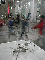
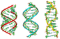
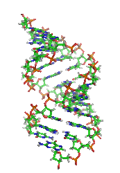
- Molecular models of DNA
-
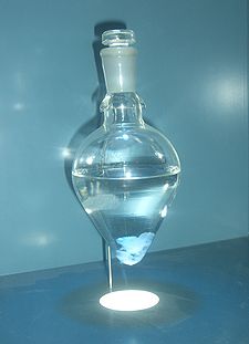
 While this purified DNA precipitated in a water jug (left) appears to be a formless mass, nucleic acids actually possess intricate structure at the nanoscale (right).
Molecular models of DNA structures are representations of the molecular geometry and topology of Deoxyribonucleic acid (DNA) molecules using one of several means, with the aim of simplifying and presenting the essential, physical and chemical, properties of DNA molecular structures either in vivo or in vitro. These representations include closely packed spheres (CPK models) made of plastic, metal wires for 'skeletal models', graphic computations and animations by computers, artistic rendering. Computer molecular models also allow animations and molecular dynamics simulations that are very important for understanding how DNA functions in vivo.
The more advanced, computer-based molecular models of DNA involve molecular dynamics simulations as well as quantum mechanical computations of vibro-rotations, delocalized molecular orbitals (MOs), electric dipole moments, hydrogen-bonding, and so on. DNA molecular dynamics modeling involves simulations of DNA molecular geometry and topology changes with time as a result of both intra- and inter- molecular interactions of DNA. Whereas molecular models of Deoxyribonucleic acid (DNA) molecules such as closely packed spheres (CPK models) made of plastic or metal wires for 'skeletal models' are useful representations of static DNA structures, their usefulness is very limited for representing complex DNA dynamics. Computer molecular modeling allows both animations and molecular dynamics simulations that are very important for understanding how DNA functions in vivo.
History
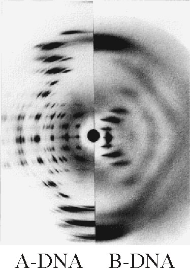
 The A-DNA double-helix molecular model of Crick and Watson (consistent with their X-ray data) for which they received a Nobel prize together with M.H.F. Wilkins.
Double Helix
Discovery
William Astbury Oswald Avery Francis Crick Erwin Chargaff Max Delbrück Jerry Donohue Rosalind Franklin Raymond Gosling Phoebus Levene Friedrich Miescher Linus Pauling Sir John Randall Erwin Schrödinger Alex Stokes James Watson Maurice Wilkins Herbert Wilson Further information: History of molecular biologyFrom the very early stages of structural studies of DNA by X-ray diffraction and biochemical means, molecular models such as the Watson-Crick double-helix model were successfully employed to solve the 'puzzle' of DNA structure, and also find how the latter relates to its key functions in living cells. The first high quality X-ray diffraction patterns of A-DNA were reported by Rosalind Franklin and Raymond Gosling in 1953.[1] The first calculations of the Fourier transform of an atomic helix were reported one year earlier by Cochran, Crick and Vand,[2] and were followed in 1953 by the computation of the Fourier transform of a coiled-coil by Crick.[3]
Structural information is generated from X-ray diffraction studies of oriented DNA fibers with the help of molecular models of DNA that are combined with crystallographic and mathematical analysis of the X-ray patterns.
The first reports of a double-helix molecular model of B-DNA structure were made by Watson and Crick in 1953.[4][5] Last-but-not-least, Maurice F. Wilkins, A. Stokes and H.R. Wilson, reported the first X-ray patterns of in vivo B-DNA in partially oriented salmon sperm heads.[6] The development of the first correct double-helix molecular model of DNA by Crick and Watson may not have been possible without the biochemical evidence for the nucleotide base-pairing ([A---T]; [C---G]), or Chargaff's rules.[7][8][9][10][11][12]Although such initial studies of DNA structures with the help of molecular models were essentially static, their consequences for explaining the in vivo functions of DNA were significant in the areas of protein biosynthesis and the quasi-universality of the genetic code. Epigenetic transformation studies of DNA in vivo were however much slower to develop in spite of their importance for embryology, morphogenesis and cancer research. Such chemical dynamics and biochemical reactions of DNA are much more complex than the molecular dynamics of DNA physical interactions with water, ions and proteins/enzymes in living cells.
Importance
Further information: Nucleic acid structureAn old standing dynamic problem is how DNA "self-replication" takes place in living cells that should involve transient uncoiling of supercoiled DNA fibers. Although DNA consists of relatively rigid, very large elongated biopolymer molecules called "fibers" or chains (that are made of repeating nucleotide units of four basic types, attached to deoxyribose and phosphate groups), its molecular structure in vivo undergoes dynamic configuration changes that involve dynamically attached water molecules and ions. Supercoiling, packing with histones in chromosome structures, and other such supramolecular aspects also involve in vivo DNA topology which is even more complex than DNA molecular geometry, thus turning molecular modeling of DNA into an especially challenging problem for both molecular biologists and biotechnologists. Like other large molecules and biopolymers, DNA often exists in multiple stable geometries (that is, it exhibits conformational isomerism) and configurational, quantum states which are close to each other in energy on the potential energy surface of the DNA molecule.
Such varying molecular geometries can also be computed, at least in principle, by employing ab initio quantum chemistry methods that can attain high accuracy for small molecules, although claims that acceptable accuracy can be also achieved for polynuclelotides, as well as DNA conformations, were recently made on the basis of VCD spectral data. Such quantum geometries define an important class of ab initio molecular models of DNA whose exploration has barely started especially in connection with results obtained by VCD in solutions. More detailed comparisons with such ab initio quantum computations are in principle obtainable through 2D-FT NMR spectroscopy and relaxation studies of polynucleotide solutions or specifically labeled DNA, as for example with deuterium labels.
In an interesting twist of roles, the DNA molecule itself was proposed to be utilized for quantum computing. Both DNA nanostructures as well as DNA 'computing' biochips have been built.
Fundamental concepts
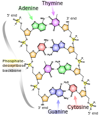
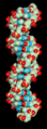
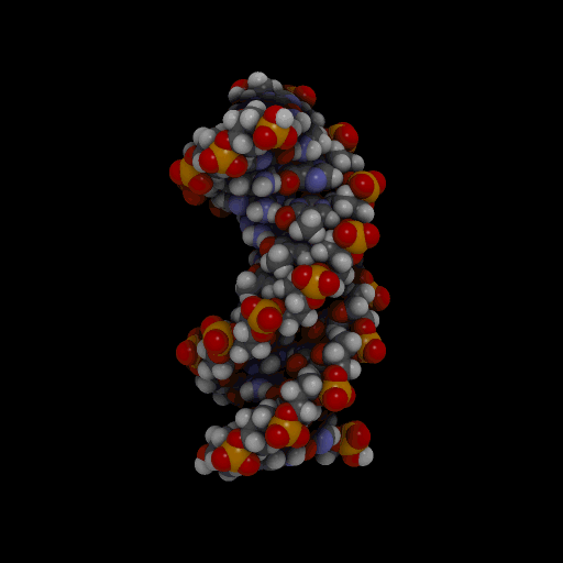
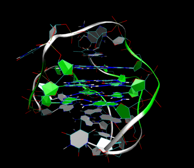
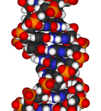 At left, the chemical structure of DNA showing the base-pairing. This depiction of a DNA duplex lacks information about the molecule's three-dimensional structure, at right.
The chemical structure of DNA is insufficient to understand the complexity of the 3D structures of DNA. On the other hand, animated molecular models allow one to visually explore the three-dimensional (3D) structure of DNA. The DNA model shown (far right) is a space-filling, or CPK, model of the DNA double-helix. Animated molecular models, such as the wire, or skeletal, type shown at the top of this article, allow one to visually explore the three-dimensional (3D) structure of DNA. Another type of DNA model is the space-filling, or CPK, model.
The hydrogen bonding dynamics and proton exchange is very different by many orders of magnitude between the two systems of fully hydrated DNA and water molecules in ice. Thus, the DNA dynamics is complex, involving nanosecond and several tens of picosecond time scales, whereas that of liquid ice is on the picosecond time scale, and that of proton exchange in ice is on the millisecond time scale. The proton exchange rates in DNA and attached proteins may vary from picosecond to nanosecond, minutes or years, depending on the exact locations of the exchanged protons in the large biopolymers.
A simple harmonic oscillator 'vibration' is only an oversimplified dynamic representation of the longitudinal vibrations of the DNA intertwined helices which were found to be anharmonic rather than harmonic as often assumed in quantum dynamic simulations of DNA.
DNA structure
The structure of DNA shows a variety of forms, both double-stranded and single-stranded. The mechanical properties of DNA, which are directly related to its structure, are a significant problem for cells. Every process which binds or reads DNA is able to use or modify the mechanical properties of DNA for purposes of recognition, packaging and modification. The extreme length (a chromosome may contain a 10 cm long DNA strand), relative rigidity and helical structure of DNA has led to the evolution of histones and of enzymes such as topoisomerases and helicases to manage a cell's DNA. The properties of DNA are closely related to its molecular structure and sequence, particularly the weakness of the hydrogen bonds and electronic interactions that hold strands of DNA together compared to the strength of the bonds within each strand.
Experimental techniques which can directly measure the mechanical properties of DNA are relatively new, and high-resolution visualization in solution is often difficult. Nevertheless, scientists have uncovered large amount of data on the mechanical properties of this polymer, and the implications of DNA's mechanical properties on cellular processes is a topic of active current research.
The DNA found in many cells can be macroscopic in length - a few centimetres long for each human chromosome. Consequently, cells must compact or "package" DNA to carry it within them. In eukaryotes this is carried by spool-like proteins known as histones, around which DNA winds. It is the further compaction of this DNA-protein complex which produces the well known mitotic eukaryotic chromosomes.
Alternate non-double-helical models have been proposed for the in vivo structure of DNA, such as in plasmids and chromatin.[13][14] Such "side-by-side" models of DNA were proposed early in the history of molecular biology, but these were set aside in favor of the double-helical model due to X-ray crystallography of DNA duplexes and later the nucleosome core particle, as well as the discovery of topoisomerases, and these non-double-helical models are not currently accepted by the mainstream scientific community.[15]
DNA structure determination using molecular modeling and DNA X-ray patterns
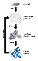
 Left, the major steps involved in DNA structure determination by X-ray crystallography showing the important role played by molecular models of DNA structure in this iterative process. Right, an image of actual A- and B- DNA X-ray patterns obtained from oriented and hydrated DNA fibers (courtesy of Dr. Herbert R. Wilson, FRS- see refs. list).Main article: Nucleic acid structure determination
After DNA has been separated and purified by standard biochemical techniques one has a sample in a jar much like in the figure at the top of this article. Below are the main steps involved in generating structural information from X-ray diffraction studies of oriented DNA fibers that are drawn from the hydrated DNA sample with the help of molecular models of DNA that are combined with crystallographic and mathematical analysis of the X-ray patterns.
Paracrystalline lattice models of B-DNA structures
 Silica glass is another example of a material which is organized into a paracrystalline lattice.
A paracrystalline lattice, or paracrystal, is a molecular or atomic lattice with significant amounts (e.g., larger than a few percent) of partial disordering of molecular arrangements. Limiting cases of the paracrystal model are nanostructures, such as glasses, liquids, etc., that may possess only local ordering and no global order. A simple example of a paracrystalline lattice is shown in the following figure for a silica glass:
Liquid crystals also have paracrystalline rather than crystalline structures.
Highly hydrated B-DNA occurs naturally in living cells in such a paracrystalline state, which is a dynamic one in spite of the relatively rigid DNA double-helix stabilized by parallel hydrogen bonds between the nucleotide base-pairs in the two complementary, helical DNA chains (see figures). For simplicity most DNA molecular models omit both water and ions dynamically bound to B-DNA, and are thus less useful for understanding the dynamic behaviors of B-DNA in vivo. The physical and mathematical analysis of X-ray[16][17] and spectroscopic data for paracrystalline B-DNA is therefore much more complicated than that of crystalline, A-DNA X-ray diffraction patterns. The paracrystal model is also important for DNA technological applications such as DNA nanotechnology. Novel techniques that combine X-ray diffraction of DNA with X-ray microscopy in hydrated living cells are now also being developed.[18]
Genomic and biotechnology applications of DNA molecular modeling
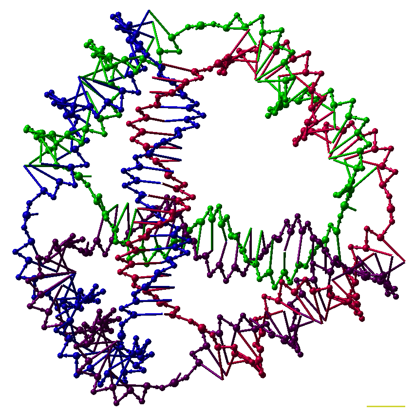
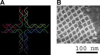 Molecular models are useful in the design of structures for DNA nanotechnology. Here, individual DNA tiles (model at left) self-assemble into a highly ordered DNA 2D-nanogrid (AFM image at right).
There are various uses of DNA molecular modeling in Genomics and Biotechnology research applications, from DNA repair to PCR and DNA nanostructures. Two-dimensional DNA junction arrays have been visualized by Atomic force microscopy.[19]
DNA molecular modeling has various uses in genomics and biotechnology, with research applications ranging from DNA repair to PCR and DNA nanostructures. These include computer molecular models of molecules as varied as RNA polymerase, an E. coli, bacterial DNA primase template suggesting very complex dynamics at the interfaces between the enzymes and the DNA template, and molecular models of the mutagenic, chemical interaction of potent carcinogen molecules with DNA. These are all represented in the gallery below.
Technological application include a DNA biochip and DNA nanostructures designed for DNA computing and other dynamic applications of DNA nanotechnology.[20][21][22][23][24][25] The image at right is of self-assembled DNA nanostructures. The DNA “tile” structure in this image consists of four branched junctions oriented at 90° angles. Each tile consists of nine DNA oligonucleotides as shown; such tiles serve as the primary “building block” for the assembly of the DNA nanogrids shown in the AFM micrograph.
Quadruplex DNA may be involved in certain cancers.[26][27] Images of quadruplex DNA are in the gallery below.
Gallery of DNA models
-
Spinning DNA generic model.]]
-

An oversimplified sketch of the double-helix structure of A-DNA.
-

A model of DNA replication based on the double-helix concept.
-

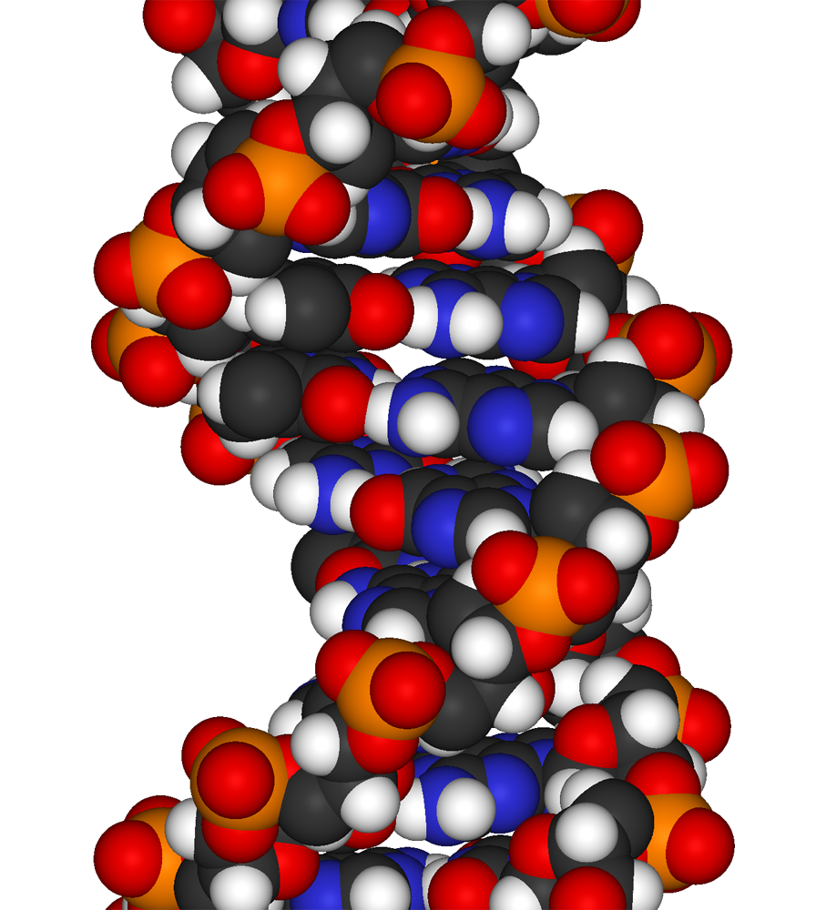
Animated, space-filling moelcular model of the A-DNA double helix
-
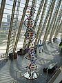
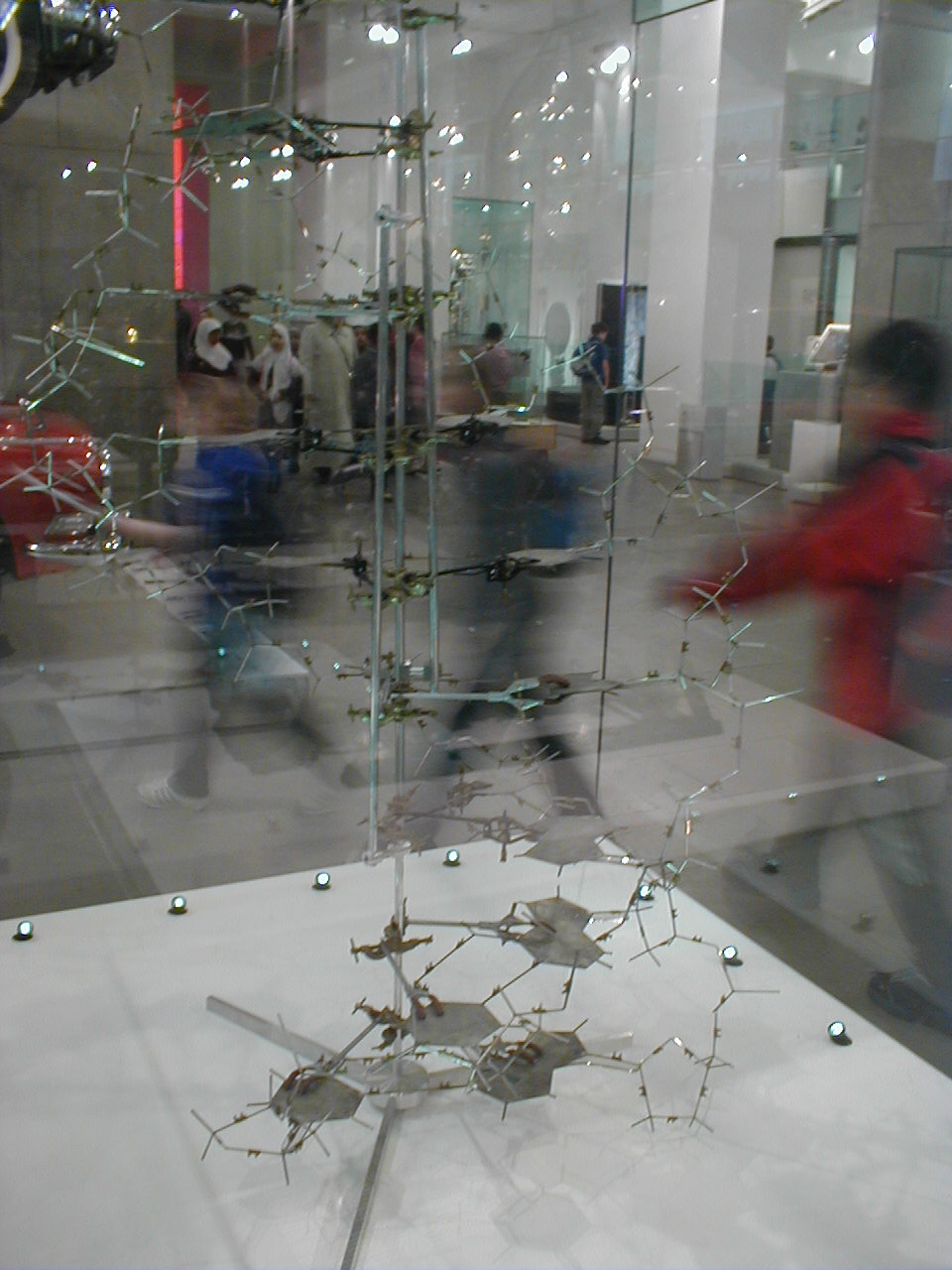
A large-scale Crick-Watson DNA Model shown in the Museum of Príncipe Felipe.
-
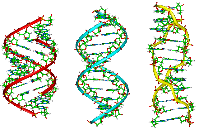
Side view of molecular models of A- , B-, Z- DNA.
-

Oversimplified model of the A-DNA double-helix.
-
Molecular modeling of RNA Polymerase.
-

Molecular modeling of a bacterial DNA Primase Template.
-
Molecular modeling of DNA interactions with the carcinogen molecule MGMT.
-
3D Molecular model of DNA damaged by carcinogenic 2-aminofluorene(AF).
-

Fig.6. Molecular modeling of DNA repair
-

Animated skeletal model of A-DNA.
-

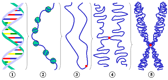
Simplified models of chromatin.
-

Simplified model of chromosome structure.
-
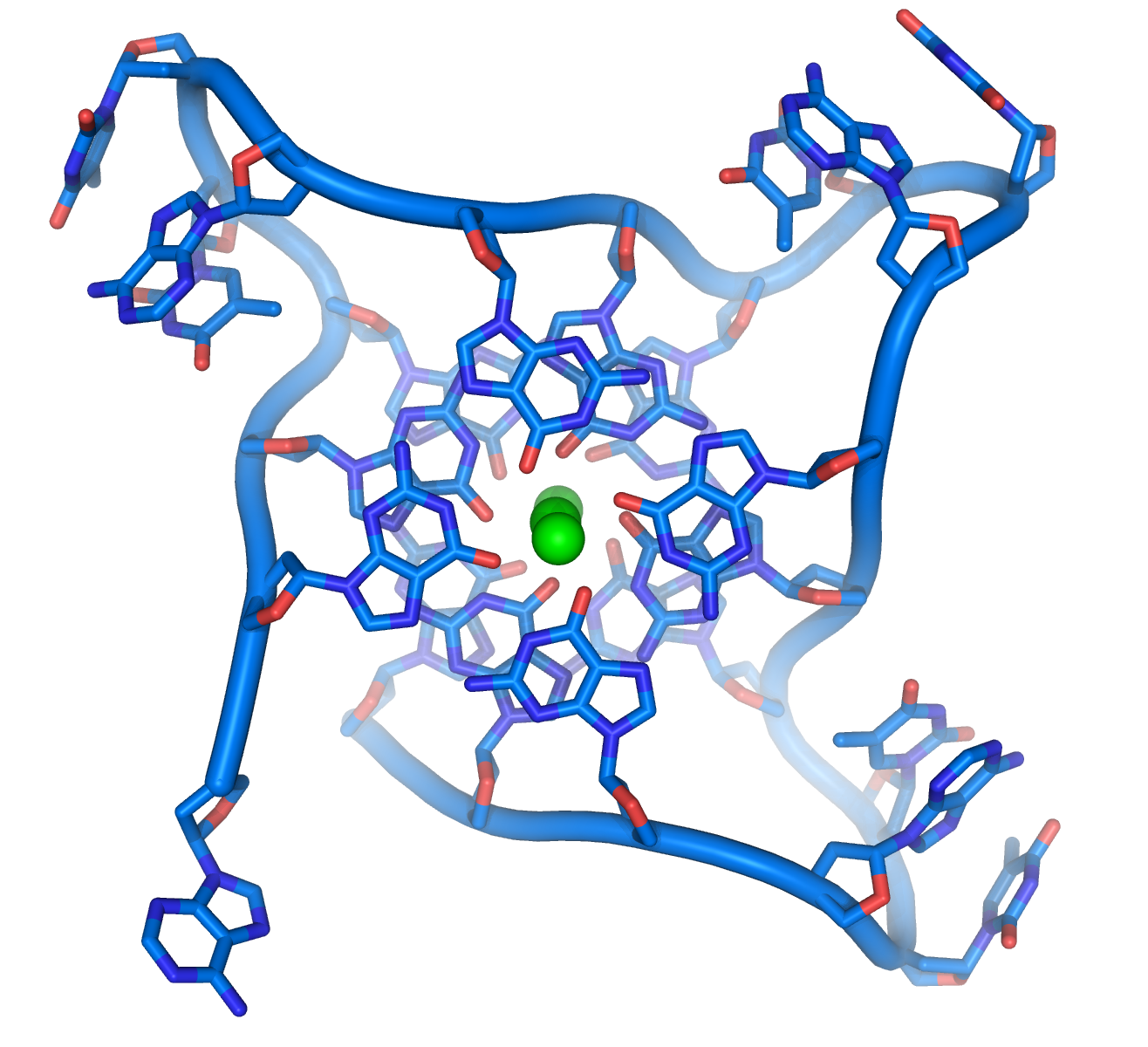
A hypothetical quadruplex of guanine-rich DNA structures that may be involved in cancers.
-
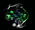
3D Molecular Structure of the intramolecular human telomeric G-quadruplex in potassium solution.
-

DNA spacefilling molecular model
-

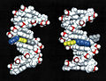
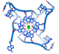
A model of a designed DNA tetrahedron.
See also
- G-quadruplex
- Crystallography
- Crystal lattices
- DiProDB Dinucleotide Property Database. The database is designed to collect and analyse thermodynamic, structural and other dinucleotide properties.
- X-ray microscopy
- X-ray scattering
- Neutron scattering
- Vibrational circular dichroism (VCD)
- Raman spectroscopy/microscopy and CARS
- Sir Lawrence Bragg, FRS
- List of nucleic acid simulation software
- AMBER
- CHARMM
- Abalone (molecular mechanics)
- Sirius visualization software
- QMC@Home
- FT-NMR
- NMR microscopy
- Microwave spectroscopy
- FT-IR
- FT-NIR
- Spectral, Hyperspectral, and Chemical imaging
- Fluorescence correlation spectroscopy
- Fluorescence cross-correlation spectroscopy and FRET
- Confocal microscopy
References
- ^ Franklin, R.E., Gosling, R.G. (6 March 1953). "The Structure of Sodium Thymonucleate Fibres I. The Influence of Water Content". Acta Cryst. 6 (8): 673. doi:10.1107/S0365110X53001939.
Franklin, R.E., Gosling, R.G. (6 March 1953). "The Structure of Sodium Thymonucleate Fibres II. The Cylindrically Symmetrical Patterson Function". Acta Cryst. 6 (8): 678. doi:10.1107/S0365110X53001940. - ^ Cochran, W., Crick, F.H.C., Vand V. (1952). "The Structure of Synthetic Polypeptides. 1. The Transform of Atoms on a Helix". Acta Cryst. 5 (5): 581–6. doi:10.1107/S0365110X52001635.
- ^ Crick, F.H.C. (1953a). "The Fourier Transform of a Coiled-Coil". Acta Cryst. 6 (8–9): 685–9. doi:10.1107/S0365110X53001952.
- ^ Watson, James D., Crick, Francis H.C. (25 April 1953). "A structure for Deoxyribose Nucleic Acid" (PDF). Nature 171 (4356): 737–8. Bibcode 1953Natur.171..737W. doi:10.1038/171737a0. PMID 13054692. http://www.nature.com/nature/dna50/watsoncrick.pdf., .
- ^ Watson, J.D; Crick F.H.C. (1953b). "The Structure of DNA". Cold Spring Harbor Symposia on Quantitative Biology 18: 123–31. PMID 13168976.
- ^ Wilkins M.H.F., A.R. Stokes A.R. & Wilson, H.R. (1953). "Molecular Structure of Deoxypentose Nucleic Acids" (PDF). Nature 171 (4356): 738–40. Bibcode 1953Natur.171..738W. doi:10.1038/171738a0. PMID 13054693. http://www.nature.com/nature/dna50/wilkins.pdf.
- ^ Elson D, Chargaff E (1952). "On the deoxyribonucleic acid content of sea urchin gametes". Experientia 8 (4): 143–5. doi:10.1007/BF02170221. PMID 14945441.
- ^ Chargaff E, Lipshitz R, Green C (1952). "Composition of the deoxypentose nucleic acids of four genera of sea-urchin". J Biol Chem 195 (1): 155–160. PMID 14938364.
- ^ Chargaff E, Lipshitz R, Green C, Hodes ME (1951). "The composition of the deoxyribonucleic acid of salmon sperm". J Biol Chem 192 (1): 223–230. PMID 14917668.
- ^ Chargaff E (1951). "Some recent studies on the composition and structure of nucleic acids". J Cell Physiol Suppl 38 (Suppl).
- ^ Magasanik B, Vischer E, Doniger R, Elson D, Chargaff E (1950). "The separation and estimation of ribonucleotides in minute quantities". J Biol Chem 186 (1): 37–50. PMID 14778802.
- ^ Chargaff E (1950). "Chemical specificity of nucleic acids and mechanism of their enzymatic degradation". Experientia 6 (6): 201–9. doi:10.1007/BF02173653. PMID 15421335.
- ^ Wu, Richard; Te Wu, Tai (1996). "A novel intact circular dsDNA supercoil". Bulletin of Mathematical Biology 58 (6): 1171–85. doi:10.1007/BF02458388. PMID 8953261.
- ^ Delmonte, Clive (2009). "Reflections on the Secondary Structure of DNA and Other Biopolymers". Journal of Chemistry, Biochemistry, and Molecular Biology (Scientific Journals International) 1 (1). http://www.scientificjournals.org/journals2009/j_of_chemistry1_2009.htm. Retrieved 1 October 2011.
- ^ Gautham, N. (25 May 2004). "Response to "Variety in DNA secondary structure"". Current Science 86 (10): 1352–1353. http://www.ias.ac.in/currsci/may252004/1352.pdf. Retrieved 22 February 2011. "However, the discovery of topoisomerases took "the sting" out of the topological objection to the plectonaemic double helix. The more recent solution of the single crystal X-ray structure of the nucleosome core particle showed nearly 150 base pairs of the DNA (i.e. about 15 complete turns), with a structure that is in all essential respects the same as the Watson–Crick model. This dealt a death blow to the idea that other forms of DNA, particularly double helical DNA, exist as anything other than local or transient structures."
- ^ Hosemann R., Bagchi R.N., Direct analysis of diffraction by matter, North-Holland Publs., Amsterdam – New York, 1962.
- ^ Baianu, I.C. (1978). "X-ray scattering by partially disordered membrane systems". Acta Cryst. A34 (5): 751–3. doi:10.1107/S0567739478001540.
- ^ Yamamoto Y, Shinohara K (October 2002). "Application of X-ray microscopy in analysis of living hydrated cells". Anat. Rec. 269 (5): 217–23. doi:10.1002/ar.10166. PMID 12379938.
- ^ Mao, Chengde; Sun, Weiqiong & Seeman, Nadrian C. (16 June 1999). "Designed Two-Dimensional DNA Holliday Junction Arrays Visualized by Atomic Force Microscopy". Journal of the American Chemical Society 121 (23): 5437–43. doi:10.1021/ja9900398.
- ^ Robinson, Bruche H.; Seeman, Nadrian C. (August 1987). "The Design of a Biochip: A Self-Assembling Molecular-Scale Memory Device". Protein Engineering 1 (4): 295–300. doi:10.1093/protein/1.4.295. ISSN 0269-2139. PMID 3508280. Link
- ^ Rothemund, Paul W. K.; Ekani-Nkodo, Axel; Papadakis, Nick; Kumar, Ashish; Fygenson, Deborah Kuchnir & Winfree, Erik (22 December 2004). "Design and Characterization of Programmable DNA Nanotubes". Journal of the American Chemical Society 126 (50): 16344–52. doi:10.1021/ja044319l. PMID 15600335.
- ^ Keren, K.; Kinneret Keren, Rotem S. Berman, Evgeny Buchstab, Uri Sivan, Erez Braun (November 2003). "DNA-Templated Carbon Nanotube Field-Effect Transistor". Science 302 (6549): 1380–2. doi:10.1126/science.1091022. PMID 14631035. http://www.sciencemag.org/cgi/content/abstract/sci;302/5649/1380.
- ^ Zheng, Jiwen; Constantinou, Pamela E.; Micheel, Christine; Alivisatos, A. Paul; Kiehl, Richard A. & Seeman Nadrian C. (2006). "2D Nanoparticle Arrays Show the Organizational Power of Robust DNA Motifs". Nano Letters 6 (7): 1502–4. doi:10.1021/nl060994c. PMID 16834438.
- ^ Cohen, Justin D.; Sadowski, John P.; Dervan, Peter B. (2007). "Addressing Single Molecules on DNA Nanostructures". Angewandte Chemie 46 (42): 7956–9. doi:10.1002/anie.200702767. PMID 17763481.
- ^ Constantinou, Pamela E.; Wang, Tong; Kopatsch, Jens; Israel, Lisa B.; Zhang, Xiaoping; Ding, Baoquan; Sherman, William B.; Wang, Xing; Zheng, Jianping; Sha, Ruojie & Seeman, Nadrian C. (2006). "Double cohesion in structural DNA nanotechnology". Organic and Biomolecular Chemistry 4 (18): 3414–9. doi:10.1039/b605212f. PMID 17036134.
- ^ http://www.phy.cam.ac.uk/research/bss/molbiophysics.php
- ^ http://planetphysics.org/encyclopedia/TheoreticalBiophysics.html
Further reading
- Applications of Novel Techniques to Health Foods, Medical and Agricultural Biotechnology.(June 2004) I. C. Baianu, P. R. Lozano, V. I. Prisecaru and H. C. Lin., q-bio/0406047.
- F. Bessel, Untersuchung des Theils der planetarischen Störungen, Berlin Abhandlungen (1824), article 14.
- Sir Lawrence Bragg, FRS. The Crystalline State, A General survey. London: G. Bells and Sons, Ltd., vols. 1 and 2., 1966., 2024 pages.
- Cantor, C. R. and Schimmel, P.R. Biophysical Chemistry, Parts I and II., San Franscisco: W.H. Freeman and Co. 1980. 1,800 pages.
- Voet, D. and J.G. Voet. Biochemistry, 2nd Edn., New York, Toronto, Singapore: John Wiley & Sons, Inc., 1995, ISBN 0-471-58651-X., 1361 pages.
- Watson, G. N. A Treatise on the Theory of Bessel Functions., (1995) Cambridge University Press. ISBN 0-521-48391-3.
- Watson, James D. Molecular Biology of the Gene. New York and Amsterdam: W.A. Benjamin, Inc. 1965., 494 pages.
- Wentworth, W.E. Physical Chemistry. A short course., Malden ( Mass.): Blackwell Science, Inc. 2000.
- Herbert R. Wilson, FRS. Diffraction of X-rays by proteins, Nucleic Acids and Viruses., London: Edward Arnold (Publishers) Ltd. 1966.
- Kurt Wuthrich. NMR of Proteins and Nucleic Acids., New York, Brisbane,Chicester, Toronto, Singapore: J. Wiley & Sons. 1986., 292 pages.
- Hallin PF, David Ussery D (2004). "CBS Genome Atlas Database: A dynamic storage for bioinformatic results and DNA sequence data". Bioinformatics 20 (18): 3682–6. doi:10.1093/bioinformatics/bth423. PMID 15256401.
- Zhang CT, Zhang R, Ou HY (2003). "The Z curve database: a graphic representation of genome sequences". Bioinformatics 19 (5): 593-599. doi:10.1093/bioinformatics/btg041 PMID 12651717
External links
- DNA the Double Helix Game From the official Nobel Prize web site
- MDDNA: Structural Bioinformatics of DNA
- Double Helix 1953–2003 National Centre for Biotechnology Education
- DNAlive: a web interface to compute DNA physical properties. Also allows cross-linking of the results with the UCSC Genome browser and DNA dynamics.
- Further details of mathematical and molecular analysis of DNA structure based on X-ray data
- Bessel functions corresponding to Fourier transforms of atomic or molecular helices.
- overview of STM/AFM/SNOM principles with educative videos
Databases for DNA molecular models and sequences
X-ray diffraction
- NDB ID: UD0017 Database
- X-ray Atlas -database
- PDB files of coordinates for nucleic acid structures from X-ray diffraction by NA (incl. DNA) crystals
- Structure factors dowloadable files in CIF format
Neutron scattering
- ISIS neutron source: ISIS pulsed neutron source:A world centre for science with neutrons & muons at Harwell, near Oxford, UK.
X-ray microscopy
Electron microscopy
NMR databases
- NMR Atlas--database
- mmcif downloadable coordinate files of nucleic acids in solution from 2D-FT NMR data
- NMR constraints files for NAs in PDB format
Genomic and structural databases
- CBS Genome Atlas Database — contains examples of base skews.
- The Z curve database of genomes — a 3-dimensional visualization and analysis tool of genomes.
- DNA and other nucleic acids' molecular models: Coordinate files of nucleic acids molecular structure models in PDB and CIF formats
Atomic force microscopy
Types of nucleic acids Constituents Nucleobases · Nucleosides · Nucleotides · DeoxynucleotidesRibonucleic acids
(coding and non-coding)translation: mRNA (pre-mRNA/hnRNA) · tRNA · rRNA · tmRNA
regulatory: miRNA · siRNA · piRNA · aRNA · RNAi ·
RNA processing: snRNA · snoRNA
other/ungrouped: gRNA · shRNA · stRNA · ta-siRNADeoxyribonucleic acids Nucleic acid analogues Cloning vectors biochemical families: prot · nucl · carb (glpr, alco, glys) · lipd (fata/i, phld, strd, gllp, eico) · amac/i · ncbs/i · ttpy/i -


Wikimedia Foundation. 2010.