- Computed tomography of the abdomen and pelvis
-
Computed tomography of the abdomen and pelvis Intervention 
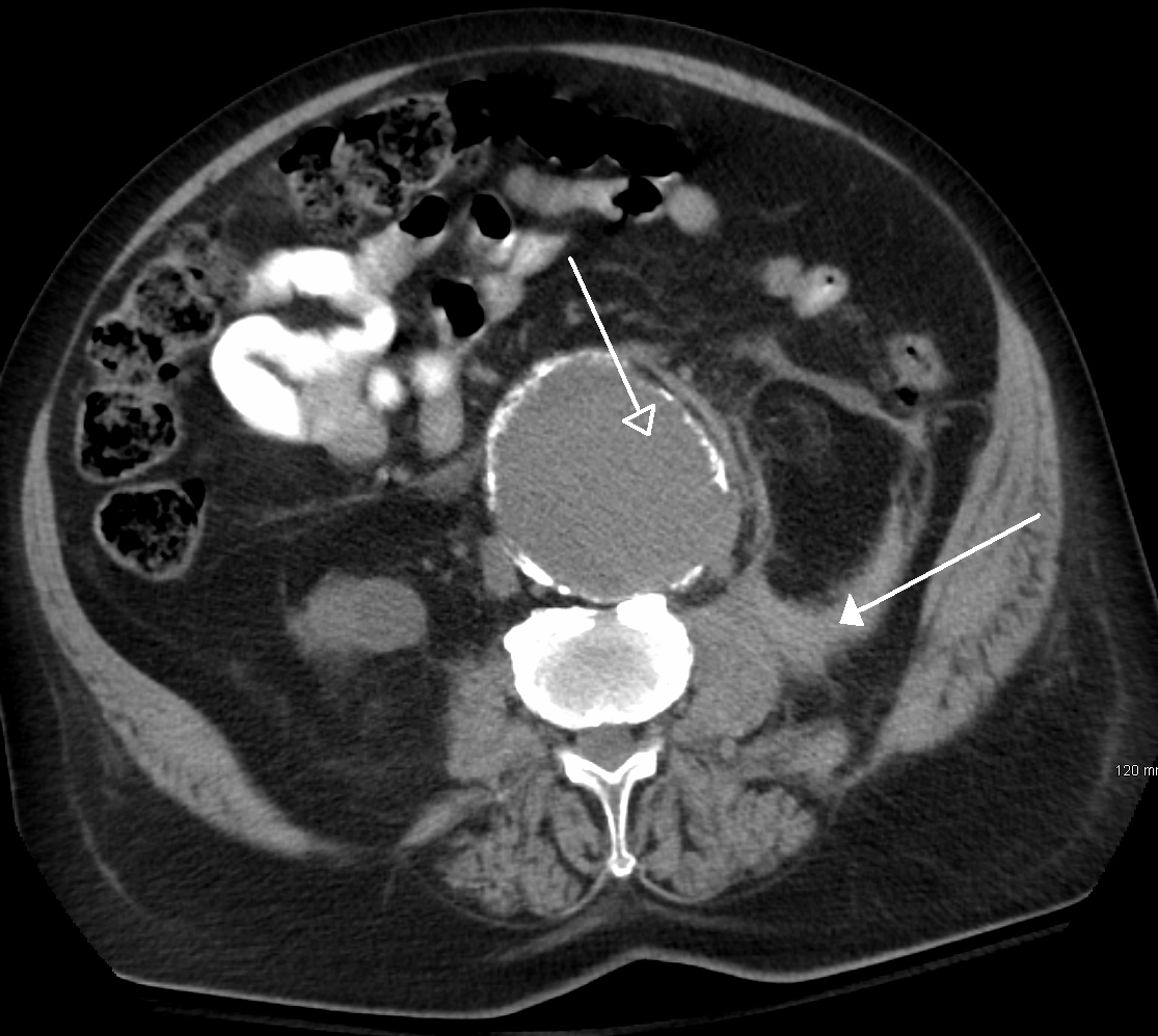
An CT scan image showing a ruptured a abdominal aortic aneurysmICD-9-CM 88.01 OPS-301 code: 3-225-3-226 CT Scan of 11 cm Wilms' tumor of right kidney in 13 month old patient.
CT is a sensitive method for diagnosis of abdominal diseases. It is used frequently to determine stage of cancer and to follow progress. It is also a useful test to investigate acute abdominal pain (especially of the lower quadrants, whereas ultrasound is the preferred first line investigation for right upper quadrant pain). Renal stones, appendicitis, pancreatitis, diverticulitis, abdominal aortic aneurysm, and bowel obstruction are conditions that are readily diagnosed and assessed with CT. CT is also the first line for detecting solid organ injury after trauma.
Multidetector CT (MDCT) can clearly delineate anatomic structures in the abdomen, which is critical in the diagnosis of internal diaphragmatic and other nonpalpable or unsuspected hernias. MDCT also offers clear detail of the abdominal wall allowing wall hernias to be identified accurately.[1]
Oral and/or rectal contrast may be used depending on the indications for the scan. A dilute (2% w/v) suspension of barium sulfate is most commonly used. The concentrated barium sulfate preparations used for fluoroscopy, e.g., barium enema, are too dense and cause severe artifacts on CT. Iodinated contrast agents may be used if barium is contraindicated (for example, suspicion of bowel injury). Other agents may be required to optimize the imaging of specific organs, such as rectally administered gas (air or carbon dioxide) or fluid (water) for a colon study, or oral water for a stomach study.
CT has limited application in the evaluation of the pelvis. For the female pelvis in particular, ultrasound and MRI are the imaging modalities of choice. Nevertheless, it may be part of abdominal scanning (e.g., for tumors), and has uses in assessing fractures.
CT is also used in osteoporosis studies and research alongside dual energy X-ray absorptiometry (DXA). Both CT and DXA can be used to assess bone mineral density (BMD), which is used to indicate bone strength, however CT results do not correlate exactly with DXA (the gold standard of BMD measurement). CT is far more expensive, and subjects patients to much higher levels of ionizing radiation, so it is used infrequently.
References
- ^ Lee HK, Park SJ, Yi BH. Multidetector CT reveals diverse variety of abdominal hernias. Diagnostic Imaging. 2010;32(5):27-31.
Categories:
Wikimedia Foundation. 2010.