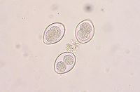
- Coccidia
-
Coccidia Coccidia oocysts Scientific classification Domain: Eukaryota Kingdom: Chromalveolata Superphylum: Alveolata Phylum: Apicomplexa Class: Conoidasida Subclass: Coccidia Order: Eucoccidiorida Suborder, Family, Genera & Species Adeleorina
- Adeleidae
- Dactylosomatidae
- Haemogregarinidae
- Hepatozoidae
- Karyolysidae
- Klossiellidae
- Legerellidae
Eimeriorina
- Aggregatidae
- Aggregata
- Merocystis
- Selysina
- Calyptosporiidae
- Calyptospora
- Cryptosporidiidae
- Eimeriidae
- Atoxoplasma
- Barrouxia
- Caryospora
- Caryotropha
- Cyclospora
- Diaspora
- Dorisa
- Dorisiella
- Eimeria
- Grasseella
- Isospora
- Mantonella
- Ovivora
- Pfeifferinella
- Pseudoklossia
- Tyzzeria
- Wenyonella
- Elleipsisomatidae
- Elleipsisoma
- Lankesterellidae
- Lankesterella
- Schellackia
- Sarcocystidae
- Sarcocystinae
- Frenkelia
- Sarcocystis
- Toxoplasmatinae
- Sarcocystinae
- Selenococcidiidae
- Selenococcidium
- Spirocystidae
- Spirocystis
Coccidia is a subclass of microscopic, spore-forming, single-celled obligate parasites belonging to the apicomplexan class Conoidasida.[1] Coccidian parasites infect the intestinal tracts of animals,[2] and are the largest group of apicomplexan protozoa.
Coccidia are obligate, intracellular parasites, which means that they must live and reproduce within an animal cell.
They form a subclass within the Conoidasida and are divided into four orders distinguished by the presence or absence of various asexual and sexual stages.
Contents
Taxonomy
Order Adelidea Ledger 1911
Order Agamococcidiida Levine 1979
Order Coccidiida Leukart 1879
Order Protococcidiida Cheissin 1956
The order Coccidia is divided into two groups. The first group (suborder Adeleorina) comprises coccidia of invertebrates and the coccidia that alternate between blood-sucking invertebrates and various vertebrates; this group includes Hemogregarina and Hepatozoon. The second group (suborder Eimeriorina) comprises coccidia of vertebrates as well as cyst-forming coccidia, including Toxoplasma and Sarcocystis. The fundamental difference between these two groups lies in their sexual development: syzygy for Adeleorina and independent gametes for Eimeriorina.
Coccidiosis
Coccidiosis is the disease caused by coccidian infection. Coccidiosis is a parasitic disease of the intestinal tract of animals, caused by coccidian protozoa. The disease spreads from one animal to another by contact with infected feces or ingestion of infected tissue. Diarrhea, which may become bloody in severe cases, is the primary symptom. Most animals infected with coccidia are asymptomatic; however, young or immuno-compromised animals may suffer severe symptoms, including death.
While coccidian organisms can infect a wide variety of animals, including humans, birds, and livestock, they are usually species-specific. One well-known exception is toxoplasmosis, caused by Toxoplasma gondii.
People often first encounter coccidia when they acquire a young puppy or kitten who is infected. The infectious organisms are canine/feline-specific and are not contagious to humans (compare to zoonotic diseases).
Coccidia in dogs
Young puppies are frequently infected with coccidia and often develop active Coccidiosis—even puppies obtained from diligent professional breeders. Infected puppies almost always have received the parasite from their mother's feces. Typically, healthy adult animals shedding the parasite's oocysts in their feces will be asymptomatic because of their developed immune systems. However, undeveloped immune systems make puppies more susceptible. Further, stressors such as new owners, travel, weather changes, and unsanitary conditions are believed to activate infections in susceptible animals.
Symptoms in young dogs are universal: at some point around 2–3 months of age, an infected dog develops persistently loose stools. This diarrhea proceeds to stool containing liquid, thick mucus, and light colored fecal matter. As the infection progresses, spots of blood may become apparent in the stool, and sudden bowel movements may surprise both dog and owner alike. Other symptoms may include poor appetite, vomiting, dehydration, and sometimes death. Coccidia infection is so common that any pup under 4 months old with these symptoms can almost surely be assumed to have coccidiosis.
Fortunately, the treatment is inexpensive, extremely effective, and routine. A veterinarian can easily diagnose the disease through low-powered microscopic examination of an affected dog's feces, which usually will be replete with oocysts. One of many easily administered and inexpensive drugs will be prescribed, and, in the course of just a few days, an infection will be eliminated or perhaps reduced to such a level that the dog's immune system can make its own progress against the infection. Even when an infection has progressed sufficiently that blood is present in feces, permanent damage to the gastrointestinal system is rare, and the dog will most likely make a complete recovery without long-lasting negative effects.
If one dog of a litter has coccidiosis, then most certainly all dogs at a breeder's kennels have active coccidia infections. Breeders should be notified if a newly-acquired pup is discovered to be infected with coccidia. Breeders can take steps to eradicate the organism from their kennels, including applying medications in bulk to an entire facility.
Genera and species that cause coccidiosis
- Genus Isospora is the most common cause of intestinal coccidiosis in dogs and cats and is usually what is meant by coccidiosis. Species of Isospora are species specific, meaning they only infect one type of species. Species that infect dogs include I. canis, I. ohioensis, I. burrowsi, and I. neorivolta. Species that infect cats include I. felis and I. rivolta. The most common symptom is diarrhea. sulfonamides are the most common treatment.[3]
- Genus Cryptosporidium contains two species known to cause cryptosporidiosis, C. parvum and C. muris. Cattle are most commonly affected by Cryptosporidium, and their feces are often assumed to be a source of infection for other mammals including humans. Recent genetic analyses of Cryptosporidium in humans have identified Cryptosporidium hominis as a new species specific for humans. Infection occurs most commonly in individuals that are immunocompromised, e.g. dogs with canine distemper, cats with feline leukemia virus infection, and humans with AIDS. Very young puppies and kittens can also become infected with Cryptosporidium, but the infection is usually eliminated without treatment.[3]
- Genus Hammondia is transmitted by ingestion of cysts found in the tissue of grazing animals and rodents. Dogs and cats are the definitive hosts, with the species H. heydorni infecting dogs and the species H. hammondi and H. pardalis infecting cats. Hammondia usually does not cause disease.[3]
- Genus Besnoitia infect cats that ingest cysts found in the tissue of rodents and opossum, but usually does not cause disease.[3]
- Genus Sarcocystis infect carnivores that ingest cysts from various intermediate hosts. It is possible for Sarcocystis to cause disease in dogs and cats.[3]
- Genus Toxoplasma has one important species, Toxoplasma gondii. Cats are the definitive host, but all mammals and some fish, reptiles, and amphibians can be intermediate hosts. Therefore, only cat feces will hold infective oocysts, but infection through ingestion of cysts can occur with the tissue of any intermediate host. Toxoplasmosis occurs in humans usually as low-grade fever or muscle pain for a few days. A normal immune system will suppress the infection but the tissue cysts will persist in that animal or human for years or the rest of its life. In immunocompromised individuals, those dormant cysts can be reactivated and cause many lesions in the brain, heart, lungs, eyes, etc. Without a competent immune system, the animal or human will most likely die from the infection. For pregnant women, the fetus is at risk if the pregnant woman becomes infected for the first time during pregnancy. If the woman had been infected during childhood or adolescence, she will have an immunity that will protect her developing fetus during pregnancy. The most important misconception about the transmission of toxoplasmosis comes from statements like 'ingestion of raw or undercooked meat, or cat feces.' Kitchen hygiene is much more important because people do tend to taste marinades or sauces before being cooked, or chop meat then vegetables without properly cleaning the knife and cutting board. Many physicians mistakenly put panic in their pregnant clients and advise them to get rid of their cat without really warning them of the likely sources of infection. Adult cats are very unlikely to shed infective oocysts. Symptoms in cats include fever, weight loss, diarrhea, vomiting, uveitis, and central nervous system signs. Disease in dogs includes a rapidly progressive form seen in dogs also infected with distemper, and a neurological form causing paralysis, tremors, and seizures. Dogs and cats are usually treated with clindamycin.[3]
- Genus Neospora has one important species, Neospora caninum, that affects dogs in a manner similar to toxoplasmosis. Neosporosis is difficult to treat.[3]
- Genus Hepatozoon contains one species that causes hepatozoonosis in dogs and cats, Hepatozoon canis. Animals become infected by ingesting an infected Rhipicephalus sanguineus, also known as the brown dog tick. Symptoms include fever, weight loss, and pain of the spine and limbs.
The most common medications used to treat coccidian infections are in the sulphonamide family. Although unusual, sulphonamides can damage the tear glands in some dogs, causing keratoconjunctivitis sicca, or "dry eye", which may have a life-long impact. Some veterinarians recommend measuring tear production prior to sulphonamide administration, and at various intervals after administration. Other veterinarians will simply avoid using sulphonamides, instead choosing another product effective against coccidia.
Left untreated, the infection may clear of its own accord, or in some cases may continue to ravage an animal and cause permanent damage or, occasionally, death.
See also
- cryptosporidiosis
- List of parasites (human)
- Zoalene is a fodder additive for poultry, used to prevent infections from coccidia.
References
- ^ "The Taxonomicon & Systema Naturae" (Website database). Taxon: Genus Cryptosporidium. Universal Taxonomic Services, Amsterdam, The Netherlands. 2000. http://www.taxonomy.nl/taxonomicon/TaxonTree.aspx?id=660.
- ^ "Biodiversity explorer: Apicomplexa (apicomplexans, sporozoans)". Iziko Museums of Cape Town. http://www.museums.org.za/bio/apicomplexa/index.htm.
- ^ a b c d e f g Ettinger, Stephen J.; Feldman, Edward C. (1995). Textbook of Veterinary Internal Medicine (4th ed.). W.B. Saunders Company. ISBN 0-7216-6795-3.
External links
- Coccidiosis treatment Coccidiosis treatment for Calves and Lambs
- Mar Vista Animal Medical Center.
- Mar Vista Animal Medical Center.
- Mar Vista Animal Medical Center.
- The Coccidia of the World, Donald W. Duszynski, Steve J. Upton, Lee Couch, Feb. 21, 2004.
- Life Cycle EIMERIA, Andreas Weck-Heimann, 1996–2005
- FarmingUK, Information about Coccidiosis
- Lillehoj, Hyun S. (October 1996). "Two Strategies for Protecting Poultry From Coccidia". Agricultural Research magazine (United States Department of Agriculture: Agricultural Research Service) (October 1996). http://www.ars.usda.gov/is/AR/archive/oct96/coccidia1096.htm. Describes using live-parasite vaccine versus a monoclonal antibody to block the sporozoite from invading a host's cell.
Ciliophora Spirotrichea (Stylonychia) · Litostomatea (Didinium, Balantidium) · Phyllopharyngea (Tokophrya) · Nassophorea (Nassula) · Colpodea (Colpoda) · Oligohymenophorea (Tetrahymena, Ichthyophthirius, Vorticella, Paramecium) · Plagiopylea (Plagiopyla) · Prostomatea (Coleps)OtherMyzozoa Plasmodiidae/Haemosporida (Plasmodium, Haemoproteus, Leucocytozoon)
Piroplasmida (Babesia, Theileria)CoccidiaAdele-Eimeri-Cryptosporidiidae (Cryptosporidium)
Eimeriidae (Isospora, Cyclospora, Eimeria)
Sarcocystidae (Toxoplasma, Sarcocystis, Besnoitia, Neospora)Agamo-Rhytidocystidae (Rhytidocystis)GregariniaGregarinasina (Gregarina)ColpodellidaeChromeridaWith a theca: Peridiniales (Pfiesteria, Peridinium) · Gonyaulacales (Ceratium, Gonyaulax) · Prorocentrales (Prorocentrum) · Dinophysiales (Dinophysis, Histioneis, Ornithocercus, Oxyphysis)
Without theca: Gymnodiniales (Gymnodinium, Karenia, Karlodinium, Amphidinium) · Suessiales (Polarella, Symbiodinium)
Noctilucales (Noctiluca)Syndiniales: Amoebophryaceae (Amoebophyra) · Duboscquellaceae (Duboscquella) · Syndiniaceae (Hematodinium, Syndinium)OtherRelatedInfectious diseases – Parasitic disease: protozoan infection: Chromalveolate and Archaeplastida (A07, B50–B54,B58, 007, 084) Chromalveolate Coccidia: Cryptosporidium hominis/Cryptosporidium parvum (Cryptosporidiosis) · Isospora belli (Isosporiasis) · Cyclospora cayetanensis (Cyclosporiasis) · Toxoplasma gondii (Toxoplasmosis)Archaeplastida Categories:- Parasites
- Apicomplexa
- Dog diseases
- Cat diseases
- Animal diseases
- Veterinary protozoology
Wikimedia Foundation. 2010.

