- Mannan-binding lectin pathway
-
The Mannan-binding lectin pathway (also known simply as the Lectin Pathway) is similar in structure to the classical complement pathway,[1] in that, after activation, it proceeds through the action of C4 and C2 to produce activated complement proteins further down the cascade. In contrast to the classical complement pathway, it is not antibody-dependent.
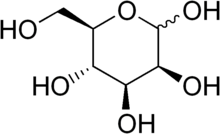
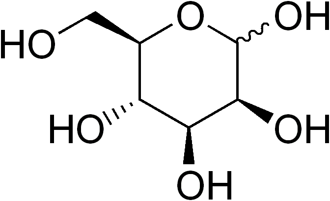
In this pathway, mannose-binding lectin binds to mannose, glucose or other sugars with 3- and 4-OH groups placed in the equatorial plane, in terminal positions on carbohydrate or glycoprotein components of microorganisms including bacteria such as Salmonella, Listeria, and Neisseria strains. Fungal pathogens such as Candida albicans and Cryptococcus neoformans as well as some viruses such as HIV-1 and Respiratory syncytial virus (RSV) are bound by MBL.
Mannan-binding lectin (MBL, also called mannose-binding lectin) is a protein belonging to the collectin family that is produced by the liver and can initiate the complement cascade by binding to pathogen surfaces.
Contents
MBL
MBL is a 6- to 18-headed molecule that forms a complex with MASP-1 (Mannan-binding lectin-Associated Serine Protease), MASP-2 and MASP-3, two protease zymogens. The MASPs are very similar to C1r and C1s molecules of the classical complement pathway, respectively, and are thought to have a common evolutionary ancestor. When the carbohydrate-recognising heads of MBL bind to specifically arranged mannose residues on the surface of a pathogen, MASP-1 and MASP-2 are activated to cleave complement components C4 and C2 into C4a, C4b, C2a, and C2b. In addition, two smaller MBL-associated proteins (MAps) are found in complex with MBL. MBL-associated protein of 19 kDa (MAp19) and MBL-associated protein of 44 kDa (Map44). MASP-1, MASP-3 and MAp44 are alternative splice products of the MASP1 gene, while MASP-2 and MAp19 are alternative splice products of the MASP-2 gene. MAp44 has been suggested to act as a competitive inhibitor of lectin pathway activation, by displacing MASP-2 from MBL, hence preventing cleavage of C4 and C2 [2]
C3 convertase
C4b and C2a combine on the surface of the pathogen to form C3 convertase (C4b and C2a), while C4a and C2a act as chemoattractants.
C3 convertase was, in classical terms, C4b2a; however, in keeping with the number and letter scheme, it was changed to C4b2b in the 1990s to reflect the enzymatic activity of the C2b fragment.
Clinical significance
It has been found that people deficient in MBL experience a substantial increase in infections during the early years of childhood.
See also
References
- ^ Wallis R, Mitchell DA, Schmid R, Schwaeble WJ, Keeble AH (2010). "Paths reunited: Initiation of the classical and lectin pathways of complement activation". Immunobiology 215 (1): 1–11. doi:10.1016/j.imbio.2009.08.006. PMC 2824237. PMID 19783065. http://linkinghub.elsevier.com/retrieve/pii/S0171-2985(09)00146-6.
- ^ Degn, Søren; Annette G. Hansen, Rudi Steffensen,Christian Jacobsen, Jens C. Jensenius and Steffen Thiel (November 2009). "MAp44, a Human Protein Associated with Pattern Recognition Molecules of the Complement System and Regulating the Lectin Pathway of Complement Activation". Journal of Immunology 183 (11): 7371–7378. doi:10.4049/jimmunol.0902388. PMID 19917686. http://www.jimmunol.org/cgi/content/full/183/11/7371.
External links
Proteins: complement system (C, L, A) Activators/enzymes EarlyMiddleLateInhibitors Complement receptors Categories:- Immunology stubs
- Complement system
Wikimedia Foundation. 2010.