- Pleomorphic undifferentiated sarcoma
-
Pleomorphic undifferentiated sarcoma Classification and external resources 
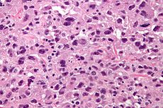
Micrograph of a pleomorphic undifferentiated sarcoma. H&E stain.ICD-10 C49 (ILDS C49.M10) OMIM 8830/3 DiseasesDB 31471 eMedicine radio/420 MeSH D051677 Pleomorphic undifferentiated sarcoma (abbreviated PUS), also undifferentiated pleomorphic sarcoma and previously malignant fibrous histiocytoma (abbreviated MFH), is a type of soft tissue sarcoma.
It is considered a diagnosis of exclusion for sarcomas that cannot been more precisely categorized.[1][2]
Contents
Epidemiology
PUS are regarded as the most common soft tissue sarcoma of late adult life. It rarely occurs in children.[3] It occurs more often in Caucasians, than those of African or Asian descent, and is a male predominant disease, afflicting two males for every female.
Presentation
PUS occurs most commonly in the extremities and retroperitoneum, but has been reported in other sites. Metastasis occur most frequently in the lungs (90%), bones (8%) and liver (1%).
In the extremities, it presents as a painless enlarging soft tissue mass.
Diagnosis
It can be detected by magnetic resonance imaging (MRI), but a biopsy is required for the definitive diagnosis. MRI findings typically shows a well-circumscribed mass that is dark on T1-weighted images and bright on T2-weighted images. Central necrosis is often present and identifiable by imaging, especially in larger masses.
Pathology
Pleomorphic undifferentiated sarcomas are, by definition, undifferentiated (as the name implies) meaning they do not bear a resemblance to any normal tissue.
The histomorphology, otherwise, is characterized by high cellularity, marked nuclear pleomorphism, usually accompanied by abundant mitotic activity (including atypical mitoses), and a spindle cell morphology. Necrosis is common and characteristic of high grade lesions.
Treatment
Treatment consists of surgical excision (the extent of which ranges from tumor excision to limb amputation depending on the tumor) and in some cases radiation. Chemotherapy remains controversial in MFH. The usual site of metastatic disease is the lungs, and metastases should be resected if possible.
Prognosis
Prognosis depends on the primary tumor grade (appearance under the microscope as judged by a pathologist), size, resectability (whether it can be cut-out completely), and presence of metastases. The five-year survival ranges from 35-60%.
Human-human transmission
There is a case report of human-human transmission, in a surgeon that injured their hand while working on a PUS case.[4]
References
- ^ Matushansky I, Charytonowicz E, Mills J, Siddiqi S, Hricik T, Cordon-Cardo C (August 2009). "MFH classification: differentiating undifferentiated pleomorphic sarcoma in the 21st Century". Expert Rev Anticancer Ther 9 (8): 1135–44. doi:10.1586/era.09.76. PMC 3000413. PMID 19671033. http://www.pubmedcentral.nih.gov/articlerender.fcgi?tool=pmcentrez&artid=3000413.
- ^ "Neoplasia". http://library.med.utah.edu/WebPath/NEOHTML/NEOPL045.html.
- ^ Alaggio R, Collini P, Randall RL, Barnette P, Million L, Coffin CM (2010). "Undifferentiated high-grade pleomorphic sarcomas in children: a clinicopathologic study of 10 cases and review of literature". Pediatr. Dev. Pathol. 13 (3): 209–17. doi:10.2350/09-07-0673-OA.1. PMID 20055602.
- ^ Gärtner, Hermine-Valeria; Christian Seidl, Christine Luckenbach, Georg Schumm, Erhard Seifried, Horst Ritter, Burkhard Bultmann (1996-11-14). "Genetic Analysis of a Sarcoma Accidentally Transplanted from a Patient to a Surgeon". N Engl J Med 335 (20): 1494–1497. doi:10.1056/NEJM199611143352004. PMID 8890100. http://content.nejm.org/cgi/content/extract/335/20/1494. Retrieved 2010-01-05.
External links
- Malignant Fibrous Histiocytoma (MFH) - Sarcoma Learning Center
Connective/soft tissue tumors and sarcomas (ICD-O 8800–9059) (C45–C49/D17–D21, 171/214–215) Not otherwise specified (8800–8809) Connective tissue neoplasm Fibromatous (8810–8839)Fibroma/fibromatosis: Aggressive infantile fibromatosis · Aponeurotic fibroma · Collagenous fibroma · Diffuse infantile fibromatosis · Familial myxovascular fibromas · Fibroma of tendon sheath · Fibromatosis colli · Infantile digital fibromatosis · Juvenile hyaline fibromatosis · Plantar fibromatosis · Pleomorphic fibroma · Oral submucous fibrosisHistiocytoma/histiocytic sarcoma: Benign fibrous histiocytoma · Malignant fibrous histiocytoma · Atypical fibroxanthomaSolitary fibrous tumorMyxomatous (8840–8849)Myxoma/myxosarcoma (Cutaneous myxoma, Superficial acral fibromyxoma) · Angiomyxoma · Ossifying fibromyxoid tumourFibroepithelial (9000–9039)Synovial-like (9040–9049)Lipomatous (8850–8889) Chondroid lipoma · Intradermal spindle cell lipoma · Pleomorphic lipoma · Benign lipoblastomatosis · Spindle cell lipoma · HibernomaMyomatous (8890–8929) general: Myoma/myosarcomaskeletal muscle: Rhabdomyoma/rhabdomyosarcoma: Embryonal rhabdomyosarcoma (Sarcoma botryoides) · Alveolar rhabdomyosarcomaLeiomyoma · Angioleiomyoma · Angiolipoleiomyoma · Genital leiomyoma · Leiomyosarcoma · Multiple cutaneous and uterine leiomyomatosis syndrome · Multiple cutaneous leiomyoma · Neural fibrolipoma · Solitary cutaneous leiomyomaComplex mixed and stromal (8930–8999) Adenomyoma · Pleomorphic adenoma · Mixed Müllerian tumor · Mesoblastic nephroma · Wilms' tumor · Rhabdoid tumour · Clear-cell sarcoma of the kidney · Hepatoblastoma · Pancreatoblastoma · CarcinosarcomaMesothelial (9050–9059) see also Template:Connective tissue
Tumors: urogenital neoplasia: urinary organs (C64–C68/D30, 188–189/223) Abdominal KidneyGlandular and epithelial neoplasm: Renal cell carcinoma · Renal oncocytomaUreterPelvic Retroperitoneum Categories:- Sarcoma
- Dermal and subcutaneous growths
Wikimedia Foundation. 2010.
