- Neuroplasticity
-
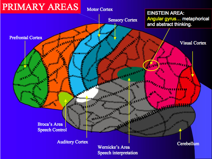
 Contrary to common ideas as expressed in this diagram, brain functions are not confined to certain fixed locations.
Neuroplasticity is a non-specific neuroscience term referring to the ability of the brain and nervous system in all species to change structurally and functionally as a result of input from the environment.[1] Plasticity occurs on a variety of levels, ranging from cellular changes involved in learning, to large-scale changes involved in cortical remapping in response to injury. The most widely recognized forms of plasticity are learning, memory, and recovery from brain damage. During most of the 20th century, the general consensus among neuroscientists was that brain structure is relatively immutable after a critical period during early childhood. This belief has been challenged by new findings, revealing that many aspects of the brain remain plastic even into adulthood.[2]
Hubel and Wiesel had demonstrated that ocular dominance columns in the lowest neocortical visual area, V1, were largely immutable after the critical period in development.[3] Critical periods also were studied with respect to language; the resulting data suggested that sensory pathways were fixed after the critical period. However, studies determined that environmental changes could alter behavior and cognition by modifying connections between existing neurons and via neurogenesis in the hippocampus and other parts of the brain, including the cerebellum.[4]
Decades of research[5] have now shown that substantial changes occur in the lowest neocortical processing areas, and that these changes can profoundly alter the pattern of neuronal activation in response to experience. Neurological research indicates that experience can actually change both the brain's physical structure (anatomy) and functional organization (physiology). Neuroscientists are currently engaged in a reconciliation of critical period studies demonstrating the immutability of the brain after development with the more recent research showing how the brain can, and does, change.[6]
Contents
Etymology
This idea was first proposed in 1890 by William James in The Principles of Psychology, though the idea was largely neglected for the next fifty years.[7] The first person to use the term neural plasticity appears to have been the Polish neuroscientist Jerzy Konorski.[8] The term has no specific scientific definition, as set out by McEachern and Shaw:[1]
Given the central importance of neuroplasticity, an outsider would be forgiven for assuming that it was well defined and that a basic and universal framework served to direct current and future hypotheses and experimentation. Sadly, however, this is not the case. While many neuroscientists use the word neuroplasticity as an umbrella term it means different things to different researchers in different subfields ... In brief, a mutually agreed upon framework does not appear to exist.Neurobiology
One of the fundamental principles of how neuroplasticity functions is linked to the concept of synaptic pruning, the idea that individual connections within the brain are constantly being removed or recreated, largely dependent upon how they are used. This concept is captured in the aphorism, "neurons that fire together, wire together"/"neurons that fire apart, wire apart." If there are two nearby neurons that often produce an impulse simultaneously, their cortical maps may become one. This idea also works in the opposite way, i.e. that neurons which do not regularly produce simultaneous impulses will form different maps.
Cortical maps
Cortical organization, especially for the sensory systems, is often described in terms of maps.[9] For example, sensory information from the foot projects to one cortical site and the projections from the hand target in another site. As the result of this somatotopic organization of sensory inputs to the cortex, cortical representation of the body resembles a map (or homunculus).
In the late 1970s and early 1980s, several groups began exploring the impacts of removing portions of the sensory inputs. Michael Merzenich and Jon Kaas and Doug Rasmusson used the cortical map as their dependent variable. They found—and this has been since corroborated by a wide range of labs—that if the cortical map is deprived of its input it will become activated at a later time in response to other, usually adjacent inputs. At least in the somatic sensory system, in which this phenomenon has been most thoroughly investigated, JT Wall and J Xu have traced the mechanisms underlying this plasticity. Re-organization is not cortically emergent, but occurs at every level in the processing hierarchy; this produces the map changes observed in the cerebral cortex.[10]
Merzenich and William Jenkins (1990) initiated studies relating sensory experience, without pathological perturbation, to cortically observed plasticity in the primate somatosensory system, with the finding that sensory sites activated in an attended operant behavior increase in their cortical representation. Shortly thereafter, Ford Ebner and colleagues (1994) made similar efforts in the rodent whisker barrel cortex (also somatic sensory system). These two groups largely diverged over the years. The rodent whisker barrel efforts became a focus for Ebner, Matthew Diamond, Michael Armstrong-James, Robert Sachdev, Kevin Fox and great inroads were made in identifying the locus of change as being at cortical synapses expressing NMDA receptors, and in implicating cholinergic inputs as necessary for normal expression. However, the rodent studies were poorly focused on the behavioral end, and Ron Frostig and Daniel Polley (1999, 2004) identified behavioral manipulations as causing a substantial impact on the cortical plasticity in that system.
Merzenich and DT Blake (2002, 2005, 2006) went on to use cortical implants to study the evolution of plasticity in both the somatosensory and auditory systems. Both systems show similar changes with respect to behavior. When a stimulus is cognitively associated with reinforcement, its cortical representation is strengthened and enlarged. In some cases, cortical representations can increase two to threefold in 1–2 days at the time at which a new sensory motor behavior is first acquired, and changes are largely finished within at most a few weeks. Control studies show that these changes are not caused by sensory experience alone: they require learning about the sensory experience, and are strongest for the stimuli that are associated with reward, and occur with equal ease in operant and classical conditioning behaviors.
An interesting phenomenon involving cortical maps is the incidence of phantom limbs. Phantom limbs are experienced by people that have undergone amputations in hands, arms, and legs, but it is not limited to extremities. Although the neurological basis of phantom limbs is still not entirely understood it is believed that cortical reorganization plays an important role. [11]
Norman Doidge, following the lead of Michael Merzenich, separates manifestations of neuroplasticity into adaptations that have positive or negative behavioral consequences. For example, if an organism can recover after a stroke to normal levels of performance, that adaptiveness could be considered an example of "positive plasticity". An excessive level of neuronal growth leading to spasticity or tonic paralysis, or an excessive release of neurotransmitters in response to injury which could kill nerve cells; this would have to be considered a "negative" plasticity. In addition, drug addiction and obsessive-compulsive disorder are deemed examples of "negative plasticity" by Dr. Doidge, as the synaptic rewiring resulting in these behaviors is also highly maladaptive.[11][12]
A 2005 study found that the effects of neuroplasticity occur even more rapidly than previously expected. Medical students' brains were imaged during the period when they were studying for their exams. In a matter of months, the students' gray matter increased significantly in the posterior and lateral parietal cortex.[13]
History
Proposal
Until around the 1970s, an accepted idea across neuroscience was that the nervous system was essentially fixed throughout adulthood, both in terms of brain functions, as well as the idea that it was impossible for new neurons to develop after birth.[14]
In 1793, Italian anatomist Michele Vicenzo Malacarne described experiments in which he paired animals, trained one of the pair extensively for years, and then dissected both. He discovered that the cerebellums of the trained animals were substantially larger. But, these findings were eventually forgotten.[15] The idea that the brain and its functions are not fixed throughout adulthood was proposed in 1890 by William James in The Principles of Psychology, though the idea was largely neglected.[7]
Research and discovery
In 1923, Karl Lashley conducted experiments on rhesus monkeys which demonstrated changes in neuronal pathways, which he concluded to be evidence of plasticity, although despite this, as well as further examples of research suggesting this, the idea of neuroplasticity was not widely accepted by neuroscientists. However, more significant evidence began to be produced in the 1960s and after, notably from scientists including Paul Bach-y-Rita, Michael Merzenich along with Jon Kaas, as well as several others.[14][16]
In the 1960s, Paul Bach-y-Rita invented a device that allowed blind people to read, perceive shadows, and distinguish between close and distant objects. This “machine was one of the first and boldest applications of neuroplasticity.”[11] The patient sat in an electrically stimulated chair that had a large camera behind it which scanned the area, sending electrical signals of the image to four hundred vibrating stimulators on the chair against the patient’s skin. The six subjects of the experiment were eventually able to recognize a picture of the supermodel Twiggy.[11]
It must be emphasized that these people were congenitally blind and had previously not been able to see. Bach-y-Rita believed in sensory substitution; if one sense is damaged, your other senses can sometimes take over. He thought skin and its touch receptors could act as a retina (using one sense for another[17]). In order for the brain to interpret tactile information and convert it into visual information, it has to learn something new and adapt to the new signals. The brain's capacity to adapt implied that it possessed plasticity. He thought, “We see with our brains, not with our eyes.”[11]
A tragic stroke that left his father paralyzed inspired Bach-y-Rita to study brain rehabilitation. His brother, a physician, worked tirelessly to develop therapeutic measures which were so successful that the father recovered complete functionality by age 68 and was able to live a normal, active life which even included mountain climbing. “His father’s story was firsthand evidence that a ‘late recovery’ could occur even with a massive lesion in an elderly person.”[11] He found more evidence of this possible brain reorganization with Shepherd Ivory Franz's work.[18] One study involved stroke patients who were able to recover through the use of brain stimulating exercises after having been paralyzed for years. “Franz understood the importance of interesting, motivating rehabilitation: ‘Under conditions of interest, such as that of competition, the resulting movement may be much more efficiently carried out than in the dull, routine training in the laboratory’(Franz, 1921, pg.93).”[19] This notion has led to motivational rehabilitation programs that are used today.
Michael Merzenich is a neuroscientist who has been one of the pioneers of brain plasticity for over three decades. He has made some of “the most ambitious claims for the field - that brain exercises may be as useful as drugs to treat diseases as severe as schizophrenia - that plasticity exists from cradle to the grave, and that radical improvements in cognitive functioning - how we learn, think, perceive, and remember are possible even in the elderly.”[11] Merzenich’s work was affected by a crucial discovery made by David Hubel and Torsten Wiesel in their work with kittens. The experiment involved sewing one eye shut and recording the cortical brain maps. Hubel and Wiesel saw that the portion of the kitten’s brain associated with the shut eye was not idle, as expected. Instead, it processed visual information from the open eye. It was“… as though the brain didn’t want to waste any ‘cortical real estate’ and had found a way to rewire itself.”[11]
This implied brain plasticity during the critical period. However, Merzenich argued that brain plasticity could occur beyond the critical period. His first encounter with adult plasticity came when he was engaged in a postdoctoral study with Clinton Woosley. The experiment was based on observation of what occurred in the brain when one peripheral nerve was cut and subsequently regenerated. The two scientists micromapped the hand maps of monkey brains before and after cutting a peripheral nerve and sewing the ends together. Afterwards, the hand map in the brain that was expected to be jumbled was nearly normal. This was a substantial breakthrough. Merzenich asserted that “if the brain map could normalize its structure in response to abnormal input, the prevailing view that we are born with a hardwired system had to be wrong. The brain had to be plastic.”[11]
Applications and examples
Treatment of brain damage
A surprising consequence of neuroplasticity is that the brain activity associated with a given function can move to a different location; this can result from normal experience and also occurs in the process of recovery from brain injury. Neuroplasticity is the fundamental issue that supports the scientific basis for treatment of acquired brain injury with goal-directed experiential therapeutic programs in the context of rehabilitation approaches to the functional consequences of the injury.
The adult brain is not "hard-wired" with fixed and immutable neuronal circuits. There are many instances of cortical and subcortical rewiring of neuronal circuits in response to training as well as in response to injury. There is solid evidence that neurogenesis (birth of brain cells) occurs in the adult, mammalian brain—and such changes can persist well into old age.[2] The evidence for neurogenesis is mainly restricted to the hippocampus and olfactory bulb, but current research has revealed that other parts of the brain, including the cerebellum, may be involved as well.[4]
In the rest of the brain, neurons can die, but they cannot be created. However, there is now ample evidence for the active, experience-dependent re-organization of the synaptic networks of the brain involving multiple inter-related structures including the cerebral cortex. The specific details of how this process occurs at the molecular and ultrastructural levels are topics of active neuroscience research. The manner in which experience can influence the synaptic organization of the brain is also the basis for a number of theories of brain function including the general theory of mind and epistemology referred to as Neural Darwinism and developed by immunologist Nobel laureate Gerald Edelman. The concept of neuroplasticity is also central to theories of memory and learning that are associated with experience-driven alteration of synaptic structure and function in studies of classical conditioning in invertebrate animal models such as Aplysia. This latter program of neuroscience research has emanated from the ground-breaking work of another Nobel laureate, Eric Kandel, and his colleagues at Columbia University College of Physicians and Surgeons.
Paul Bach-y-Rita, deceased in 2006, was the “father of sensory substitution and brain plasticity.”[20] In working with a patient whose vestibular system had been damaged he developed BrainPort,[21] a machine that “replaces her vestibular apparatus and [will] send balance signals to her brain from her tongue.”[11] After she had used this machine for some time it was no longer necessary, as she regained the ability to function normally. Her balancing act days were over.[22]
Plasticity is the major explanation for the phenomenon. Because her vestibular system was “disorganized” and sending random rather than coherent signals, the apparatus found new pathways around the damaged or blocked neural pathways, helping to reinforce the signals that were sent by remaining healthy tissues. Bach-y-Rita explained plasticity by saying, “If you are driving from here to Milwaukee and the main bridge goes out, first you are paralyzed. Then you take old secondary roads through the farmland. Then you use these roads more; you find shorter paths to use to get where you want to go, and you start to get there faster. These “secondary” neural pathways are “unmasked” or exposed and strengthened as they are used. The “unmasking” process is generally thought to be one of the principal ways in which the plastic brain reorganizes itself.”[11]
Randy Nudo's group found that if a small stroke (an infarction) is induced by obstruction of blood flow to a portion of a monkey’s motor cortex, the part of the body that responds by movement will move when areas adjacent to the damaged brain area are stimulated. In one study, intracortical microstimulation (ICMS) mapping techniques were used in nine normal monkeys. Some underwent ischemic infarction procedures and the others, ICMS procedures. The monkeys with ischemic infarctions retained more finger flexion during food retrieval and after several months this deficit returned to preoperative levels.[23] With respect to the distal forelimb representation, “postinfarction mapping procedures revealed that movement representations underwent reorganization throughout the adjacent, undamaged cortex.”[23] Understanding of interaction between the damaged and undamaged areas provides a basis for better treatment plans in stroke patients. Current research includes the tracking of changes that occur in the motor areas of the cerebral cortex as a result of a stroke. Thus, events that occur in the reorganization process of the brain can be ascertained. Nudo is also involved in studying the treatment plans that may enhance recovery from strokes, such as physiotherapy, pharmacotherapy and electrical stimulation therapy.
Jon Kaas, a professor at Vanderbilt University, has been able to show “how somatosensory area 3b and ventroposterior (VP) nucleus of the thalamus are affected by long standing unilateral dorsal column lesions at cervical levels in macaque monkeys.”[24] Adult brains have the ability to change as a result of injury but the extent of the reorganization depends on the extent of the injury. His recent research focuses on the somatosensory system, which involves a sense of the body and its movements using many senses. Usually when people damage the somatosensory cortex, impairment of the body perceptions are experienced. He is trying to see how these systems (somatosensory, cognitive, motor systems) are plastic as a result of injury.[24]
One of the most recent applications of neuroplasticity involves work done by a team of doctors and researchers at Emory University, specifically Dr. Donald Stein (who has been in the field for over three decades)[25] and Dr. David Wright. This is the first treatment in 40 years that has significant results in treating traumatic brain injuries while also incurring no known side effects and being cheap to administer.[26] Dr. Stein noticed that female mice seemed to recover from brain injuries better than male mice. Also in females, he noticed that at certain points in the estrus cycle females recovered even more. After lots of research, they attributed this difference due to the levels of progesterone. The highest level of progesterone present led to the fastest recovery of brain injury in these mice.
They developed a treatment that includes increased levels of progesterone injections to give to brain injured patients. “Administration of progesterone after traumatic brain injury[27] (TBI) and stroke reduces edema, inflammation, and neuronal cell death, and enhance spatial reference memory and sensory motor recovery.”[28] In their clinical trials, they had a group of severely injured patients that after the three days of progesterone injections had a 60% reduction in mortality.[26] Sam* was in a horrific car accident that left him with marginal brain activity; according to the doctors, he was one point away from being brain dead. His parents decided to have him participate in Dr. Stein’s clinical trial and he was given the three-day progesterone treatment. Three years after the accident, he had achieved an inspiring recovery with no brain complications and the ability to live a healthy, normal life.[26]
Stein has done some studies in which beneficial effects have been seen to be similar in aged rats to those seen in youthful rats. As there are physiological differences in the two age groups, the model was tweaked for the elderly animals by reducing their stress levels with increased physical contact. During surgery, anesthesia was kept at a higher oxygen level with lower overall isoflurane percentage and “the aged animals were give subcutaneous lactated ringers solution post-surgery to replace fluids lost through increased bleeding.”[29] The promising results of progesterone treatments “could have a significant impact on the clinical management of TBI.”[29] These treatments have been shown to work on human patients who receive treatment soon after the TBI. However, Dr. Stein now focuses his research on those persons who have longstanding traumatic brain injury in order to determine if progesterone treatments will assist them in the recovery of lost functions as well.
Treatment of learning difficulties
Michael Merzenich developed a series of “plasticity-based computer programs known as Fast ForWord.” FastForWord offers seven brain exercises to help with the language and learning deficits of dyslexia. In a recent study, experimental training was done in adults to see if it would help to counteract the negative plasticity that results from age-related cognitive decline (ARCD). The ET design included six exercises designed to reverse the dysfunctions caused by ARCD in cognition, memory, motor control, and so on [9]. After use of the ET program for 8–10 weeks, there was a “significant increase in task-specific performance.”[9] The data collected from the study indicated that a brain plasticity-based program could notably improve cognitive function and memory in adults with ARCD.
Brain plasticity during operation of brain-machine interfaces
Brain-machine interface (BMI) is a rapidly developing field of neuroscience. According to the results obtained by Mikhail Lebedev, Miguel Nicolelis and their colleagues,[30] operation of BMIs results in incorporation of artificial actuators into brain representations. The scientists showed that modifications in neuronal representation of the monkey's hand and the actuator that was controlled by the monkey brain occurred in multiple cortical areas while the monkey operated a BMI. In these single day experiments, monkeys initially moved the actuator by pushing a joystick. After mapping out the motor neuron ensembles, control of the actuator was switched to the model of the ensembles so that the brain activity, and not the hand, directly controlled the actuator. The activity of individual neurons and neuronal populations became less representative of the animal's hand movements while representing the movements of the actuator. Presumably as a result of this adaptation, the animals could eventually stop moving their hands yet continue to operate the actuator. Thus, during BMI control, cortical ensembles plastically adapt, within tens of minutes, to represent behaviorally significant motor parameters, even if these are not associated with movements of the animal's own limb.
Active laboratory groups include those of John Donoghue at Brown, Richard Andersen at Caltech, Krishna Shenoy at Stanford, Nicholas Hatsopoulos of University of Chicago, Andy Schwartz at University of Pittsburgh, Sandro Mussa-Ivaldi at Northwestern and Miguel Nicolelis at Duke. Donoghue and Nicolelis' groups have independently shown that animals can control external interfaces in tasks requiring feedback, with models based on activity of cortical neurons, and that animals can adaptively change their minds to make the models work better. Donoghue's group took the implants from Richard Normann's lab at Utah (the "Utah" array), and improved it by changing the insulation from polyimide to parylene-c, and commercialized it through the company Cyberkinetics. These efforts are the leading candidate for the first human trials on a broad scale for motor cortical implants to help quadriplegic or locked-in patients communicate with the outside world.
Phantom limbs
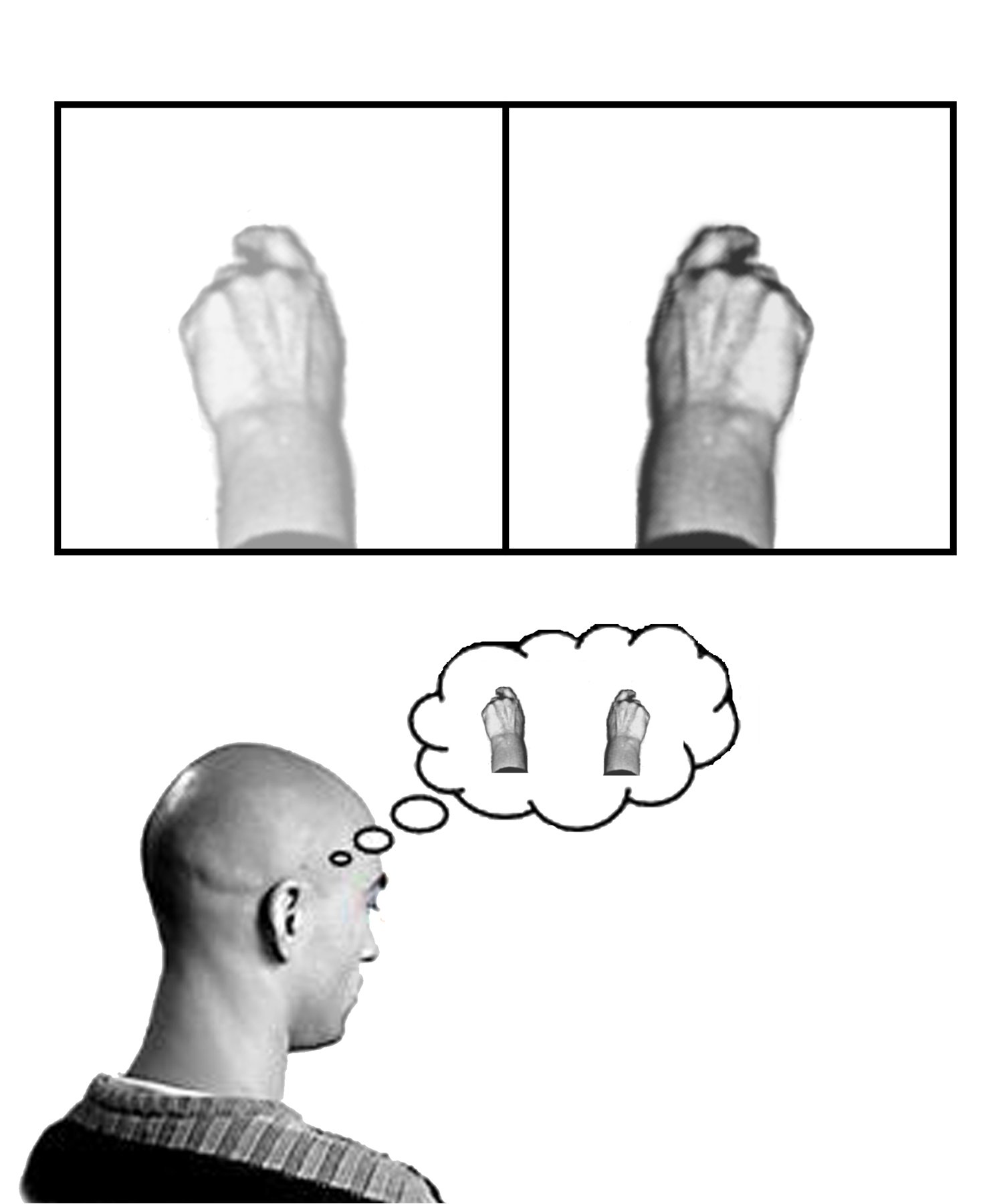
 A diagrammatic explanation of the mirror box. The patient places the good limb into one side of the box (in this case the right hand) and the amputated limb into the other side. Due to the mirror, the patient sees a reflection of the good hand where the missing limb would be (indicated in lower contrast). The patient thus receives artificial visual feedback that the "resurrected" limb is now moving when they move the good hand.Main articles: Phantom limb and Mirror box
The experience of Phantom limbs is a phenomenon in which a person continues to feel pain or sensation within a part of their body which has been amputated. An explanation for this refers to the concept of neuroplasticity, as the cortical maps of the removed limbs are believed to have become engaged with the area around them in the postcentral gyrus. This results in activity within the surrounding area of the cortex being misinterpreted by the area of the cortex formerly responsible for the amputated limb.
The relationship between phantom limbs and neuroplasticity is a complex one. In the early 1990s V.S. Ramachandran theorized that phantom limbs were the result of cortical remapping. However, in 1995 Herta Flor and her colleagues demonstrated that cortical remapping occurs only in patients who have phantom pain.[31] Her research showed that phantom limb pain (rather than referred sensations) was the perceptual correlate of cortical reorganization.[32] This phenomenon is sometimes referred to as maladaptive plasticity.
In 2009 Lorimer Moseley and Peter Brugger carried out a remarkable experiment in which they encouraged arm amputee subjects to use visual imagery to contort their phantom limbs into impossible configurations. Four of the seven subjects succeeded in performing impossible movements of the phantom limb. This experiment suggests that the subjects had modified the neural representation of their phantom limbs and generated the motor commands needed to execute impossible movements in the absence of feedback from the body.[33] The authors stated that:"In fact, this finding extends our understanding of the brain's plasticity because it is evidence that profound changes in the mental representation of the body can be induced purely by internal brain mechanisms--the brain truly does change itself."
Meditation
Main article: Research on meditationA number of studies have linked meditation practice to differences in cortical thickness or density of gray matter. One of the most well-known studies to demonstrate this was led by Sara Lazar, from Harvard University, in 2000.[34] Richard Davidson, a neuroscientist at the University of Wisconsin, has led experiments in cooperation with the Dalai Lama on effects of meditation on the brain. His results suggest that long-term, or short-term practice of meditation results in different levels of activity in brain regions associated with such qualities as attention, anxiety, depression, fear, anger, the ability of the body to heal itself, and so on. These functional changes may be caused by changes in the physical structure of the brain.[35][36][37][38]
Fitness and Exercise
In a 2009 study, scientists made two groups of mice swim a water maze, and then in a separate trial subjected them to an unpleasant stimulus to see how quickly they would learn to move away from it. Then, over the next four weeks they allowed one group of mice to run inside their rodent wheels, an activity most mice enjoy, while they forced the other group to work harder on minitreadmills at a speed and duration controlled by the scientists. They then tested both groups again to track their learning skills and memory. Both groups of mice improved their performances in the water maze from the earlier trial. But only the extra-worked treadmill runners were better in the avoidance task, a skill that, according to neuroscientists, demands a more complicated cognitive response.[39]
The mice who were forced to run on the treadmills showed evidence of molecular changes in several portions of their brains when viewed under a microscope, while the voluntary wheel-runners had changes in only one area. “Our results support the notion that different forms of exercise induce neuroplasticity changes in different brain regions,” Chauying J. Jen, a professor of physiology and an author of the study, said.[40]
Human Echolocation
Human echolocation is a learned ability for humans to sense their environment from echoes. This ability is used by some blind people to navigate their environment and sense their surroundings in detail. Studies in 2010 [41] and 2011 [42] using Functional magnetic resonance imaging techniques have shown that parts of the brain associated with visual processing are adapted for the new skill of echolocation.
See also
- Activity-dependent plasticity
- Arrowsmith School
- Brain fitness
- Edward Taub
- Environmental enrichment (neural)
- Hebbian theory
- Malleable intelligence
- Metaplasticity
- Michael M. Merzenich
- Neuroconstructivism
- Neuroplastic effects of pollution
- Synaptic plasticity
- Non-synaptic plasticity
- Vision restoration therapy
Trauma:
References
- ^ a b Shaw, Christopher; McEachern, Jill, eds (2001). Toward a theory of neuroplasticity. London, England: Psychology Press. ISBN 9781841690216.
- ^ a b Rakic, P. (January 2002). "Neurogenesis in adult primate neocortex: an evaluation of the evidence". Nature Reviews Neuroscience 3 (1): 65–71. doi:10.1038/nrn700. PMID 11823806.
- ^ Hubel, D.H.; Wiesel, T.N. (February 1, 1970). "The period of susceptibility to the physiological effects of unilateral eye closure in kittens". The Journal of Physiology 206 (2): 419–436. PMC 1348655. PMID 5498493. http://www.pubmedcentral.nih.gov/articlerender.fcgi?tool=pmcentrez&artid=1348655. Retrieved 2010-02-01.
- ^ a b Ponti, Giovanna; Peretto, Paolo; Bonfanti, Luca; Reh, Thomas A. (2008). "Genesis of Neuronal and Glial Progenitors in the Cerebellar Cortex of Peripuberal and Adult Rabbits". PLoS ONE 3 (6): e2366. doi:10.1371/journal.pone.0002366. PMC 2396292. PMID 18523645. http://www.pubmedcentral.nih.gov/articlerender.fcgi?tool=pmcentrez&artid=2396292. Retrieved 2010-02-01.
- ^ Chaney, Warren, Dynamic Mind, 2007, Las Vegas, Houghton-Brace Publishing, pp 33-35, ISBN 0-9793392-0-0 [1]
- ^ Chaney, Warren, Workbook for a Dynamic Mind, 2006, Las Vegas, Houghton-Brace Publishing, page 44, ISBN 0 0979339219 [2]
- ^ a b "The Principles of Psychology", William James 1890, Chapter IV, Habits
- ^ LeDoux, Joseph E. (2002). Synaptic self: how our brains become who we are. New York, United States: Viking. p. 137. ISBN 0670030287.
- ^ Buonomano, Dean V.; Merzenich, Michael M. (March 1998). "CORTICAL PLASTICITY: From Synapses to Maps". Annual Review of Neuroscience 21: 149–186. doi:10.1146/annurev.neuro.21.1.149. PMID 9530495.
- ^ Wall, J.T.; Xu, J.; Wang, X. (September 2002). "Human brain plasticity: an emerging view of the multiple substrates and mechanisms that cause cortical changes and related sensory dysfunctions after injuries of sensory inputs from the body". Brain Research Reviews (Elsevier Science B.V.) 39 (2-3): 181–215. doi:10.1016/S0165-0173(02)00192-3. PMID 12423766.
- ^ a b c d e f g h i j k Doidge, Norman (2007). The Brain That Changes Itself: Stories of Personal Triumph from the frontiers of brain science. New York: Viking. ISBN 9780670038305.
- ^ Interview with Merzenich, 2004
- ^ Draganski et al. "Temporal and Spatial Dynamics of Brain Structure Changes during Extensive Learning" The Journal of Neuroscience, June 7, 2006, 26(23):6314-6317
- ^ a b Meghan O'Rourke Train Your Brain April 25, 2007
- ^ Rosenzweig, Mark R. (1996). "Aspects of the search for neural mechanisms of memory". Annual Review of Psychology 47: 1–32. doi:10.1146/annurev.psych.47.1.1.
- ^ Brain Science Podcast Episode #10, "Neuroplasticity"
- ^ "Wired Science . Video: Mixed Feelings". PBS. http://www.pbs.org/kcet/wiredscience/video/286-mixed_feelings.html. Retrieved 2010-06-12.
- ^ "Shepherd Ivory Franz". Rkthomas.myweb.uga.edu. http://rkthomas.myweb.uga.edu/Franz.htm. Retrieved 2010-06-12.
- ^ Colotla, Victor A.; Bach-y-Rita, Paul (2002). "Shepherd Ivory Franz: His contributions to neuropsychology and rehabilitation". Cognitive, Affective & Behavioral Neuroscience 2 (2): 141–148. doi:10.3758/CABN.2.2.141. http://htpprints.yorku.ca/archive/00000236/01/Colotla_Bach-y-Rita_2002.pdf.
- ^ "Remembering Leaders in the Field of Blindness and Visual Impairment." National Center for Leadership in Visual Impairment. Salus University. 20 Nov. 2008
- ^ "BrainPort, Dr. Paul Bach-y-Rita, and ... - Mind States - tribe.net". Mindstates.tribe.net. 2005-03-30. http://mindstates.tribe.net/thread/a8b9f33f-7a6f-4af8-9c0c-588719606271. Retrieved 2010-06-12.
- ^ "Wisconsin Alumni Association - Balancing Act". Uwalumni.com. http://www.uwalumni.com/home/onwisconsin/archives/spring2007/balancingact.aspx. Retrieved 2010-06-12.
- ^ a b Frost, S.B.; Barbay, S.; Friel, K.M.; Plautz, E.J.; Nudo, R.J. (2003). "Reorganization of Remote Cortical Regions After Ischemic Brain Injury: A Potential Substrate for Stroke Recovery". Journal of Neurophysiology 89 (6): 3205–3214. doi:10.1152/jn.01143.2002. PMID 12783955. http://jn.physiology.org/cgi/reprint/89/6/3205.pdf.
- ^ a b Jain, Neeraj; Qi, HX; Collins, CE; Kaas, JH (October 22, 2008). "Large-Scale Reorganization in the Somatosensory Cortex and Thalamus after Sensory Loss in Macaque Monkeys". The Journal of Neuroscience 28 (43): 11042–11060. doi:10.1523/JNEUROSCI.2334-08.2008. PMC 2613515. PMID 18945912. http://www.pubmedcentral.nih.gov/articlerender.fcgi?tool=pmcentrez&artid=2613515. Retrieved 2010-02-01.
- ^ "Coulter Department of Biomedical Engineering: BME Faculty". Bme.gatech.edu. http://www.bme.gatech.edu/facultystaff/faculty_record.php?id=31. Retrieved 2010-06-12.
- ^ a b c Stein, Donald. "Plasticity." Personal interview. Alyssa Walz. 19 Nov. 2008.
- ^ Traumatic Brain Injury (a story of TBI and the results of ProTECT using progesterone treatments) Emory University News Archives
- ^ Cutler, Sarah M.; Hoffman, Stuart W.; Pettus, Edward H.; Stein, Donald G. (October 2005). "Tapered progesterone withdrawal enhances behavioral and molecular recovery after traumatic brain injury". Experimental Neurology (Elsevier) 195 (2): 423–429. doi:10.1016/j.expneurol.2005.06.003. PMID 16039652.
- ^ a b Cutler, Sarah M.; Cekic, Milos; Miller, Darren M.; Wali, Bushra; VanLandingham, Jacob W.; Stein, Donald G. (September 24, 2007). "Progesterone Improves Acute Recovery after Traumatic Brain Injury in the Aged Rats". Journal of Neurotrauma 24 (9): 1475–1486. doi:10.1089/neu.2007.0294. PMID 17892409.
- ^ Lebedev, Mikhail A.; Carmena, Jose M.; O'Doherty, Joseph E.; Zacksenhouse, Miriam; Henriquez, Craig S.; Principe, Jose C.; Nicolelis, Miguel A. L. (May 11, 2005). "Cortical Ensemble Adaptation to Represent Velocity of an Artificial Actuator Controlled by a Brain-Machine Interface". The Journal of Neuroscience 25 (19): 4681–4693. doi:10.1523/JNEUROSCI.4088-04.2005. PMID 15888644. http://www.jneurosci.org/cgi/content/full/25/19/4681. Retrieved 2010-01-31.
- ^ Flor H, Elbert T, Knecht S, Wienbruch C, Pantev C, Birbaumer N, et al. Phantom-limb pain as a perceptual correlate of cortical reorganization following arm amputation. Nature 1995; 375: 482–484.
- ^ Flor H, Cortical Reorganization And Chronic Pain: Implications For Rehabilitation, J Rehabil Med, 2003, Suppl.41:66-72
- ^ Moseley, Brugger, Interdependence of movement and anatomy persists when amputees learn a physiologically impossible movement of their phantom limb, PNAS, Sept 16, 2009,[3]
- ^ Lazar, S.; Kerr, C.; Wasserman, R.; Gray, J.; Greve, D. (2005-11-28). "Meditation experience is associated with increased cortical thickness". Neuroreport 16 (17): 1893–97. doi:10.1097/01.wnr.0000186598.66243.19. PMC 1361002. PMID 16272874. http://www.pubmedcentral.nih.gov/articlerender.fcgi?tool=pmcentrez&artid=1361002. Retrieved 2011-01-15.
- ^ Lutz, A.; Greischar, L.L.; Rawlings, N.B.; Ricard, M.; Davidson, R. J. (2004-11-16). "Long-term meditators self-induce high-amplitude gamma synchrony during mental practice". PNAS 101 (46): 16369–73. doi:10.1073/pnas.0407401101. PMC 526201. PMID 15534199. http://www.pnas.org/cgi/content/full/101/46/16369. Retrieved 2007-07-08
- ^ Sharon Begley (20 Jan 2007). "How Thinking Can Change the Brain". Wall Street Journal. http://www.dalailama.com/news.112.htm.
- ^ Davidson, Richard; Lutz, Antoine (January 2008). "Buddha’s Brain: Neuroplasticity and Meditation". IEEE Signal Processing Magazine. http://brainimaging.waisman.wisc.edu/publications/2008/DavidsonBuddhaIEEE.pdf
- ^ Chris Frith (17 February 2007). "Stop meditating, start interacting". New Scientist. http://www.newscientist.com/article/mg19325912.400-stop-meditating-start-interacting.html.
- ^ Liu, Yu-Fan; Chen, Hsuin-ing; Wul, Chao-Liang; Kuol, Yu-Min; Yu, Lung; Huang, A-Min; Wu, Fong-Sen; Chuang, Jih-Ing; and Jen, Chauying J. (2009). "Differential effects of treadmill running and wheel running on spatial or aversive learning and memory: Roles of amygdalar brain-derived neurotrophic factor and synaptotagmin I." Journal of Physiology 587(13): 3221-3231. doi:10.1113/jphysiol.2009.173088.
- ^ Gretchen Reynolds (16 September 2009). "Phys Ed: What Sort of Exercise Can Make You Smarter?". New York Times. http://well.blogs.nytimes.com/2009/09/16/what-sort-of-exercise-can-make-you-smarter/.
- ^ Human Echolocation, Journal of Vision August 13, 2010 vol. 10 no. 7 article 1050 http://www.journalofvision.org/content/10/7/1050.abstract
- ^ Neural Correlates of Natural Human Echolocation in Early and Late Blind Echolocation Experts,PLoS One, May 25, 2011, doi:10.1371/journal.pone.0020162, http://www.plosone.org/article/info:doi%2F10.1371%2Fjournal.pone.0020162
Further reading
- Pinaud, Raphael; Tremere, Liisa A.; De Weerd, Peter, eds (2006). Plasticity in the visual system: from genes to circuits. New York: Springer. ISBN 9780387281902.
- Pinaud, Raphael; Tremere, Liisa A., eds (2006). Immediate early genes in sensory processing, cognitive performance and neurological disorders. New York: Springer. ISBN 9780387336039.
- Begley, Sharon (November 5, 2004). "Scans of Monks' Brains Show Meditation Alters Structure, Functioning". The Wall Street Journal (Washington D.C.): p. B1. http://psyphz.psych.wisc.edu/web/News/Meditation_Alters_Brain_WSJ_11-04.htm.
- Donoghue, John P. (2002). "Connecting cortex to machines: recent advances in brain interfaces". Nature Neuroscience 5: 1085–1088. doi:10.1038/nn947. PMID 12403992. http://www.smpp.northwestern.edu/savedLiterature/Donoghue2002NatureNeurosci5p1085.pdf. Retrieved 2010-02-01.
- Flor, H. (July 2002). "Phantom-limb pain: characteristics, causes, and treatment". The Lancet Neurology (Elsevier) 1 (3): 182–189. doi:10.1016/S1474-4422(02)00074-1.
- Ramachandran, Vilayanur S.; Hirstein, William (1998). "The perception of phantom limbs. The D. O. Hebb lecture" (PDF). Brain 121 (9): 1603–1630. doi:10.1093/brain/121.9.1603. PMID 9762952. http://brain.oxfordjournals.org/cgi/reprint/121/9/1603.pdf. Retrieved 2010-01-31.
- Cohen, Wendy; Hodson, Ann; O'Hare, Anne; Boyle, James; Durrani, Tariq; McCartney, Elspeth; Mattey, Mike; Naftalin, Lionel et al. (June 2005). "Effects of Computer-Based Intervention Through Acoustically Modified Speech (Fast ForWord) in Severe Mixed Receptive-Expressive Language Impairment: Outcomes From a Randomized Controlled Trial". Journal of Speech, Language, and Hearing Research 48: 715–729. doi:10.1044/1092-4388(2005/049).
- Giszter, Simon F. (January 2008). "Spinal Cord Injury: Present and Future Therapeutic Devices and Prostheses". Neurotherapeutics (Elsevier) 5 (1): 147–162. doi:10.1016/j.nurt.2007.10.062. PMC 2390875. PMID 18164494. http://www.pubmedcentral.nih.gov/articlerender.fcgi?tool=pmcentrez&artid=2390875. Retrieved 2010-02-01.
- Mahncke, Henry W.; Connor, Bonnie B.; Appelman, Jed; Ahsanuddin, Omar N.; Hardy, Joseph L.; Wood, Richard A.; Joyce, Nicholas M.; Boniske, Tania et al. (August 15, 2006). "Memory enhancement in healthy older adults using a brain plasticity-based training program: a randomized, controlled study". Proceedings of the National Academy of Sciences of the USA 103 (33): 12523–12528. doi:10.1073/pnas.0605194103. PMC 1526649. PMID 16888038. http://www.pubmedcentral.nih.gov/articlerender.fcgi?tool=pmcentrez&artid=1526649.
- Stein, Donald G.; Hoffman, Stuart W. (July/August 2003). "Concepts of CNS Plasticity in the Context of Brain Damage and Repair". Journal of Head Trauma Rehabilitation 18 (4): 317–341. doi:10.1097/00001199-200307000-00004. PMID 16222128. http://journals.lww.com/headtraumarehab/Fulltext/2003/07000/Concepts_of_CNS_Plasticity_in_the_Context_of_Brain.4.aspx.
- Nudo, Randolph J.; Milliken, Garrett W. (1996). "Reorganization of Movement Representations in Primary Motor Cortex Following Focal Ischemic Infarct in Adult Squirrel Monkeys". Journal of Neurophysiology 75 (5): 2144–149. PMID 8734610.
- Wieloch, Tadeusz; Nikolich, Karoly (June 2006). "Mechanisms of neural plasticity following brain injury". Current Opinion in Neurobiology 16 (3): 258–264. doi:10.1016/j.conb.2006.05.011. PMID 16713245.
- Videos
- Ramachandran. Phantom Limb Syndrome. http://neurophilosophy.wordpress.com/2006/10/05/ramachandran-on-concsiousness-mirror-neurons-phantom-limb-sydrome/. about consciousness, mirror neurons, and phantom limb syndrome
- Other readings
- Chorost, Michael (2005). Rebuilt: how becoming part computer made me more human. Boston: Houghton Mifflin. ISBN 0618378294.
External links
- Neuroscience for Kids - Brain Plasticity
- The PBS Program About Neuroplasticity -- Suggested Practices for Improving Brain Function
- the nature of things with David Suzuki:The Brain that Changes Itself -- program about neuroplasticity
- Phantom Limb Pain and Neuroplasticity -- Additional Information
- Mammalian Neurogenesis in the Cerebellum
- Scientists Pinpoint Molecules That Generate Synapses in the Cerebellum -- Molecular Basis of Neuroplasticity
- What Can Change in the Brain? Electrical Synapses, Research Shows -- Synaptogenesis and Brain Plasticity
- Long-Term Changes in Experience Cause Neurons to Sprout New Long-Lasting Connections -- Synaptogenesis Resulting from Novel Sensory Experience
- Scientists Rewire the Brain through the Tongue -- The Work of Neuroscientist Paul Bach y Rita
- The importance of brain-derived neurotrophic factor (BDNF) in memory and learning; Synaptogenesis -- Molecular Basis of Neuroplasticity
- Finnerty lab, MRC Centre for Neurodegeneration Research, London
- PositScience
- Brain Plasticity: You Can Teach an Old Dog New Tricks A simplified explanation of the recent findings and personal benefits of neuroplasticity research
- MeSH Neuroplasticity
Nervous system physiology: neurophysiology / clinical neurophysiology Primarily CNS Primarily PNS Both Bereitschaftspotential · P300 · Auditory evoked potential · Somatosensory evoked potentials · Somatosensory evoked potentials · Visual evoked potentialOther short termLong termOtherCategories:- Memory processes
- Neuroscience
- Neurology
- Neurophysiology
- Neurotrauma
- Neural networks
Wikimedia Foundation. 2010.