- Neurotechnology
-
Neurotechnology is any technology that has a fundamental influence on how people understand the brain and various aspects of consciousness, thought, and higher order activities in the brain. It also includes technologies that are designed to improve and repair brain function and allow researchers and clinicians to visualize the brain.
Contents
Background
The field of neurotechnology has been around for nearly half a century but has only reached maturity in the last twenty years. The advent of brain imaging revolutionized the field, allowing researchers to directly monitor the brain’s activities during experiments. Neurotechnology has made significant impact on society, though its presence is so commonplace that many do not realize its ubiquity. From pharmaceutical drugs to brain scanning, neurotechnology affects nearly all industrialized people either directly or indirectly, be it from drugs for depression, sleep, ADD, or anti-neurotics to cancer scanning, stroke rehabilitation, and much more.
As the field’s depth increases it will potentially allow society to control and harness more of what the brain does and how it influences lifestyles and personalities. Commonplace technologies already attempt to do this; games like BrainAge,[1] and programs like Fast ForWord[2] that aim to improve brain function, are neurotechnologies.
Currently, modern science can image nearly all aspects of the brain as well as control a degree of the function of the brain. It can help control depression, over-activation, sleep deprivation, and many other conditions. Therapeutically it can help improve stroke victims’ motor coordination, improve brain function, reduce epileptic episodes (see epilepsy), improve patients with degenerative motor diseases (Parkinson’s Disease, Huntington’s Disease, ALS), and can even help alleviate phantom pain perception.[3] Advances in the field promise many new enhancements and rehabilitation methods for patients suffering from neurological problems. The neurotechnology revolution has given rise to the Decade of the Mind initiative, which was started in 2007.[4] It also offers the possibility of revealing the mechanisms by which mind and consciousness emerge from the brain.
Current technologies
Imaging
Magnetic resonance imaging (MRI) is used for scanning the brain for topological and landmark structure in the brain, but can also be used for imaging activation in the brain.[5] While detail about how MRI works is reserved for the actual MRI article, the uses of MRI are far reaching in the study of neuroscience. It is a cornerstone technology in studying the mind, especially with the advent of functional MRI (fMRI).[6] Functional MRI measures the oxygen levels in the brain upon activation (higher oxygen content = neural activation) and allows researchers to understand what loci are responsible for activation under a given stimulus. This technology is a large improvement to single cell or loci activation by means of exposing the brain and contact stimulation. Functional MRI allows researchers to draw associative relationships between different loci and regions of the brain and provides a large amount of knowledge in establishing new landmarks and loci in the brain.[7]
Computed tomography (CT) is another technology used for scanning the brain. It has been used since the 1970s and is another tool used by neuroscientists to track brain structure and activation.[5] While many of the functions of CT scans are now done using MRI, CT can still be used as the mode by which brain activation and brain injury are detected. Using an X-ray, researchers can detect radioactive markers in the brain that indicate brain activation as a tool to establish relationships in the brain as well as detect many injuries/diseases that can cause lasting damage to the brain such as aneurysms, degeneration, and cancer.
Positron emission tomography (PET) is another imaging technology that aids researchers. Instead of using magnetic resonance or X-rays, PET scans rely on positron emitting markers that are bound to a biologically relevant marker such as glucose.[8] The more activation in the brain the more that region requires nutrients, so higher activation appears more brightly on an image of the brain. PET scans are becoming more frequently used by researchers because PET scans are activated due to metabolism whereas MRI is activated on a more physiological basis (sugar activation versus oxygen activation).
Transcranial magnetic stimulation
Transcranial magnetic stimulation (TMS) is essentially direct magnetic stimulation to the brain. Because electric currents and magnetic fields are intrinsically related, by stimulating the brain with magnetic pulses it is possible to interfere with specific loci in the brain to produce a predictable effect.[9] This field of study is currently receiving a large amount of attention due to the potential benefits that could come out of better understanding this technology.[10]
Cranial surface measurements
Electroencephalography (EEG) is a method of measuring brainwave activity non-invasively. A number of electrodes are placed around the head and scalp and electrical signals are measured. Typically EEGs are used when dealing with sleep, as there are characteristic wave patterns associated with different stages of sleep.[11] Clinically EEGs are used to study epilepsy as well as stroke and tumor presence in the brain. EEGs are a different method to understand the electrical signaling in the brain during activation.
Magnetoencephalography (MEG) is another method of measuring activity in the brain by measuring the magnetic fields that arise from electrical currents in the brain.[12] The benefit to using MEG instead of EEG is that these fields are highly localized and give rise to better understanding of how specific loci react to stimulation or if these regions over-activate (as in epileptic seizures).
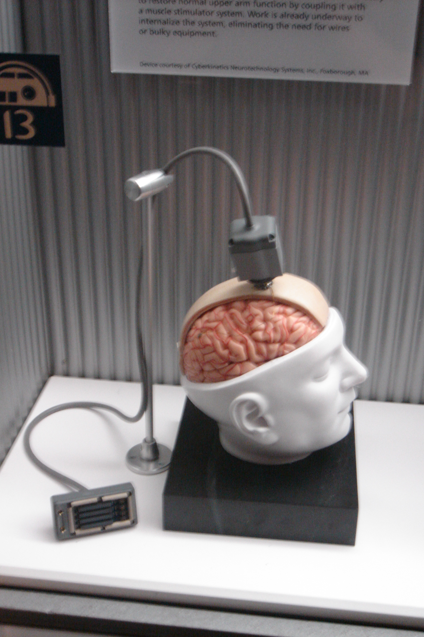
Implant technologies
Neurodevices are any devices used to monitor or regulate brain activity. Currently there are a few available for clinical use as a treatment for Parkinson’s disease. The most common neurodevices are deep brain stimulators (DBS) that are used to give electrical stimulation to areas stricken by inactivity.[13] Parkinson’s disease is known to be caused by an inactivation of the basal ganglia (nuclei) and recently DBS has become the more preferred form of treatment for Parkinson’s disease, although current research questions the efficiency of DBS for movement disorders.[13]
Neuromodulation is a relatively new field that combines the use of neurodevices and neurochemistry. The basis of this field is that the brain can be regulated using a number of different factors (metabolic, electrical stimulation, physiological) and that all these can be modulated by devices implanted in the neural network. While currently this field is still in the researcher phase, it represents a new type of technological integration in the field of neurotechnology. Neurotechnology
Cell therapy
Researchers have begun looking at uses for stem cells in the brain, which recently have been found in a few loci. A large number of studies are being done to determine if this form of therapy could be used in a large scale. Experiments have successfully used stem cells in the brains of children who suffered from injuries in gestation and elderly people with degenerative diseases in order to induce the brain to produce new cells and to make more connections between neurons.
Pharmaceuticals
Pharmaceuticals play a vital role in maintaining stable brain chemistry, and are the most commonly used neurotechnology by the general public and medicine. Drugs like sertraline, methylphenidate, and zolpidem act as chemical modulators in the brain, and they allow for normal activity in many people whose brains cannot act normally under physiological conditions. While pharmaceuticals are usually not mentioned and have their own field, the role of pharmaceuticals is perhaps the most far-reaching and commonplace in modern society (the focus on this article will largely ignore neuropharmaceuticals, for more information, see neuropsychopharmacology).
How these help study the brain
Magnetic resonance imaging is a vital tool in neurological research in showing activation in the brain as well as providing a comprehensive image of the brain being studied. While MRIs are used clinically for showing brain size, it still has relevance in the study of brains because it can be used to determine extent of injuries or deformation. These can have a significant effect on personality, sense perception, memory, higher order thinking, movement, and spatial understanding. However, current research tends to focus more so on fMRI or real-time functional MRI (rtfMRI).[14] These two methods allow the scientist or the participant, respectively, to view activation in the brain. This is incredibly vital in understanding how a person thinks and how their brain reacts to a person’s environment, as well as understanding how the brain works under various stressors or dysfunctions. Real-time functional MRI is a revolutionary tool available to neurologists and neuroscientists because patients can see how their brain reacts to stressors and can perceive visual feedback.[7] CT scans are very similar to MRI in their academic use because they can be used to image the brain upon injury, but they are more limited in perceptual feedback.[5] CTs are generally used in clinical studies far more than in academic studies, and are found far more often in a hospital than a research facility. PET scans are also finding more relevance in academia because they can be used to observe metabolic uptake of neurons, giving researchers a wider perspective about neural activity in the brain for a given condition.[8] Combinations of these methods can provide researchers with knowledge of both physiological and metabolic behaviors of loci in the brain and can be used to explain activation and deactivation of parts of the brain under specific conditions.
Transcranial magnetic stimulation is a relatively new method of studying how the brain functions and is used in many research labs focused on behavioral disorders and hallucinations. What makes TMS research so interesting in the neuroscience community is that it can target specific regions of the brain and shut them down or activate temporarily; thereby changing the way the brain behaves. Personality disorders can stem from a variety of external factors, but when the disorder stems from the circuitry of the brain TMS can be used to deactivate the circuitry. This can give rise to a number of responses, ranging from “normality” to something more unexpected, but current research is based on the theory that use of TMS could radically change treatment and perhaps act as a cure for personality disorders and hallucinations.[10] Currently, repetitive transcranial magnetic stimulation (rTMS) is being researched to see if this deactivation effect can be made more permanent in patients suffering from these disorders. Some techniques combine TMS and another scanning method such as EEG to get additional information about brain activity such as cortical response.[15]
Both EEG and MEG are currently being used to study the brain’s activity under different conditions. Each uses similar principles but allows researchers to examine individual regions of the brain, allowing isolation and potentially specific classification of active regions. As mentioned above, EEG is very useful in analysis of immobile patients, typically during the sleep cycle. While there are other types of research that utilize EEG,[15] EEG has been fundamental in understanding the resting brain during sleep.[11] There are other potential uses for EEG and MEG such as charting rehabilitation and improvement after trauma as well as testing neural conductivity in specific regions of epileptics or patients with personality disorders.
Neuromodulation can involve numerous technologies combined or used independently to achieve a desired effect in the brain. Gene and cell therapy are becoming more prevalent in research and clinical trials and these technologies could help stunt or even reverse disease progression in the central nervous system. Deep brain stimulation is currently used in many patients with movement disorders and is used to improve the quality of life in patients.[13] While deep brain stimulation is a method to study how the brain functions per se, it provides both surgeons and neurologists important information about how the brain works when certain small regions of the basal ganglia (nuclei) are stimulated by electrical currents.
Future technologies
The future of neurotechnologies lies in how they are fundamentally applied, and not so much on what new versions will be developed. Current technologies give a large amount of insight into the mind and how the brain functions, but basic research is still needed to demonstrate the more applied functions of these technologies. Currently, rtfMRI is being researched as a method for pain therapy. deCharms et al. have shown that there is a significant improvement in the way people perceive pain if they are made aware of how their brain is functioning while in pain. By providing direct and understandable feedback, researchers can help patients with chronic pain decrease their symptoms. This new type of bio/mechanical-feedback is a new development in pain therapy.[7] Functional MRI is also being considered for a number of more applicable uses outside of the clinic. Research has been done on testing the efficiency of mapping the brain in the case when someone lies as a new way to detect lying.[16] Along the same vein, EEG has been considered for use in lie detection as well.[17] TMS is being used in a variety of potential therapies for patients with personality disorders, epilepsy, PTSD, migraine, and other brain-firing disorders, but has been found to have varying clinical success for each condition.[10] The end result of such research would be to develop a method to alter the brain’s perception and firing and train patients’ brains to rewire permanently under inhibiting conditions (for more information see rTMS).[10] In addition, PET scans have been found to be 93% accurate in detecting Alzheimer’s disease nearly 3 years before conventional diagnosis, indicating that PET scanning is becoming more useful in both the laboratory and the clinic.[18]
Stem cell technologies are always salient both in the minds of the general public and scientists because of their large potential. Recent advances in stem cell research have allowed researchers to ethically pursue studies in nearly every facet of the body, which includes the brain. Research has shown that while most of the brain does not regenerate and is typically a very difficult environment to foster regeneration,[19] there are portions of the brain with regenerative capabilities (specifically the hippocampus and the olfactory bulbs).[20] Much of the research in central nervous system regeneration is how to overcome this poor regenerative quality of the brain. It is important to note that there are therapies that improve cognition and increase the amount of neural pathways,[2] but this does not mean that there is a proliferation of neural cells in the brain. Rather, it is called a plastic rewiring of the brain (plastic because it indicates malleability) and is considered a vital part of growth. Nevertheless, many problems in patients stem from death of neurons in the brain, and researchers in the field are striving to produce technologies that enable regeneration in patients with stroke, Parkinson’s diseases, severe trauma, and Alzheimer’s disease, as well as many others. While still in fledgling stages of development, researchers have recently begun making very interesting progress in attempting to treat these diseases. Researchers have recently successfully produced dopaminergic neurons for transplant in patients with Parkinson’s diseases with the hopes that they will be able to move again with a more steady supply of dopamine.[21][not in citation given] Many researchers are building scaffolds that could be transplanted into a patient with spinal cord trauma to present an environment that promotes growth of axons (portions of the cell attributed with transmission of electrical signals) so that patients unable to move or feel might be able to do so again.[22] The potentials are wide-ranging, but it is important to note that many of these therapies are still in the laboratory phase and are slowly being adapted in the clinic.[23] Some scientists remain skeptical with the development of the field, and warn that there is a much larger chance that electrical prosthesis will be developed to solve clinical problems such as hearing loss or paralysis before cell therapy is used in a clinic.[24][Need quotation to verify]
Novel drug delivery systems are being researched in order to improve the lives of those who struggle with brain disorders that might not be treated with stem cells, modulation, or rehabilitation. Pharmaceuticals play a very important role in society, and the brain has a very selective barrier that prevents some drugs from going from the blood to the brain. There are some diseases of the brain such as meningitis that require doctors to directly inject medicine into the spinal cord because the drug cannot cross the blood-brain barrier.[25] Research is being conducted to investigate new methods of targeting the brain using the blood supply, as it is much easier to inject into the blood than the spine. New technologies such as nanotechnology are being researched for selective drug delivery, but these technologies have problems as with any other. One of the major setbacks is that when a particle is too large, the patient’s liver will take up the particle and degrade it for excretion, but if the particle is too small there will not be enough drug in the particle to take effect.[26] In addition, the size of the capillary pore is important because too large a particle might not fit or even plug up the hole, preventing adequate supply of the drug to the brain.[26] Other research is involved in integrating a protein device between the layers to create a free-flowing gate that is unimpeded by the limitations of the body. Another direction is receptor-mediated transport, where receptors in the brain used to transport nutrients are manipulated to transport drugs across the blood-brain barrier.[27] Some have even suggested that focused ultrasound opens the blood-brain barrier momentarily and allows free passage of chemicals into the brain.[28] Ultimately the goal for drug delivery is to develop a method that maximizes the amount of drug in the loci with as little degraded in the blood stream as possible.
Neuromodulation is a technology currently used for patients with movement disorders, although research is currently being done to apply this technology to other disorders. Recently, a study was done on if DBS could improve depression with positive results, indicating that this technology might have potential as a therapy for multiple disorders in the brain.[24][Need quotation to verify] DBS is limited by its high cost however, and in developing countries the availability of DBS is very limited.[13] A new version of DBS is under investigation and has developed into the novel field, optogenetics.[23] Optogenetics is the combination of deep brain stimulation with fiber optics and gene therapy. Essentially, the fiber optic cables are designed to light up under electrical stimulation, and a protein would be added to a neuron via gene therapy to excite it under light stimuli.[29] So by combining these three independent fields, a surgeon could excite a single and specific neuron in order to help treat a patient with some disorder. Neuromodulation offers a wide degree of therapy for many patients, but due to the nature of the disorders it is currently used to treat its effects are often temporary. Future goals in the field hope to alleviate that problem by increasing the years of effect until DBS can be used for the remainder of the patient’s life. Another use for neuromodulation would be in building neuro-interface prosthetic devices that would allow quadriplegics the ability to maneuver a cursor on a screen with their thoughts, thereby increasing their ability to interact with others around them. By understanding the motor cortex and understanding how the brain signals motion, it is possible to emulate this response on a computer screen.[30]
Ethics
Stem cells
The ethical debate about use of embryonic stem cells has stirred controversy both in the United States and abroad; although more recently these debates have lessened due to modern advances in creating induced pluripotent stem cells from adult cells. The greatest advantage for use of embryonic stem cells is the fact that they can differentiate (become) nearly any type of cell provided the right conditions and signals. However, recent advances by Shinya Yamanaka et al. have found ways to create pluripotent cells without the use of such controversial cell cultures.[31] Using the patient’s own cells and re-differentiating them into the desired cell type bypasses not only the fear of patient rejection of the cells but also gives researchers a more ethical (and larger) supply of available cells. Induced pluripotent cells are by no means perfect though, they still have the potential to form teratomas, or benign (though they can be potentially malignant in effect) tumors, and tend to have poor survivability in vivo (in the living body) on damaged tissue.[32] Much of the ethics concerning use of stem cells has subsided from the embryonic/adult stem cell debate due to its rendered moot, but now societies find themselves debating whether or not this technology can be ethically used. Enhancements of traits, use of animals for tissue scaffolding, and even arguments for moral degeneration have been made with the fears that if this technology reaches its full potential a new paradigm shift will occur in human behavior.
Military application
New neurotechnologies have always garnered the appeal of governments, from lie detection technology and virtual reality to rehabilitation and understanding the psyche. Due to the Iraq War and War on Terror, American soldiers coming back from Iraq and Afghanistan are reported to have percentages up to 12% with PTSD.[33] There are many researchers hoping to improve these peoples’ conditions by implementing new strategies for recovery. By combining pharmaceuticals and neurotechnologies, some researchers how found ways to low the fear response and theorize that it may be applicable to PTSD.[34] Virtual reality is also a technology that has found a large amount of attention in the military. If improved upon, it would be possible to train soldiers in complex situations in times of peace in order to better prepare and train a modern army. But some people raise questions about how the military would use these improvements, and while the scope of this article is about the technologies it is important to look into how these new technologies would be used.
Privacy
Finally, when these technologies are being developed society must understand that these neurotechnologies could reveal the one thing that people can always keep secret, what they are thinking. While there are large amounts of benefits associated with these technologies, it is necessary for scientists and policy makers alike to consider implications about “cognitive liberty.”[35] This term is important in many ethical circles concerned with the state and goals of progress in the field of neurotechnology (see Neuroethics). Current improvements such as “brain fingerprinting” or lie detection using EEG or fMRI could give rise to a set fixure of loci/emotional relationships in the brain, although these technologies are still years away from full application.[35] It is important to consider how all these neurotechnologies might affect the future of society, and it is suggested that political, scientific, and civil debates are heard about the implementation of these newer technologies that potentially offer a new wealth of once-private information.[35] Some ethicists are also concerned with the use of TMS and fear that the technique could be used to alter patients in ways that are undesired by the patient.[10]
See also
- Neuroscience
- Neuroengineering
Footnotes
- ^ Nintendo Company of America. BrainAge (2006). Based on the work of Ryuta Kawashima, M.D.
- ^ a b Sarah H. Broman; Jack Fletcher (1999). The changing nervous system: neurobehavioral consequences of early brain disorders. Oxford University Press US. ISBN 9780195121933. http://books.google.com/books?id=paxE8PFrsLEC.
- ^ Doidge, Norman (2007). The Brain That Changes Itself: Stories of Personal Triumph from the Frontiers of Brain Science. Viking Adult. ISBN 978-0670038305.
- ^ "The Decade of the Mind". http://www.dom2009.de/.
- ^ a b c Purves, Dale (2007). Neuroscience, Fourth Edition. Sinauer Associates, Inc.. p. 19. ISBN 978-0878936977.
- ^ Purves, Dale (2007). Neuroscience, Fourth Edition. Sinauer Associates, Inc.. p. 24. ISBN 978-0878936977.
- ^ a b c Decharms, R. C.; Maeda, F.; Glover, G. H.; Ludlow, D.; Pauly, J. M.; Soneji, D.; Gabrieli, J. D. E.; MacKey, S. C. (2005). "Control over brain activation and pain learned by using real-time functional MRI". Proceedings of the National Academy of Sciences 102 (51): 18626–31. Bibcode 2005PNAS..10218626D. doi:10.1073/pnas.0505210102. PMC 1311906. PMID 16352728. http://www.pubmedcentral.nih.gov/articlerender.fcgi?tool=pmcentrez&artid=1311906.
- ^ a b Purves, Dale (2007). Neuroscience, Fourth Edition. Sinauer Associates, Inc.. p. 20. ISBN 978-0878936977.
- ^ Wasserman, E.M. (1996)
- ^ a b c d e Illes, J; Gallo, M; Kirschen, MP (2006). "An ethics perspective on transcranial magnetic stimulation (TMS) and human neuromodulation". Behavioural neurology 17 (3-4): 149–57. PMID 17148834.
- ^ a b Purves, Dale (2007). Neuroscience, Fourth Edition. Sinauer Associates, Inc.. p. 715. ISBN 978-0878936977.
- ^ Hämäläinen, M. (November 2007). "Magnetoencephalography (MEG)". Athinoula A. Martinos Center for Biomedical Imaging. http://www.nmr.mgh.harvard.edu/martinos/research/technologiesMEG.
- ^ a b c d Gross, R. (2008). "What Happened to Posteroventral Pallidotomy for Parkinson’s Disease and Dystonia?". Neurotherapeutics 5 (2): 281–293. doi:10.1016/j.nurt.2008.02.001. PMID 18394570.
- ^ Cox, RW; Jesmanowicz, A; Hyde, JS (1995). "Real-time functional magnetic resonance imaging". Magnetic resonance in medicine : official journal of the Society of Magnetic Resonance in Medicine / Society of Magnetic Resonance in Medicine 33 (2): 230–6. PMID 7707914.
- ^ a b Veniero, D.; Bortoletto, M.; Miniussi, C. (2009). "TMS-EEG co-registration: on TMS-induced artifact". Clinical Neurophysiology 120 (7): 1392. doi:10.1016/j.clinph.2009.04.023. PMID 19535291.
- ^ Langleben, D.; Schroeder, L.; Maldjian, J.; Gur, R.; McDonald, S.; Ragland, J.; O'Brien, C.; Childress, A. (2002). "Brain Activity during Simulated Deception: an Event-Related Functional Magnetic Resonance Study". NeuroImage 15 (3): 727–732. doi:10.1006/nimg.2001.1003. PMID 11848716.
- ^ Farwell, LA; Smith, SS (2001). "Using brain MERMER testing to detect knowledge despite efforts to conceal". Journal of forensic sciences 46 (1): 135–43. PMID 11210899.
- ^ Mosconi, L., et al. (2005)
- ^ Sur, M.; Rubenstein, J. L. R. (2005). "Patterning and Plasticity of the Cerebral Cortex". Science 310 (5749): 805. Bibcode 2005Sci...310..805S. doi:10.1126/science.1112070. PMID 16272112.
- ^ Eriksson, P. S.; Perfilieva, E.; Björk-Eriksson, T.; Alborn, A. M.; Nordborg, C.; Peterson, D. A.; Gage, F. H. (1998). "Neurogenesis in the adult human hippocampus". Nature Medicine 4 (11): 1313. doi:10.1038/3305. PMID 9809557.
- ^ Sacchetti, P.; Sousa, K. M.; Hall, A. C.; Liste, I.; Steffensen, K. R.; Theofilopoulos, S.; Parish, C. L.; Hazenberg, C. et al. (2009). "Liver X Receptors and Oxysterols Promote Ventral Midbrain Neurogenesis in Vivo and in Human Embryonic Stem Cells". Cell Stem Cell 5 (4): 409. doi:10.1016/j.stem.2009.08.019. PMID 19796621.
- ^ Sharp, J.; Keirstead, H.; University of California, Irvine (November 10, 2009). "Embryonic Stem Cell Therapy Restores Walking Ability In Rats With Neck Injuries". ScienceDaily. http://www.sciencedaily.com/releases/2009/11/091109121345.htm. Retrieved November 24, 2009.
- ^ a b Lynch, Z. (2009). "The future of neurotechnology innovation". Epilepsy & Behavior 15: 120–127. doi:10.1016/j.yebeh.2009.03.030.
- ^ a b Personal correspondence with Dr. Robert Gross
- ^ Ala'Aldeen, D.; University of Nottingham (May 15, 2009). "Breakthrough In The Treatment Of Bacterial Meningitis". ScienceDaily. http://www.sciencedaily.com/releases/2009/05/090513121048.htm. Retrieved November 24, 2009.
- ^ a b Tsuji, J. S.; Maynard, A. D.; Howard, P. C.; James, J. T.; Lam, C. -W.; Warheit, D. B.; Santamaria, A. B. (2005). "Research Strategies for Safety Evaluation of Nanomaterials, Part IV: Risk Assessment of Nanoparticles". Toxicological Sciences 89 (1): 42. doi:10.1093/toxsci/kfi339. PMID 16177233.
- ^ Demeule, M.; Currie, J. C.; Bertrand, Y.; Ché, C.; Nguyen, T.; Régina, A.; Gabathuler, R.; Castaigne, J. P. et al. (2008). "Involvement of the low-density lipoprotein receptor-related protein in the transcytosis of the brain delivery vector Angiopep-2". Journal of Neurochemistry 106 (4): 1534. doi:10.1111/j.1471-4159.2008.05492.x. PMID 18489712.
- ^ Hynynen, K.; McDannold, N.; Vykhodtseva, N.; Raymond, S.; Weissleder, R.; Jolesz, F. A.; Sheikov, N. (2006). "Focal disruption of the blood–brain barrier due to 260-kHz ultrasound bursts: a method for molecular imaging and targeted drug delivery". Journal of Neurosurgery 105 (3): 445. doi:10.3171/jns.2006.105.3.445. PMID 16961141.
- ^ Adamantidis, A. R.; Zhang, F.; Aravanis, A. M.; Deisseroth, K.; De Lecea, L. (2007). "Neural substrates of awakening probed with optogenetic control of hypocretin neurons". Nature 450 (7168): 420–4. Bibcode 2007Natur.450..420A. doi:10.1038/nature06310. PMID 17943086.
- ^ Hochberg, L. R.; Serruya, M. D.; Friehs, G. M.; Mukand, J. A.; Saleh, M.; Caplan, A. H.; Branner, A.; Chen, D. et al. (2006). "Neuronal ensemble control of prosthetic devices by a human with tetraplegia". Nature 442 (7099): 164–71. Bibcode 2006Natur.442..164H. doi:10.1038/nature04970. PMID 16838014.
- ^ Takahashi, K.; Yamanaka, S. (2006). "Induction of Pluripotent Stem Cells from Mouse Embryonic and Adult Fibroblast Cultures by Defined Factors". Cell 126 (4): 663. doi:10.1016/j.cell.2006.07.024. PMID 16904174.
- ^ Laflamme, M. A.; Chen, K. Y.; Naumova, A. V.; Muskheli, V.; Fugate, J. A.; Dupras, S. K.; Reinecke, H.; Xu, C. et al. (2007). "Cardiomyocytes derived from human embryonic stem cells in pro-survival factors enhance function of infarcted rat hearts". Nature Biotechnology 25 (9): 1015. doi:10.1038/nbt1327. PMID 17721512.
- ^ "National Center for PTSD Home". National Center for PTSD. http://www.ptsd.va.gov/.
- ^ Ressler, K. J.; Rothbaum, B. O.; Tannenbaum, L.; Anderson, P.; Graap, K.; Zimand, E.; Hodges, L.; Davis, M. (2004). "Cognitive Enhancers as Adjuncts to Psychotherapy: Use of D-Cycloserine in Phobic Individuals to Facilitate Extinction of Fear". Archives of General Psychiatry 61 (11): 1136. doi:10.1001/archpsyc.61.11.1136. PMID 15520361.
- ^ a b c Wolpe, P.; Foster, K.; Langleben, D. (2005). "Emerging Neurotechnologies for Lie-Detection: Promises and Perils". American Journal of Bioethics 5 (2): 39. doi:10.1080/15265160590923367. PMID 16036700.
References
- Mosconi, L.; Brys, M.; Glodziksobanska, L.; Desanti, S.; Rusinek, H.; Deleon, M. (2007). "Early detection of Alzheimer’s disease using neuroimaging". Experimental Gerontology 42 (1-2): 129. doi:10.1016/j.exger.2006.05.016. PMID 16839732.
- Wassermann, EM (1998). "Risk and safety of repetitive transcranial magnetic stimulation: report and suggested guidelines from the International Workshop on the Safety of Repetitive Transcranial Magnetic Stimulation, June 5-7, 1996". Electroencephalography and clinical neurophysiology 108 (1): 1–16. doi:10.1016/S0168-5597(97)00096-8. PMID 9474057.
- Gross, R., Emory University Dept. of Neurosurgery, interviewed by C. Stone, Oct. 6, 2009.
Brain–computer interface Technologies Biomechatronics · Brain implant · BrainGate · Brainport · Cyberware · Exocortex · Intelligence amplification · Isolated brain · Neuroprosthetics · Neurotechnology · Optogenetics · Sensory substitution · Synthetic telepathy
Scientific phenomena Disciplines Speculative People Charles Stross · Douglas Engelbart · Hugh Herr · J. C. R. Licklider · Kevin Warwick · Matt Nagle · Merlin Donald · Miguel Nicolelis · Peter Kyberd · Steve Mann · Vernor Vinge · Yoky Matsuoka · Edward BoydenOther Categories:
Wikimedia Foundation. 2010.